Abdominal nuclear medicine emergencies
Applied Radiology — Vol. 35 , Issue 2 , pp. 25 -28
DOI: 10.37549/AR1405
Published: February 1, 2006
Categories
Generally, there are 4 major nuclear medicine applications in the setting of abdominal emergency, as listed in Table 1. Nuclear medicine has certain unique applications that can help solve emergency problems that may be difficult to resolve with radiologic imaging. After first encountering such cases, a radiologist must keep in mind potential nuclear medicine applications and inform the technologists, as some nuclear tests require early patient preparation and in vitro work.
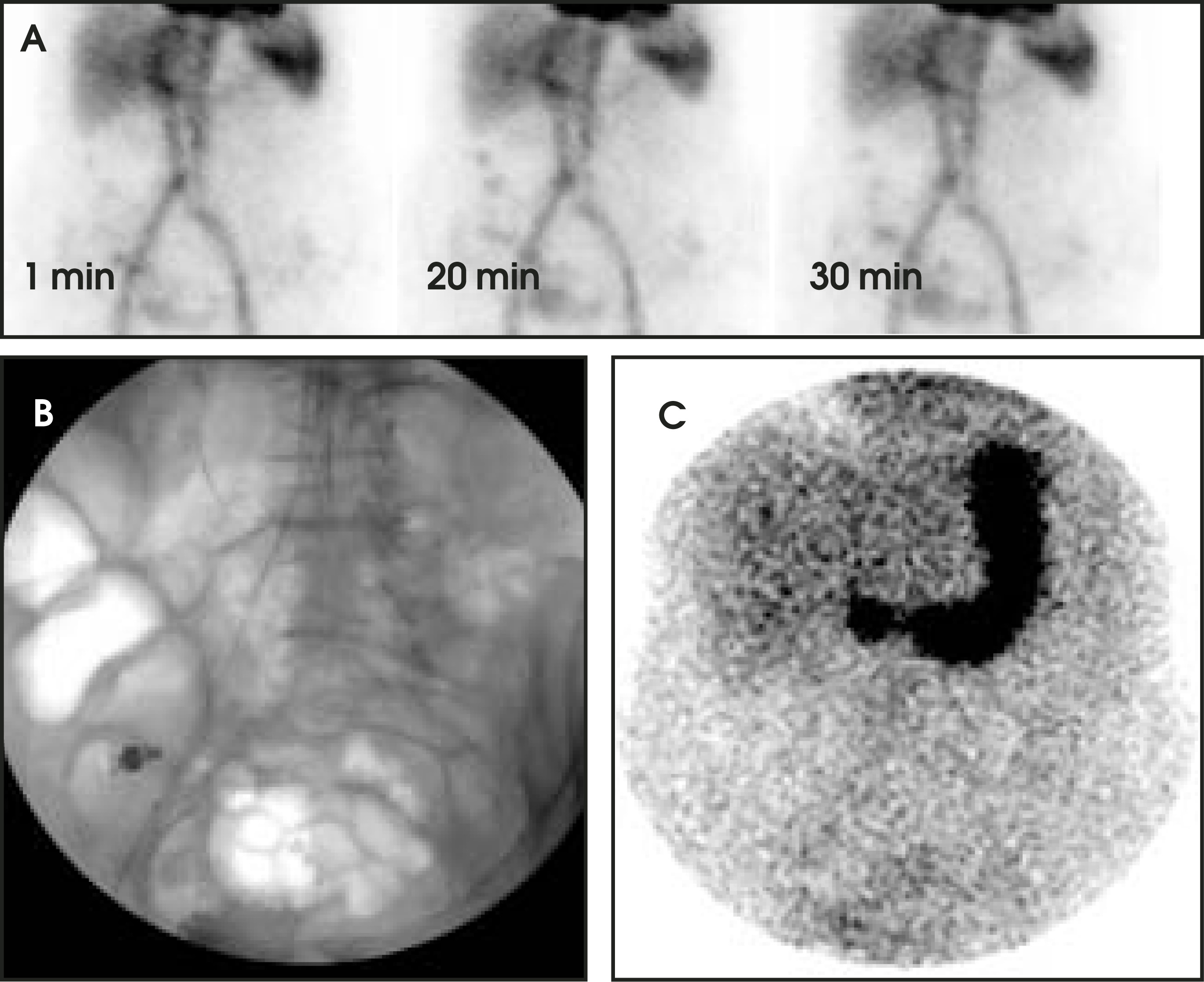
RBC GI bleeding study Angiographic and scintigraphic GI bleeding study
Gastrointestinal (GI) bleeding is a serious emergency, which carries a 2% to 10% mortality rate, depending on the site and nature of hemorrhage. Angiography can be diagnostic and therapeutic for GI bleeding but only if there is an active hemorrhage at the time of imaging. Because GI bleeding is often intermittent and the clinical signs of active bleeding are often not reliable or may develop after the hemorrhage has ceased, the tagged red cell scintigram has become an important prescreening tool before angiography. The scintigram has the advantage of being noninvasive and offering flexible monitoring time, which is more likely to catch the active bleeding, even if it is intermittent.1 In addition, imaging at delayed times can be performed, since the red blood cell (RBC) label is stable for up to 24 hours. It has been reported that a blood urea nitrogen (BUN)/creatinine (Cr) ratio ≥25 (in people without renal insufficiency) may indicate a delayed image is more likely to be positive than it is in those patients with lower ratios.2
The interventional radiologist may request a nuclear study before angiography is performed to confirm active bleeding and to determine the general location and severity of the hemorrhage, which can facilitate the angiography procedure. A nuclear GI bleeding scan can detect approximately 0.1 mL/min bleeding,3 which is approximately a 5-fold lower rate than that of angiography (0.5 mL/min). The yield of positive angiographic findngs after a positive scintigram is approximately 50%.4
It is difficult to compare the 2 tests directly, since it is impossible to perform the tests simultaneously and bleeding is often intermittent. W h e n the nuclear scan is barely positive, the angiography is less likely to be positive; but it can be, occasionally, when there is another episode of bleeding. A positive scintigram increases the likelihood of a positive angiogram from 22% to 53%.4 For hemodynamically unstable patients, such as those with systolic blood pressure <100 mm Hg,5 angiography should be done immediately. Otherwise, all patients should initially be considered to undergo nuclear medicine imaging.
Pearls and pitfalls in nuclear GI bleeding study
Pearl: When initial imaging is negative, the patient can be reimaged for up to 24 hours after the initial study without the need to relabel RBCs, which can save up to 1 hour of labeling time. It is also a convenient technique to monitor slow intermittent bleeding.
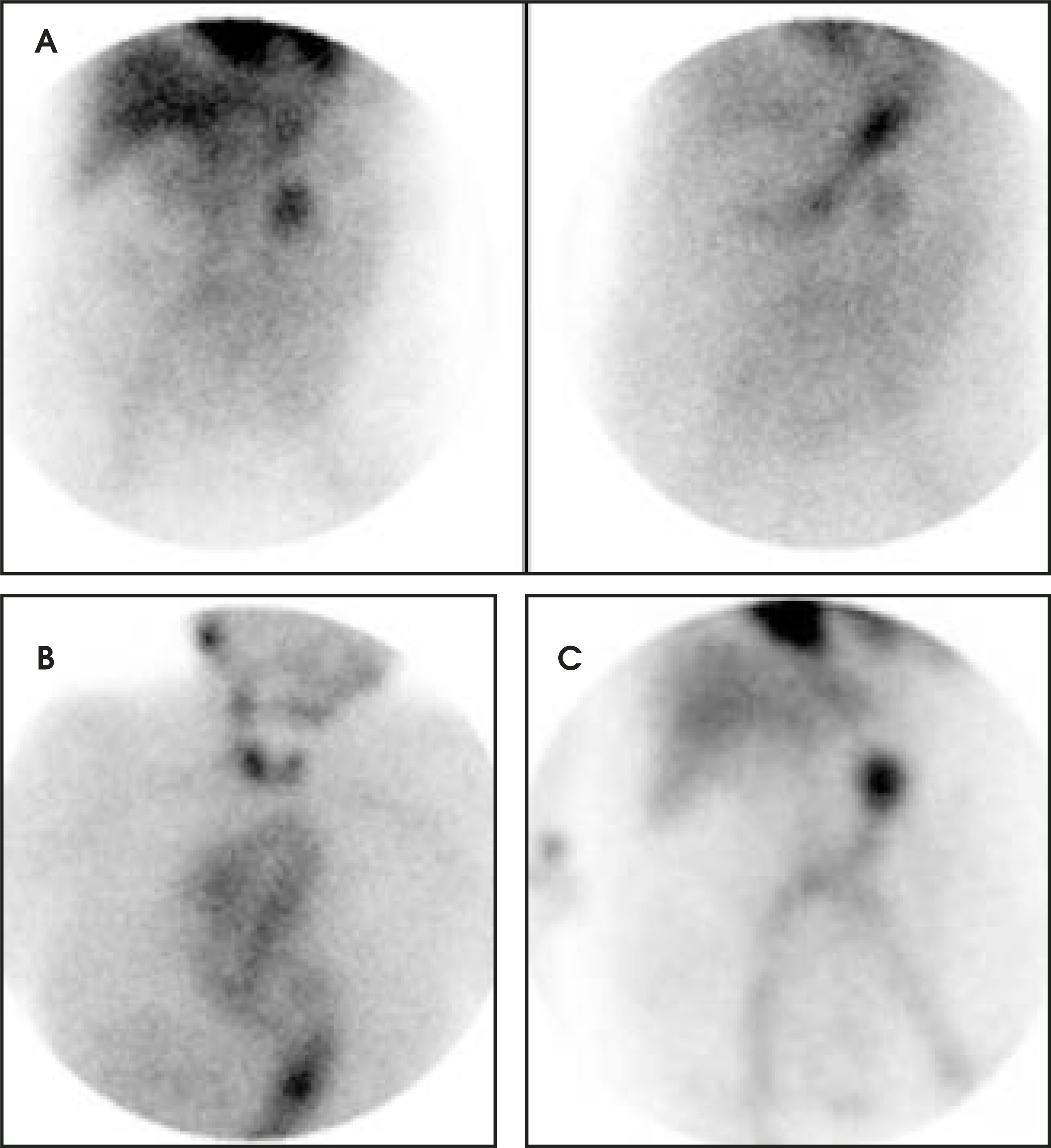
Pitfall: Free pertechnetate contamination in the RBC labeling (Figure 2) is a potential pitfall of this study. When there is gastric tracer uptake, there is a possibility of free technetium (Tc)-99m labeling of the stomach, which may mimic the appearance of gastric bleeding. Since free Tc 99m physiologically labels both the thyroid and stomach, a spot view of the neck should be obtained to evaluate for thyroid uptake. If there is thyroid uptake, then free Tc-99m is the cause of the gastric uptake.
WBC labeling for appendicitis
White blood cell (WBC) labeling with Tc-99m-labeled hexamethylpropylene amine oxime (HMPAO) had proved to be highly sensitive for excluding appendicitis.6 However, clinically, the test is unpopular because it is time-consuming (4 to 24 hours) and requires blood-sample handling.
NeutroSpec (Tyco Healthcare/ Mallinckrodt, St. Louis, MO) overcame the disadvantage of Tc - 99m-HMPAO but maintained specificity and sensitivity.7 NeutroSpec is a monoclonal antibody that radiolabels WBCs. Until it was voluntarily suspended from the market on December 19, 2005 because of Food and Drug Administration safety concerns, NeutroSpec was used for scintigraphic imaging in cases of equivocal appendicitis. The target antigen (CD15) is expressed on the surface of neutrophils, eosinophils, and monocytes. The antibody is labeled with Tc-99m, the most common nuclear medicine agent. At the time this article went to press, it was unknown whether NeutroSpec might be reintroduced to the market in the future or if another agent for this application might be in development.
HIDA scan for acute cholecystitis
The iminodiacetic acid derivatives labeled with Tc-99m have high extraction efficiency in the liver, even with elevated levels of bilirubin. The hepatobiliary iminodiacetic acid (HIDA) scan remains the gold standard test for acute cholecystitis.
Acute cholecystitis usually occurs with right-upper-quadrant pain and tenderness. Fever is seldom higher than 38°C. Ultrasound is usually the first imaging choice, but the main features of cholecystitis on ultrasonography are all nonspecific. Before the HIDA scan is performed, 3 important questions must be asked:
-
1
What was the patient’s last meal and when was it eaten?
-
2
Is the patient receiving narcotics for pain?
-
3
Are the symptoms acute or chronic?
The HIDA scan is very sensitive with very few false-negative results, but false-positive results (lack of gallbladder visualization) are possible with the following circumstances:
-
Meal eaten within 4 hours of the study (completely empty gallbladder)8
-
Prolonged fasting of >24 hours before the study (full gallbladder)8
-
Total parenteral nutrition (full gallbladder)9
-
Acute pancreatitis10
-
Hepatitis9
-
Chronic cholecystitis11
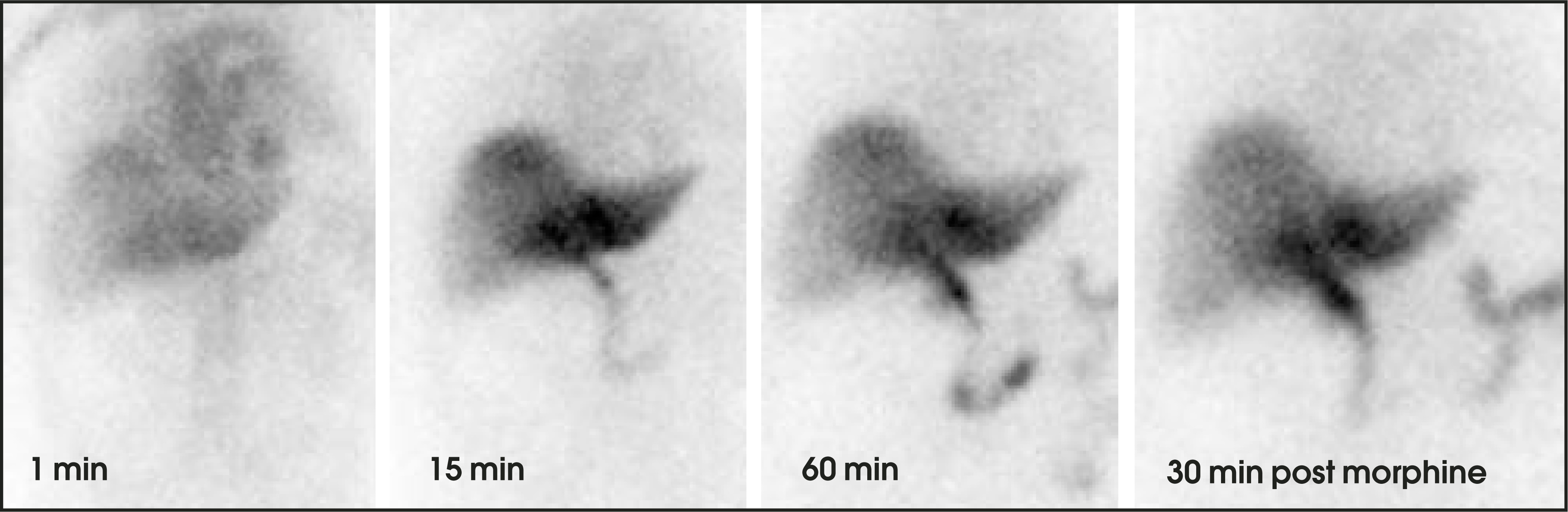
Acalculous cholecystitis (AC) may also cause false-negative findings (gallbladder visualized).12 Acalculous cholecystitis occurs more often in children (50%), in the elderly, and in patients who are critically ill with severe trauma, burns, or following surgery. Ultrasound and other anatomic imaging techniques are not sensitive for the diagnosis. When clinical suspicion of chronic AC is high, cholecystokinin (CCK)-augmented cholescintigraphy with measurement of gallbladder ejection fraction at 1 hour is the technique of choice. These patients are also treated by means of elective cholecystectomy.Augmentation with CCK before the emergency scan (to empty a sludge-filled gallbladder) in patients with acute pain suggestive of cholecystitis who have had a long nothing-by-mouth status or who have been on total parenteral alimentation and/or who have been injected with morphine sulfate during the scan at approximately 1 hour can improve the sensitivity and specificity of imaging by manipulating the sphincter of Oddi (which tightens with morphine and relaxes with CCK) and contractility (Figure 3).
Renal scan for urinary obstruction
If early in its course, acute urinary obstruction may still show a normal collecting system on ultrasound and CT. Also, many patients who have dilated systems may not be functionally obstructed. When clinical suspicion is high and CT or ultrasound has failed to show a “culprit” stone or other convincing evidence to explain the symptoms, emergency renal scans should be performed to assess differential renal function and functional obstruction.
Tc-99m mercaptoacetyltriglycine (MAG3) is the most popular radiotracer used for this purpose. Frequent use of diuretic enhancement also assesses for functional obstruction under conditions of high urinary flo w. Adequate oral or intravenous hydration is usually key to a successful renal scan. Even people with modest renal insufficiency can be studied with diuretic renography, although the furosemide dose may need to be altered in the setting of an elevated serum creatinine level.
Conclusion
As nuclear medicine emphasizes function over anatomy in both physiology and pathophysiology, it plays a complementary role to other diagnostic imaging methods in acute abdominal emergencies. This article has briefly covered the following uses: GI bleeding scintigraphy with Tc 99m RBCs, appendicitis imaging with Tc-99m HMPAO WBCs, and cholescintigraphy with Tc-99m MAG3 for acute urnary obstruction. Pharmacologic interventions with drugs (such as morphine, CCK, and furosemide) enhance the sensitivity and specificity of certain tests. The unfortunate fate of NeutroSpec has recently been noted, but the authors are hopeful that new radiopharmaceutical development will lead to safe, sensitive, and cost-effective scans for acute abdominal processes. Nuclear medicine can help fulfill the needs of a 24-7-365 hospital.
References
- O’Neill B, Gosnell J, Lull R. Cinematic nuclear scintigraphy reliably directs surgical intervention for patients with gastrointestinal bleeding. Arch Surg. 2000;135.
- Lewis D, Jacoboson A. BUN/creatinine ratios: Aid to decision making about delayed imaging in Tc-99m red blood cell scans for gastrointestinal hemorrhage. Clin Ncul Med. 1998;23:201-204.
- Smith R, Copely D, Bolen F. 99mTc RBC scintigraphy: Correlation of gastrointestinal bleeding rates with scintigraphic findings. AJR Am J Roentgenol. 1987;148:869-874.
- Gunderman R, Leef J, Ong K. Scintigraphic screening prior to visceral arteriography in acute lower gastrointestinal bleeding. J Nucl Med. 1998;39:1081-1083.
- Nicholson A, Ettles D, Hartley J. Transcatheter coil embolotherapy: A safe and effective option for major colonic hemorrhage. Gut. 1998;43:79-84.
- Rypins E, Evans D, Hinrichs W, Kipper S. Tc-99m-HMPAO white blood cell scan for diagnosis of acute appendicitis in patients with equivocal clinical presentation. Ann Surg. 1997;226:58-65.
- Rypins E, Kipper S, Weiland F. 99m Tc anti-CD 15 monoclonal antibody (LeuTech) imaging improves diagnostic accuracy and clinical management in patients with equivocal presentation of appendicitis. Ann Surg. 2002;235:232-239.
- Balon H, Fink-Bennett D, Brill D. Procedure guideline for hepatobiliary scintigraphy. Society of Nuclear Medicine. J Nucl Med. 1997;38:1654-1657.
- Shuman W, Gibbs P, Rudd T, Mack L. PIPIDA scintigraphy for cholecystitis: False positives in alcoholism and total parenteral nutrition. AJR Am J Roentgenol. 1982;138:1-5.
- Edlund G, Kempi V, van der Linden W. Transient nonvisualization of the gallbladder by Tc-99m HIDA cholescintigraphy in acute pancreatitis: Concise communication. J Nucl Med. 1982;23:117-20.
- Klingensmith W, Turner W. Cholescintigraphy for acute cholecystitis: False positive results caused by chronic cholecystitis. Gastrointest Radiol. 1990;15:129-132.
- Mariat G, Mahul P, Prevt N. Contribution of ultrasonography and cholescintigraphy to the diagnosis of acute acalculous cholecystitis in intensive care unit patients. Intensive Care Med. 2000;26:1658-1663.
Citation
. Abdominal nuclear medicine emergencies. Applied Radiology. 2006;35(2):25-28. doi:10.37549/AR1405.