Adult Cervical Embryonal Rhabdomyosarcoma
Applied Radiology — Vol. 55 , Issue 4
Published: January 29, 2026
1 Department of Radiology, Walter Reed National Military Medical Center, Bethesda, Maryland
2 Nova Southeastern University Dr. Kiran C. Patel College of Osteopathic Medicine, Clearwater, Florida
3 American College of Radiology Institute for Radiologic Pathology, Silver Spring, Maryland
* Corresponding author: M. Grant Liska (M.grant.liska1@gmail.com)
Categories
Case Summary
An adult patient presented with complaints of irregular vaginal bleeding with pelvic fullness and pain. Physical examination demonstrated a friable endovaginal soft-tissue mass, clinically presumed to be uterine prolapse.
The patient underwent a total abdominal hysterectomy and bilateral oophorectomy. Surgical pathology revealed cervical and uterine myoinvasion with no evidence of metastatic disease, consistent with stage IC according to the International Federation of Gynecology and Obstetrics (FIGO) Staging System for Adenosarcoma of Uterine Corpus, an unofficial staging system in this case. Following gross-total resection, the patient underwent four cycles of systemic chemotherapy, and the most recent postoperative imaging showed no evidence of disease recurrence.
Imaging Findings
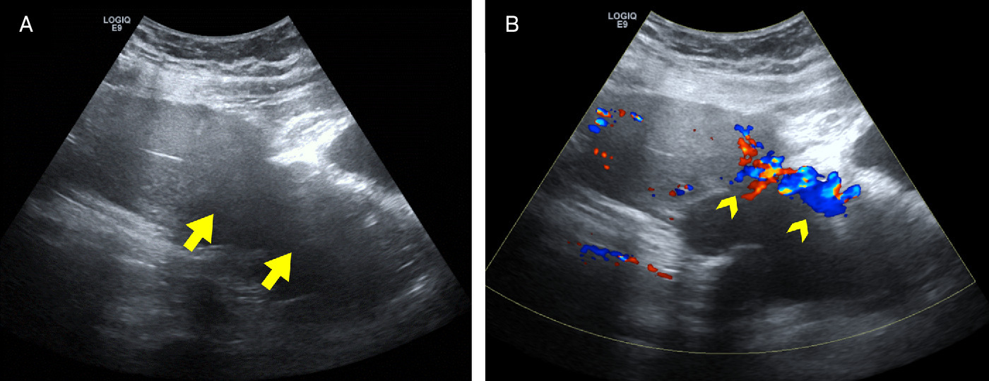
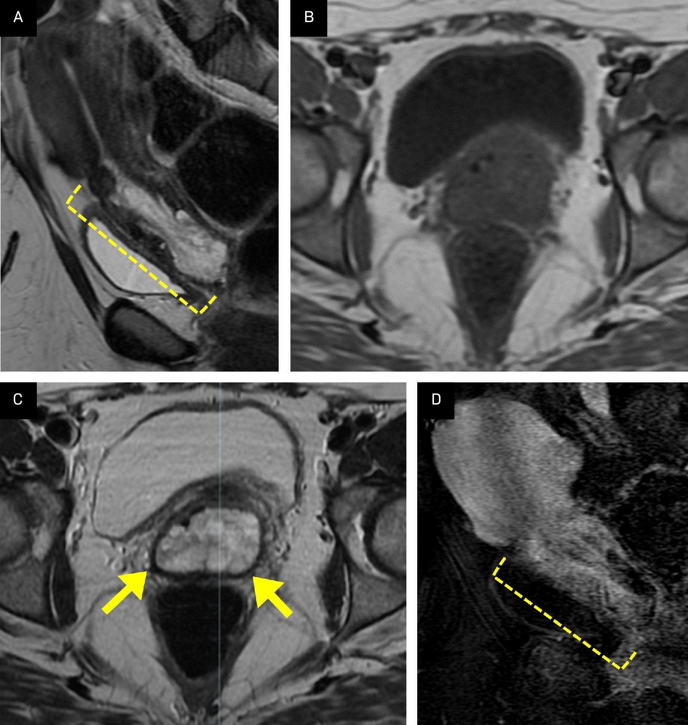
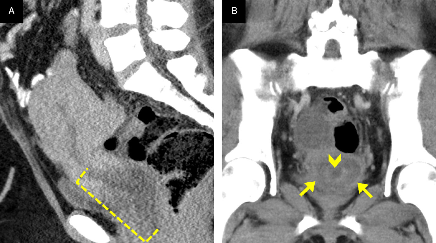
Initial imaging with transabdominal US demonstrated an amorphous hypoechoic endocervical mass with significant vascularity (Figure 1). A follow-up MRI revealed a 9.0 cm lobular T1 hypointense, T2 hyperintense mass originating from the upper cervix/lower uterine segment border and protruding into the vaginal cavity (Figure 2). The mass demonstrated heterogeneous enhancement on T1 post-contrast images (Figure 2), as well as significant diffusion restriction (not shown). There was no convincing evidence of local invasion. On preoperative CT images, the mass appeared as an ill-defined hypoattenuating region causing cervical canal dilation and filling of the vaginal cavity (Figure 3).
Diagnosis
Cervical embryonal rhabdomyosarcoma (ERMS), stage IC.
Differential considerations include uterine prolapse, pedunculated cervical leiomyoma, endometrial polyp, cervical adenoma malignum, and other primary cervical malignancies.
Discussion
Rhabdomyosarcoma is a malignant mesenchymal tumor characterized by the presence of rhabdomyoblasts, immature skeletal muscle cells that fail to fully differentiate.1 Among the various histological subtypes, ERMS is the most prevalent, accounting for roughly 50% of cases and affecting the genitourinary tract in 18-22% of cases.2 ERMS typically affects infants, particularly in the vaginal area.3 Cervical ERMS is a rare form of the disease, more commonly seen in adolescents during the 2nd decade of life.4 Presentations of this entity in adulthood are even less common and are linked to somatic mutations in the DICER1 gene. Germline mutations in the DICER1 gene are frequently seen in pediatric patients with ERMS, which is associated with a number of other solid tumors.5
Cervical ERMS tumors in adults tend to be quite large at diagnosis, likely owing to their similar appearance to more common, benign entities such as uterine prolapse, endocervical fibroids, and cervical polyps on pelvic exam and US. Distinctive imaging features of this entity are helpful in making the correct diagnosis, with tumors having high water content (hypoattenuating on CT, T2 hyperintense on MRI) and often assuming a polypoid morphology likened to a bunch of grapes (sarcoma botryoides).5,6 These features are not pathognomonic, though, and ultimately, tissue sampling is necessary for a definitive diagnosis.
There is no official classification of cervical ERMS; however, based on the largest study of cervical ERMS, the FIGO uterine adenosarcoma staging system was found to be superior to the uterine corpus sarcoma staging system for predicting survival.5,7 Using this system, this patient’s disease was classified as stage IC owing to surgical pathology’s demonstration of greater than 50% myometrial/cervical invasion without extrauterine extension or metastatic disease.
Treatment guidelines are based on data from pediatric populations but have demonstrated similar efficacy in case reports of adult patients with cervical ERMS.8 The prognosis for localized ERMS, including cervical cases, is generally favorable, particularly when early diagnosis and an aggressive surgical approach are achieved.3-5,9,10 Following gross total resection, this patient underwent 4 cycles of vincristine, actinomycin, and cyclophosphamide therapy, with no imaging evidence of disease recurrence to date.
Conclusion
ERMS is a malignancy typically seen in children. Infrequently, this tumor can occur in the cervix of adolescents and, even more rarely, present as an adult cervical cancer. Familiarity with this entity and its distinctive imaging features on MRI is critical to accurate, timely diagnosis in the adult population.
References
- Liptak J, Forrest L, Withrow S, Vail D, Page R. Withrow and MacEwen’s small animal clinical oncology. 2013:356-380. doi:10.1016/B978-1-4377-2362-5.00021-9.
- Castle J, Levy B, Allison D, Rodeberg D, Rellinger E. Pediatric rhabdomyosarcomas of the genitourinary tract. Cancers. 2023;15(10):2864. doi:10.3390/cancers15102864.
- Kaserer K, Kainz C, Reinthaller A. Sarcoma botryoides (embryonales rhabdomyosarkom) als ungewöhnliche diagnose eines zervixpolypen [sarcoma botryoides (embryonal rhabdomyosarcoma) as an unusual diagnosis of cervix polyps]. Pathologe. 1995;16(4):287-291. doi:10.1007/s002920050104.
- Zeisler H, Mayerhofer K, Joura E. Embryonal rhabdomyosarcoma of the uterine cervix: case report and review of the literature. Gynecol Oncol. 1998;69(1):78-83. doi:10.1006/gyno.1998.4962.
- Devins K, Young R, Ghioni M. Embryonal rhabdomyosarcoma of the uterine cervix: a clinicopathologic study of 94 cases emphasizing issues in differential diagnosis staging, and prognostic factors. Am J Surg Pathol. 2022;46(11):1477-1489. doi:10.1097/PAS.0000000000001933.
- Dehner L, Jarzembowski J, Hill D. Embryonal rhabdomyosarcoma of the uterine cervix: a report of 14 cases and a discussion of its unusual clinicopathological associations. Mod Pathol. 2012;25(4):602-614. doi:10.1038/modpathol.2011.185.
- .
- Ibrahim U, Saqib A, Mohammad F. Embryonal rhabdomyosarcoma of the cervix: a rare disease at an uncommon age. Cureus;9(11). doi:10.7759/cureus.1864.
- Ricker C, Crawford K, Matlock K. Defining an embryonal rhabdomyosarcoma endotype. Cold Spring Harb Mol Case Stud. 2020;6(2). doi:10.1101/mcs.a00506610.1097/PAS.0000000000001933.
- Smrkolj S, Rakar S, Malić S, Sinkovec J, Kobal B. Advanced embryonal rhabdomyosarcoma of the uterine cervix: a case report. Eur J Gynaecol Oncol. 2011;32(2):203-205. doi:10.12892/ejgo201102203.
Disclosures
The authors have no conflicts of interest to disclose. None of the authors received outside funding for the production of this original manuscript, and no part of this article has been previously published elsewhere.
Citation
. Adult Cervical Embryonal Rhabdomyosarcoma. Applied Radiology. 2026;55(4). doi:10.37549/AR-D-25-0131.