Advanced CT imaging in the evaluation and triage of acute stroke patients
Applied Radiology — Vol. 31 , Issue 2 , pp. 18 -22
DOI: 10.37549/AR1075
Published: February 1, 2002
Categories
The introduction of computed tomography angiography (CTA) and CT perfusion (CTP) imaging in acute stroke patients provides useful information for therapeutic decision making in the early stages of stroke evaluation.1 These modalities are used in conjunction with non-contrast CT (NCCT) of the brain.2 Non-contrast CT is performed initially to exclude hemorrhage; an absolute contraindication to thrombolytic therapy. Large parenchymal hypodensity, typically indicating irreversible “core” of infarction, is a relative contraindication to thrombolysis.
Computed tomography angiography demonstrates the anatomical details of the neurovasculature, from the great vessel origins at the aortic arch to their intracranial termination. This technique has been shown to be highly accurate in the identification of proximal large vessel circle of Willis occlusions, and, therefore, in the rapid triage of patients to intra-arterial (IA) or intravenous (IV) thrombolytic therapy.3
An additional modality, CTP, can produce qualitative or quantitative information used to predict the stroke end state. Whole-brain perfused blood volume imaging data is acquired simultaneously with the CTA data, without the need for additional contrast administration or scanning.4,5 Indeed, coregistration and subtraction of the NCCT images from the post-contrast whole-brain source images provides quantitative maps of cerebral blood volume (CBV).4,5 The whole-brain perfused blood volume source images alone, however, without subtraction, can provide important prognostic information regarding final infarct size and clinical outcome in acute (<6 hours post onset) stroke patients.6
Moreover, using cine imaging and dynamic contrast-enhanced bolus tracking techniques, quantitative “first-pass” CT perfusion maps of cerebral blood flow (CBF), CBV, and mean transit time (MTT) can be obtained.7-9 Performing NCCT, CTA, and CTP in acute stroke patients upon arrival to the emergency department can be a quick and useful method in guiding the use of IV and/or IA thrombolytic therapy (figure 1).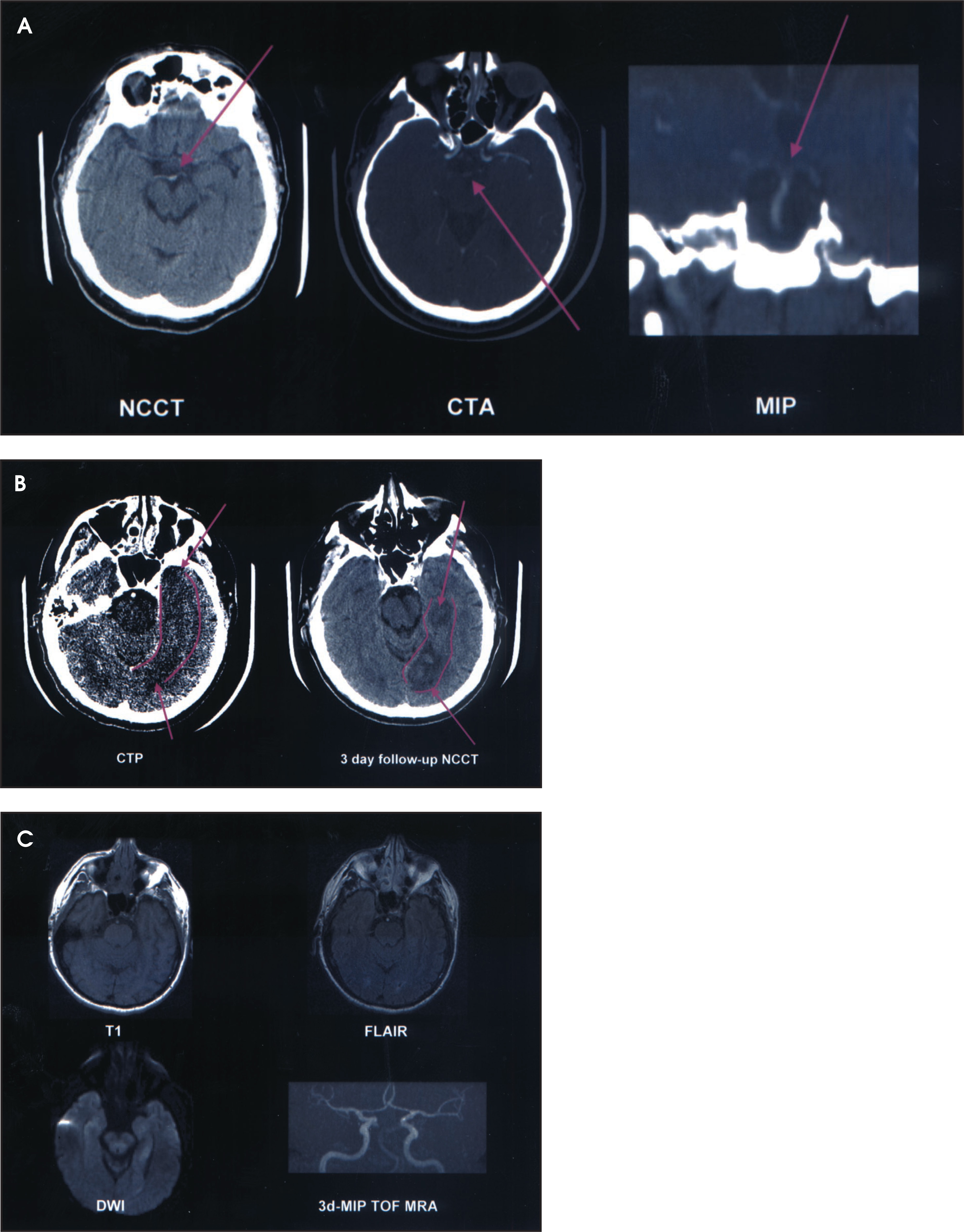
Background
Computed tomography angiography provides anatomical assessment of the vessel lumen regarding the presence of an acute embolus, occlusion, or severe stenosis. The entire course of the carotid and vertebral arteries can be imaged using multislice CT technology (MSCT), including the intracranial and extracranial segments. This data can be used to determine if a patient having an acute stroke has an arterial blockage (embolus) requiring thrombolytic therapy. Most acute emboli occur in the proximal segment of the middle cerebral artery.
Quantitative dynamic cine CTP is a contrast-enhanced study performed at selected image levels. Coverage is typically limited by the maximum detector length in the z-axis direction—approximately 2 cm for MSCT scanners. The selected levels are imaged repeatedly during contrast bolus administration, which reveals serial changes in attenuation of the brain parenchyma. The relationship between tissue attenuation, measured in Hounsfield units (HU), and the tissue concentration of iodinated contrast material, at constant mAs and kV settings, is assumed to be linear.10 Therefore, models for quantitative analysis of this perfusion dataset can be applied conveniently. Such analysis requires computer postprocessing to generate CBF, CBV, and MTT maps, typically using a mathematical process known as deconvolution.9 The actual quantification of flow values is based on the arterial input and venous outflow curves, which can also be reconstructed from the cine CTP dataset. According to the central volume principle, CBF (in mL/100g/min), CBV (in mL/100g), and MTT (in minutes) are related according to the formula CBF = CBV/MTT.9
Clinical applications
To date, the only FDA-approved treatment for acute stroke is IV thrombolysis with recombinant tissue plasminogen activator (IV rt-PA), administered within 3 hours of stroke onset.11 If thrombolysis is applied beyond this time window, the increased probability of intracranial hemorrhage is considered unacceptable.
The time window for treatment with IA agents is twice as long for the anterior circulation, and indefinite for the posterior circulation (depending on risk-to-benefit ratios); however, IA thrombolysis—although of proven benefit in preliminary trials—has not yet received FDA approval.12 For thrombosis localized to the posterior circulation, the time window for treatment may be extended beyond 6 hours due to the extreme consequences of loss of blood flow to the brainstem, despite the risk of hemorrhage.
Because of these narrow windows of opportunity, a goal of advanced imaging is therefore to select patients for treatment more rationally, potentially expanding the time window for thrombolysis by identifying patients for whom rt-PA will be both safe and effective.
Advanced CTA/CTP imaging of acute stroke has the potential to not only help exclude from thrombolysis patients at high risk for hemorrhage, but to identify those patients most likely to benefit from thrombolysis. Even without hemorrhage, treatment failure with thrombolytics is not uncommon. Many hospitals do not have the facilities to use IA rt-PA, and it is important to be able to pinpoint the state of evolution and cause of stroke in order to select the most effective treatment. It may sometimes be necessary to transfer patients to a tertiary care center with specialized expertise in the clinical assessment, imaging, and treatment of acute stroke.
At most institutions the current standard of care is to perform an NCCT exam to exclude hemorrhage; at this point, the imaging evaluation of acute stroke patients typically stops. In some centers, advanced imaging of stroke patients is obtained with diffusion-weighted magnetic resonance imaging (MRI) and MR perfusion transit time maps, but these techniques are often time-consuming, expensive, or unavailable.13,14 Computed tomography angiography with CTP can offer a more convenient advanced imaging alternative.15 Clearly, given the potential severe consequences of stroke, better methods of identifying its presence and etiology are required, and deriving reliable predictors of the extent and degree of tissue damage is desirable.
In the acute stroke setting CTA with CTP has the potential to be useful for: 1) fast and reliable identification of stroke signatures; 2) improved choice of treatment modality, including exclusion of patients from thrombolytic therapy; 3) pinpointing the vascular origin of the ischemic insult; and 4) determination of neurological consequences of the stroke, including final infarct size, clinical outcome, and hemorrhagic risk.
Available data suggests that CTA, when combined with NCCT, is significantly more sensitive than NCCT alone in delineation of intravascular clot.3,16 The choice between IA or IV thrombolysis depends on a variety of factors, including the time post-ictus, the clinical status of the patient, and whether the clot is proximal (IA) or distal (IV). When typical findings of occlusive thrombus on CTA and decreased tissue enhancement on CTP are not present, the differential diagnoses include lacunar infarct, early small distal embolic infarct, transient ischemic attack, complex migraine headaches, and seizure.1
At our institution, immediately following data acquisition for CTA/CTP, rapid “collapsed” maximum intensity projection (MIP) reconstructions (superior view) of the intracranial circulation are created, for visualization of acute circle of Willis embolus. Typically, these can be obtained in under a minute. Additional reconstructions, not typically obtained urgently but prior to endovascular treatment, are obtained displaying the aortic arch, great vessels origins, and carotid bifurcations. The cine CTP dataset, when obtained, can also be manipulated to generate quantitative CBF, CBV, and MTT maps at selected levels (figure 2).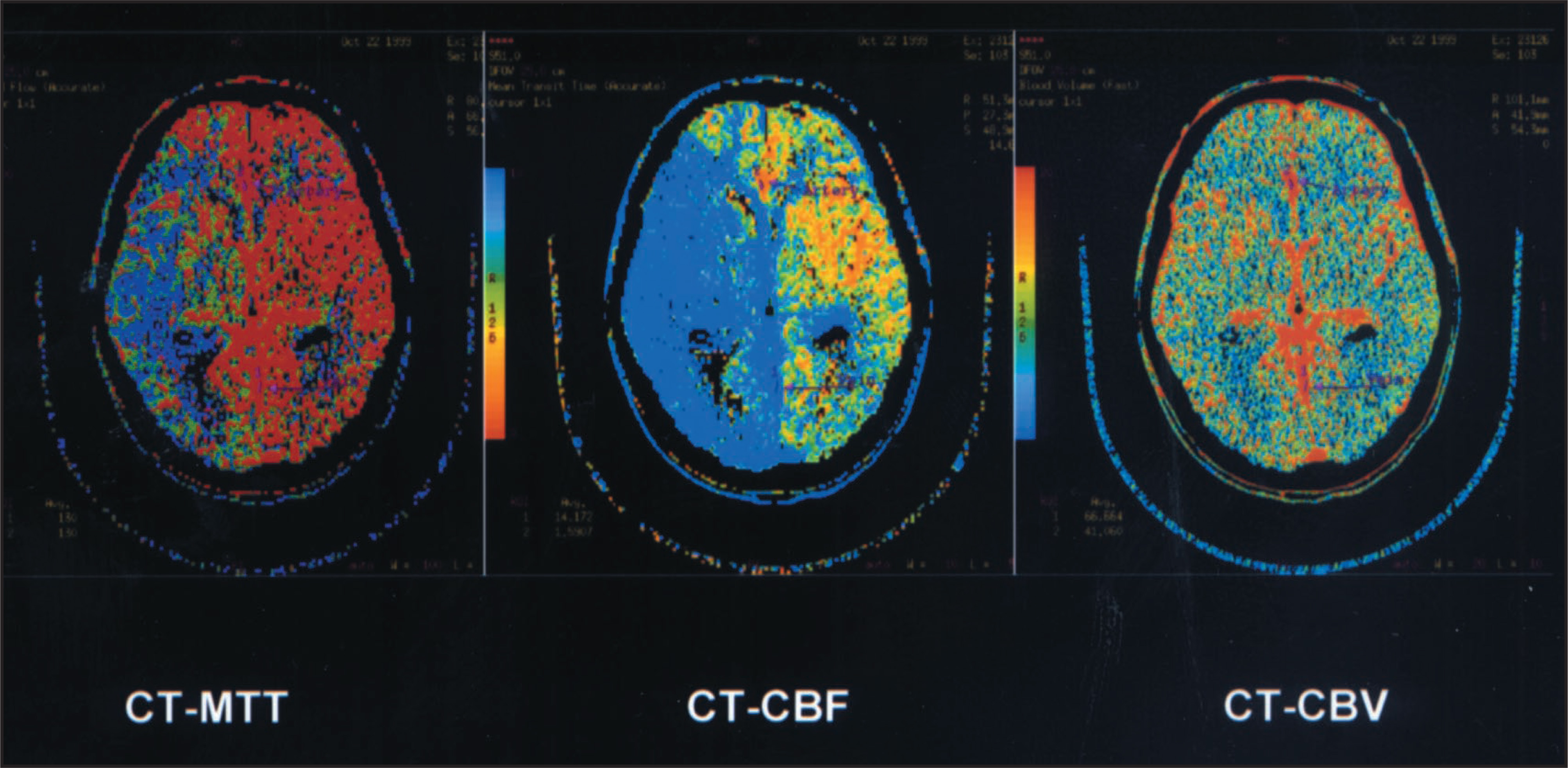
The CBF, CBV, and MTT values are dynamic—they vary as the stroke evolves. “Core” values for irreversible infarction are typically considered to be in the CBF <10 to 12 mL/100g/min range. Areas of less severe CBF reduction, with preserved CBV values, are considered to represent “ischemic penumbra”—tissue that is at high risk for infarction if circulation is not soon restored, but not yet irreversibly infarcted. The larger the ischemic penumbra relative to the core, the more likely the patient would benefit from early thrombolytic therapy. If both CBV and CBF are already reduced dramatically, however, the tissue is considered irreversibly infarcted, and successful recanalization may have a higher likelihood of causing hemorrhage.17
Imaging protocol
At our institution, CTA/CTP imaging is performed on a multislice scanner in the emergency department (GE LightSpeed, GE Medical Systems, Milwaukee, WI), which enables us to acquire imaging data from entire vascular territories in <1 minute. The initial NCCT scan is performed at 140 kV, 170 mA, pitch = “high quality” (3:1), and a table speed of 7.5 mm/sec. Images are obtained from the skull base to the vertex. Slice thickness can be set at 2.5 mm, at 5 mm, or at both. We typically use the thicker, 5-mm slices for visual image review, and the thinner, 2.5-mm slices for 3D-reconstruction. Initial image review is in “real-time,” directly at the CT console. The use of narrow window-width settings, with a center level of about 30 HU (width of 5 to 30 HU), facilitates the detection of early, subtle, ischemic changes contiguous with normal parenchyma.2
Immediately following NCCT, CTA with simultaneous whole brain perfused blood volume CTP is performed without repositioning the patient. Approximately 90 to 120 mL of nonionic, iso-osmolar contrast is used for CTA from the skull base to the vertex. The injection rate is 3 mL/sec, with a 25-second scan delay. Initial scan parameters are as per the NCCT scan described above. A second phase of scanning is performed immediately, with minimal possible delay, from the aortic arch to the skull base, with similar scan parameters except for an increase in the table speed to 15 mm/sec. Major advantages of first scanning the intracranial circulation include: 1) obtaining the most important data first, which can be reviewed during subsequent acquisition; and 2) allowing time for clearance of dense, IV contrast from the subclavian, axillary, and other veins at the thoracic inlet, reducing streak artifact. Immediate assessment of the vasculature occurs at the scanner console, where the source images are reviewed while scanning is taking place.
The axial source images from the CTA/CTP exam are typically networked to a freestanding image analysis workstation. MIP reconstructions of the intracranial circulation are performed to identify acute embolus and the extent of the clot burden. Reconstructions of the aortic arch, great vessels, and carotid bifurcations are also performed to assess for additional stenoses or occlusions of the neurovasculature prior to endovascular treatment.
Depending on the clinical circumstances—or as an entirely separate exam—quantitative cine dynamic CT perfusion maps can also be acquired. The level to be scanned is determined from the initial NCCT findings, as well as from the involved vascular distribution seen on CTA. Our MSCT scanner allows for the selection of four 5-mm or two 10-mm thick slices in axial cine mode. Approximately 45 mL of nonionic contrast is injected at 4 to 5 mL/sec, with a 5-second preparation delay. In order to generate CTP maps, the operator must select the arterial input and venous outflow parameters for the deconvolution software.
Conclusion
Potential current and future benefits of CTA/CTP include convenient, reliable, acute stroke imaging, which may improve clinicians’ ability to assess hemorrhagic risk and determine prognosis, therefore facilitating triage of acute stroke patients to appropriate treatment.15 Quantitative CTP has shown further potential to characterize the evolving status of stroke through the measurement of CBV, CBF, and MTT. We encourage the use of CTA/CTP imaging for the emergency evaluation and management of suspected stroke. AR
References
- Lev M, Nichols S. Computed tomographic angiography and computed tomographic perfusion imaging of hyperacute stroke. Top Magn Reson Imaging. 2000;11(5):273-287.
- Lev M, Farkas J, Gemmete J. Acute stroke: Improved nonenhanced CT detection—Benefits of soft-copy interpretation by using variable window width and center level settings. Radiology. 1999;213:150-155.
- Lev M, Farkas J, Rodriguez V. CT angiography in the rapid triage of patients with hyperacute stroke to intraarterial thrombolysis: Accuracy in the detection of large vessel thrombus. J Comput Assist Tomogr. 2001;25:520-528.
- Hunter G, Hamberg L, Ponzo J. Assessment of cerebral perfusion and arterial anatomy in hyperacute stroke with three-dimensional functional CT: Early clinical results. AJNR Am J Neuroradiol. 1998;19:29-37.
- Hamberg L, Hunter G, Kierstead D. Measurement of cerebral blood volume with subtraction three-dimensional functional CT. AJNR Am J Neuroradiol. 1996;17:1861-1869.
- Lev M, Segal A, Farkas J. Utility of perfusion-weighted CT imaging in acute middle cerebral artery stroke treated with intra-arterial thrombolysis: Prediction of final infarct volume and clinical outcome. Stroke. 2001;32:2021-2028.
- Nabavi D, Cenic A, Craen R. CT assessment of cerebral perfusion: Experimental validation and initial clinical experience. Radiology. 1999;213:141-149.
- Hamberg L, Hunter G, Halpern E. Quantitative high resolution measurement of cerebrovascular physiology with slip-ring CT. AJNR Am J Neuroradiol. 1996;17:639-650.
- Aksoy F, Lev M. Dynamic contrast-enhanced brain perfusion imaging: Technique and clinical applications. Semin Ultrasound CT MR. 2000;21:462-477.
- Koenig M, Klotz E, Luka B. Perfusion CT of the brain: Diagnostic approach for early detection of ischemic stroke. Radiology. 1998;209:85-93.
- Tissue plasminogen activator for acute ischemic stroke. N Engl J Med. 1995;333:1581-1587.
- Furlan A, Higashida R, Wechsler L. Intra-arterial prourokinase for acute ischemic stroke. JAMA. 1999;282:2003-2011.
- Sunshine J, Tarr R, Lanzieri C. Hyperacute stroke: Ultrafast MR imaging to triage patients prior to therapy. Radiology. 1999;212:325-332.
- Sunshine J, Bambakidis N, Tarr R. Benefits of perfusion MR imaging relative to diffusion MR imaging in the diagnosis and treatment of hyperacute stroke. AJNR Am J Neuroradiol. 2001;22:915-921.
- Koroshetz W, Gonzales R. Imaging stroke in progress: Magnetic resonance advances but computed tomography is poised for counterattack. Ann Neurol. 1999;46:556-558.
- Wildermuth S, Knauth M, Brandt T. Role of CT angiography in patient selection for thrombolytic therapy in acute hemispheric stroke. Stroke. 1998;29:935-938.
- Ueda T, Sakaki S, Yuh W. Outcome in acute stroke with successful intra-arterial thrombolysis procedure and predictive value of initial single-photon emission-computed tomography. J Cereb Blood Flow Metab. 1999;19:99-108.
Citation
. Advanced CT imaging in the evaluation and triage of acute stroke patients. Applied Radiology. 2002;31(2):18-22. doi:10.37549/AR1075.