Amyand Hernia
Applied Radiology — Vol. 1 , Issue 1 , pp. 1 -2
Published: November 1, 2025
1 University of Cincinnati College of Medicine, Cincinnati, Ohio
2 Department of Radiology, Cincinnati Children’s Hospital, University of Cincinnati College of Medicine, Cincinnati, Ohio
3 Department of Radiology, Phoenix Children’s Hospital, Phoenix, Arizona
* Corresponding author: Alexander J. Towbin (Alexander.Towbin@cchmc.org)
Abstract
Amyand hernias are a rare version of inguinal hernias containing the appendix. They are typically diagnosed intraoperatively, but imaging can provide substantial information important preoperatively, sufficient for diagnosis. Patients are typically asymptomatic. However, acute inflammation of the appendix within the hernia can rarely occur.
Keywords
gastrointestinal, hernia, neonate
Categories
Case Summary
A neonate presented with trisomy 18, subglottic stenosis, and feeding intolerance. An upper gastrointestinal (GI) examination was performed prior to expected G-tube placement.
Imaging Findings
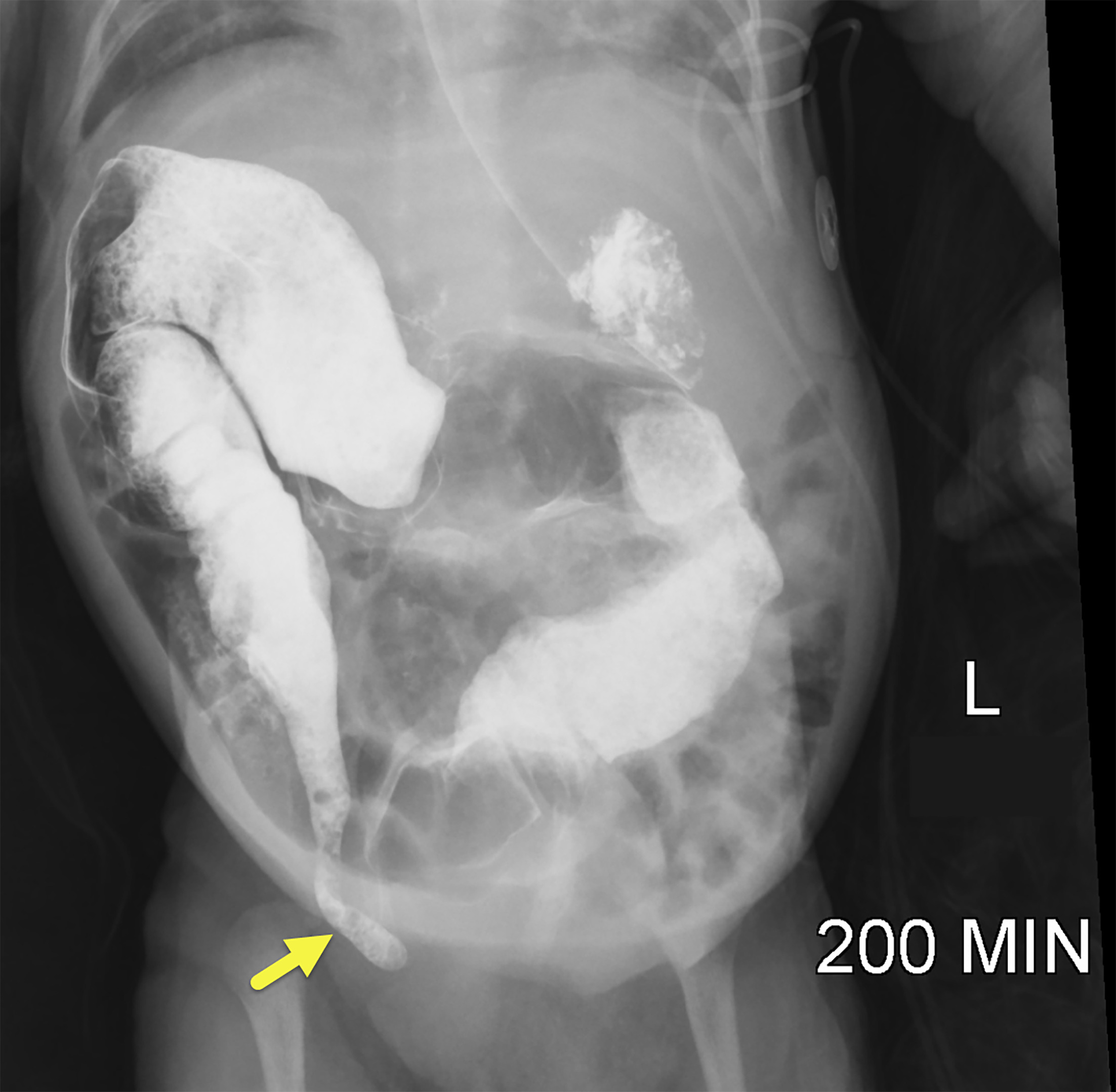
An upper GI with small bowel follow-through (Figure 1) was performed and demonstrated the appendix intermittently located within the right inguinal canal.
Diagnosis
Asymptomatic malrotation and an Amyand hernia
Discussion
Amyand hernia is a rare inguinal hernia in which the hernia sac contains the vermiform appendix. This occurs in approximately 1% of all inguinal hernias. 1 Given that inguinal hernias are typically caused by a persistent patent processus vaginalis, Amyand hernias are more common in childhood. They are more frequent in males due to the greater incidence of inguinal hernias. Amyand hernias tend to be right-sided due to the natural location of the appendix, but left-sided Amyand hernias can be observed in patients with situs inversus, intestinal malrotation, a mobile cecum, or a large appendix. 1 In Amyand hernias, the appendix can rarely be accompanied by the cecum, bladder, ovary, fallopian tube, omentum, or Meckel diverticulum. 1 Of these, the cecum is the most commonly found with the appendix in the hernia sac, especially in older patients. 2
In 0.13% of patients with an Amyand hernia, the appendix is inflamed. 3 Mortality because of an Amyand hernia has been reported to range from 5.5% to as high as 30% and has mostly been linked to peritoneal spread of sepsis. 4 Appendiceal perforation in all cases of appendicitis leads to a significant increase in mortality rate due to abdominal sepsis. The exact mechanism of appendiceal inflammation in an inguinal hernia is not fully understood. Prevailing theories include incarceration and inflammation of the appendix, 5 development of adhesions between the appendiceal serous membrane and the hernia sac, 6 and functional irreducibility of the appendix once it is inflamed and swollen, leading to impaired microcirculation of the appendiceal wall and eventual bacterial overgrowth. 7
Amyand hernias are typically asymptomatic, and if symptomatic, present as a reducible bulge with local discomfort. 1 Patients with concurrent appendicitis present with an irreducible inguinal hernia, pain in the right lower quadrant, anorexia, nausea, and vomiting.
On CT, Amyand hernias are visualized as a blind-ending, tubular structure arising from the base of the cecum and extending into the inguinal sac. 8 On US, the Amyand hernia appears as a small blind-ending tubular structure within the hernia sac. Findings of inflammation on CT include a dilated lumen, wall enhancement and thickening, and peri-appendiceal fat stranding. On US, findings of inflammation include a dilated, noncompressible appendix with wall thickening and hyperemia. 1
While Amyand hernias are typically diagnosed intraoperatively, US or CT imaging can provide substantial information for a preoperative diagnosis. In a study comparing 21 patients with an Amyand hernia, a preoperative US was performed in 12 patients and the hernia was diagnosed in 9 of them. 9 Imaging can aid in the preoperative diagnosis of complicated Amyand hernias.
The treatment and management of an Amyand hernia depend on its classification. Losanoff and Basson divided Amyand’s hernias into 4 types depending on their clinical presentation: type 1, normal appendix within an inguinal hernia; type 2, appendicitis within an inguinal hernia with no abdominal sepsis; type 3, acute appendicitis within an inguinal hernia with abdominal wall infection or peritoneal sepsis; and type 4, acute appendicitis within an inguinal hernia with related or unrelated abdominal pathology. 10 Rikki described a 5th type of Amyand hernia with 3 subtypes. This type of hernia describes the appendix within an incisional hernia: type 5a describes normal appendix within an incisional hernia, type 5b describes acute appendicitis within an incisional hernia without abdominal or peritoneal sepsis, and type 5c describes acute appendicitis within an incisional hernia with abdominal wall or peritoneal sepsis or in relation to previous surgery. 11
Amyand hernia with a noninflamed appendix is usually treated via hernia repair without appendectomy. 6 This practice is preferred as it decreases the risk of postoperative infection, decreases the recurrence rate of inguinal hernia, and preserves the appendix for potential future use. 1 Manipulation of the appendix is avoided as it could lead to secondary acute inflammation, 1 in which case appendectomy would become indicated.
Our patient passed away due to cardiopulmonary failure before surgical repair of the hernia could be performed.
Conclusion
Amyand hernias are a rare version of inguinal hernias containing the appendix. They are typically diagnosed intraoperatively, but imaging can provide substantial information important preoperatively, sufficient for diagnosis. Patients are typically asymptomatic. However, acute inflammation of the appendix within the hernia can rarely occur.
References
- Patoulias D, Kalogirou M, Patoulias I. Amyand’s hernia: an up-to-date review of the literature. Acta Medica Cordoba. 2018;60(3):131-134.
- Michalinos A, Moris D, Vernadakis S. Amyand’s hernia: a review. Am J Surg. 2014;207(6):989-995. doi:10.1016/j.amjsurg.2013.07.043.
- Morales-Cárdenas A, Ploneda-Valencia C, Sainz-Escárrega V. Amyand hernia: case report and review of the literature. Annals of Medicine & Surgery. 2015;4(2):113-115. doi:10.1016/j.amsu.2015.03.007.
- Sharma H, Gupta A, Shekhawat N, Memon B, Memon M. Amyand’s hernia: a report of 18 consecutive patients over a 15-year period. Hernia. 2007;11(1):31-35. doi:10.1007/s10029-006-0153-8.
- Weber R, Hunt Z, Kral J. Amyand’s hernia. Etiologic and therapeutic implications of two complications. Surg Rounds. 1999;22:552-556.
- Hiatt J, Hiatt N. Amyand’s hernia. N Engl J Med. 1988;318(21). doi:10.1056/NEJM198805263182120.
- Singhal S, Singhal A, Negi S. Amyand’s hernia: rare presentation of a common ailment. Case Rep Gastrointest Med. 2015;2015:629127. doi:10.1155/2015/629127.
- Annamaria G, Francesco M, Carlo S. Amyand’s hernia. Journal of Radiological Review. 2021;8(2):168-171.
- Okur M, Arslan M, Zeytun H, Otcu S. Amyand’s hernia complicated with acute appendicitis: a case report and literature review. Ped Urol Case Rep. 2015;2(4):7-12.
- Losanoff J, Basson M. Amyand hernia: a classification to improve management. Hernia. 2008;12(3):325-326. doi:10.1007/s10029-008-0331-y.
- Singal R, Gupta S. “Amyand’s hernia” – pathophysiology, role of investigations and treatment. Maedica (Buchar). 2011;6(4).
Disclosures
The authors have no conflicts of interest to disclose. None of the authors received outside funding for the production of this original manuscript and no part of this article has been previously published elsewhere.
Citation
. Amyand Hernia. Applied Radiology. 2025;1(1):1-2. doi:10.37549/JPCR-25-0022.