An algorithmic approach to neuroimaging in AIDS
Applied Radiology — Vol. 34 , Issue 12 , pp. 18 -32
DOI: 10.37549/AR1391
Published: December 1, 2005
Categories
Since the recognition of the human immunodeficiency virus (HIV) in the 1980s, awareness of the neurologic complications of the disease has been increasing. The increased longevity of patients with the disease, in part secondary to multidrug regimens, as well as the increased incidence of the disease has furthered the opportunity for radiologic assistance in the diagnosis and management of neurologic complications from HIV. It is estimated that 7% to 10% of patients with acquired immune deficiency syndrome (AIDS) present with neurologic symptoms as their initial manifestation of illness and that nearly 75% have central nervous system (CNS) involvement on postmortem examination.1-3
The clinical diagnosis of neurologic illness in patients with AIDS can be a daunting task. Multiple illnesses affecting the CNS have overlapping clinical presentations without specific diagnostic laboratory findings. A number of diseases––including HIV encephalitis, progressive multifocal leukoencephalopathy (PML), toxoplasmosis, lymphoma, and cryptococcal infection––have clinical presentations ranging from subtle personality and mental status changes and difficulty with concentration, depression, and memory loss to profound confusion that progresses to dementia. Headaches, motor and sensory neurologic deficits, and new-onset seizures are also common findings. Laboratory findings, such as cerebrospinal fluid (CSF) leukocytosis, may indicate an infectious etiology but are nonspecific, as are measurements of CSF protein and glucose counts.1
The authors propose a simple algorithmic approach to the neuroradiologic imaging findings that may assist the clinician and radiologist in the diagnosis of the neurologic complications of HIV-positive (HIV+) patients.
Algorithmic approach
Neuroimaging findings can be divided into 4 general categories that are useful in the differential diagnosis of patients with AIDS: 1) normal, 2) atrophy, 3) intra-axial lesion without mass or mass effect, and 4) mass lesion (Figure 1). Normal studies do not, however, preclude a diagnosis of infection or neoplasm.1,2,4 Close clinical follow-up with periodic reimaging may be helpful in recognition of initially radiologic occult processes in patients with persistent neurologic symptoms.
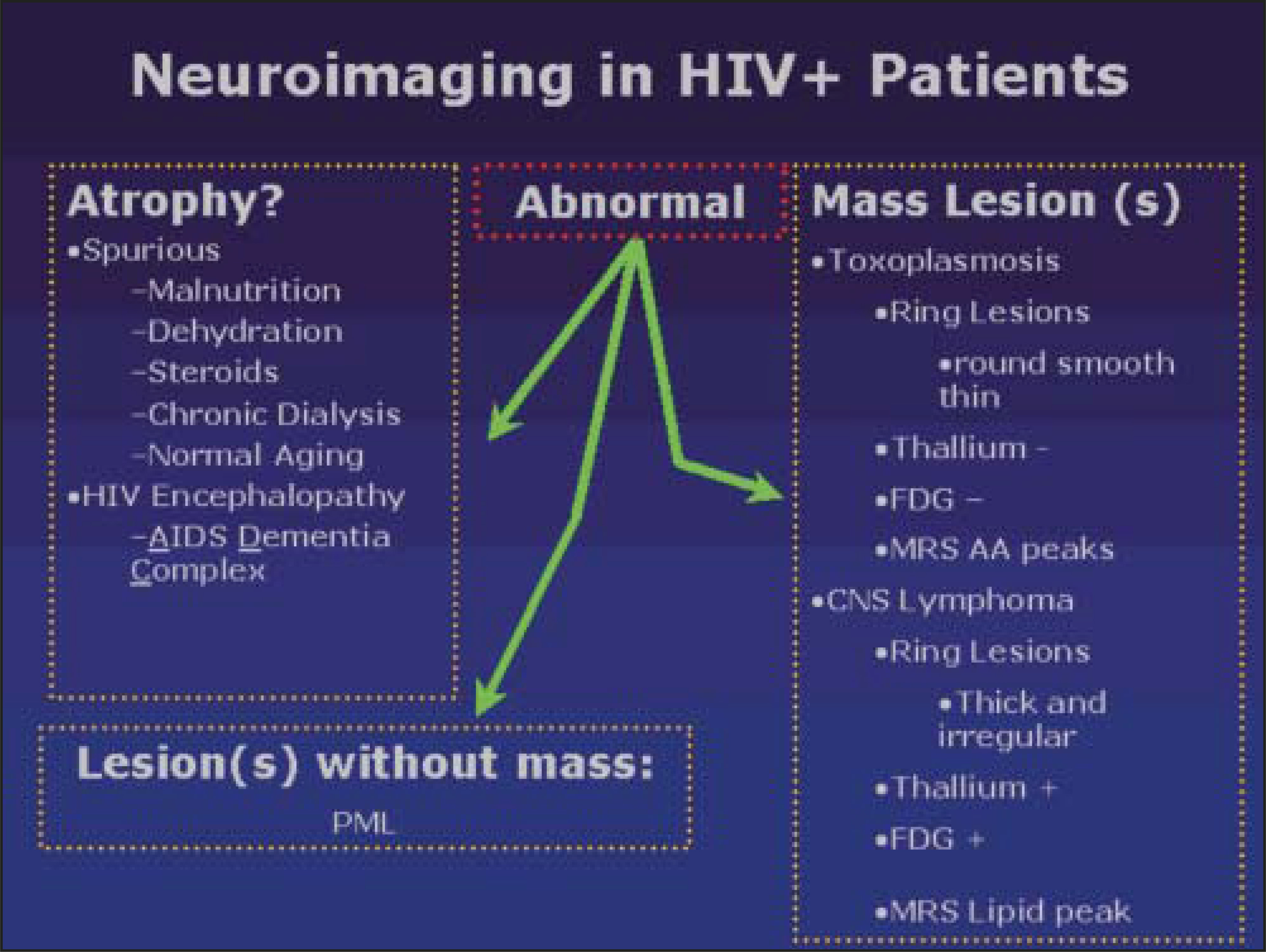
Atrophy may be related to normal aging or pathologic processes. After the exclusion of malnutrition, dehydration, and iatrogenic causes (such as steroids), HIV encephalopathy/AIDS dementia complex (ADC) must be considered. When abnormal parenchymal attenuation on computed tomography (CT) studies or abnormal signal intensity on magnetic resonance imaging (MRI) studies is seen in the absence of a mass lesion or significant mass effect, PML should be the primary consideration. An intra-axial mass lesion suggests an infectious or neoplastic etiology, most commonly toxoplasmosis or lymphoma.4-6 Since prompt clinical diagnosis is necessary for appropriate treatment, the finding of a mass lesion that is clinically suggestive of toxoplasmosis is treated empirically, with consideration of biopsy in nonresponders for further clarification of diagnosis. Cystic-appearing masses in the region of the basal ganglia are pathognomonic for cryptococcal involvement.
This algorithm provides a simple approach to the diagnosis of the most common neurologic complications of AIDS. These entities will be discussed in further detail below.
Clinical entities
While the CNS complications from AIDS are broad, understanding the most common pathologic processes and opportunistic infections and using this simple algorithmic approach may clarify an otherwise complicated clinical presentation of disease. If the imaging findings cannot be placed within one of the categories of the algorithm, further evaluation should be conducted into an uncommon manifestation of a common process or other, less common opportunistic processes.
Pathologic atrophy: AIDS dementia complex, HIV encephalopathy, HIV subacute encephalitis,
Subacute encephalitis, clinically referred to as the AIDS dementia complex (ADC), is a common neurologic complication seen in patients with AIDS. The prevalence of HIV dementia ranges from 7% to 27% of patients.1,7,8 AIDS dementia complex is characterized by cognitive disturbances that progress to dementia. The etiology is direct infection of HIV-1 in the CNS.8-10 While HIV-1 does not damage neurons directly, it causes indirect injury by infected CNS macrophages that generate neurotoxic factors.7,10 Two distinct histopathologic patterns have been identified in this disorder: HIV encephalitis (HIVE), which is predominantly perivascular, and HIV leukoencephalopathy, which is characterized by diffuse myelin loss and infiltration by macrophages. The diagnosis of ADC is made according to clinical, neuropsychological, and imaging findings.
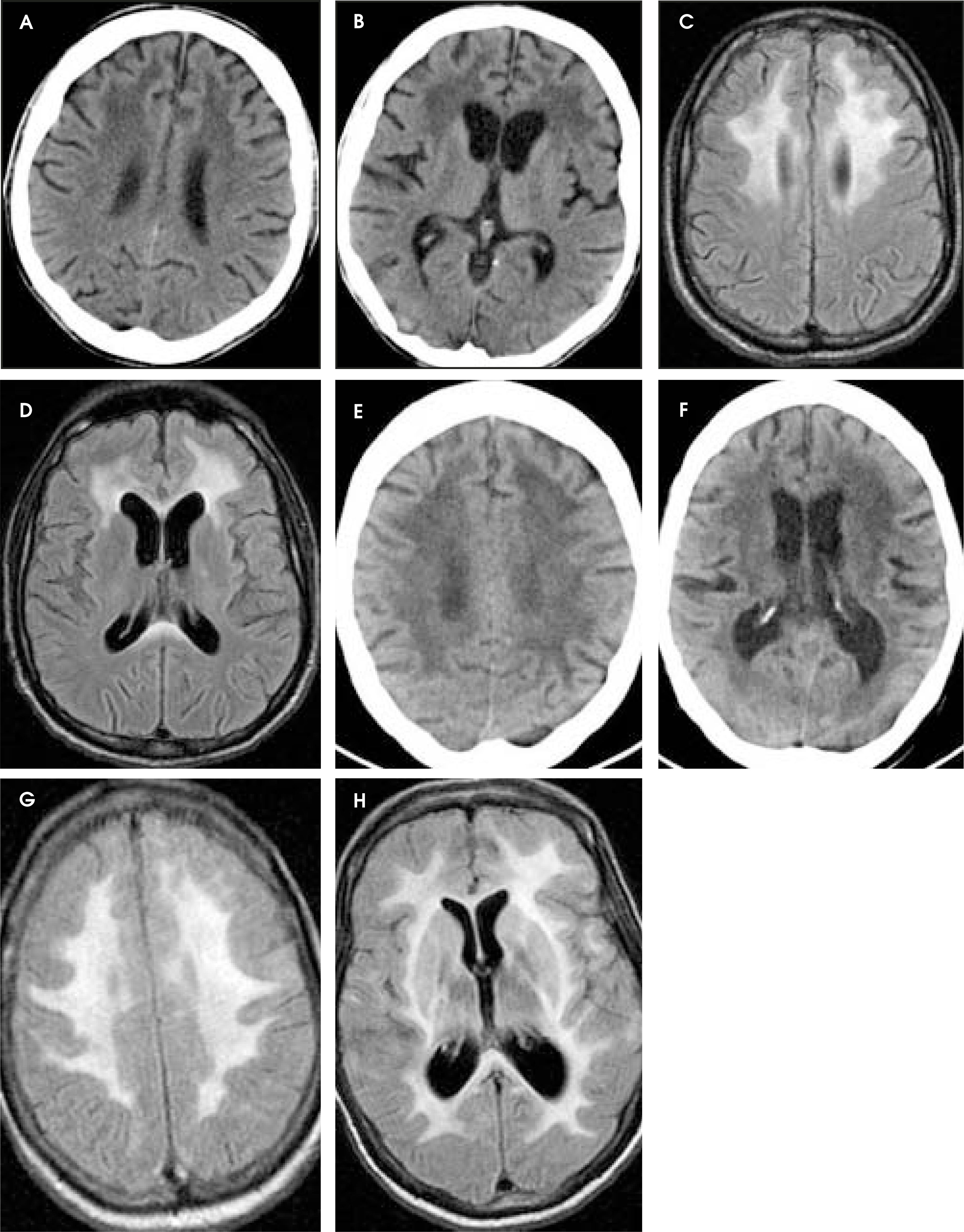
The most commonly reported imaging finding in HIVE is cerebral atrophy.8-10 The appearance of atrophy varies from mild (Figure 2A) to severe (Figure 2B), which may include ex vacuo dilatation of the ventricles. Some patients may have subtle white matter hypoattenuation on CT (Figures 2A and B), or corresponding increased T2 or fluid-attenuated inversion-recovery (FLAIR)–weighted signal intensity of the supratentorial white matter on MRI, which does not exhibit mass effect (Figures 2C and D).9,10 The white matter changes are nonspecific, occurring as focal or diffuse, symmetric or asymmetric, reversible or nonreversible. Severe HIVE is reflected by severe atrophy and white matter hypoattenuation on CT (Figures 2E and F) and corresponding FLAIR hyperintensity on MRI (Figures 2G and H). While the findings of atrophy are more specific for HIV encephalopathy, the finding of white matter lesions in addition to atrophy should not exclude the diagnosis. Neuropathologic evaluation suggests that white matter abnormalities in ADC reflect leaky capillaries and a subsequent increased water content, which explains the reversible nature of the findings.10
HIV protease inhibitor therapy and highly active antiretroviral therapy (HAART) have been reported to result in the regression or stabilization of the periventricular and subcortical white matter signal intensity abnormalities seen in HIV encephalopathy.11 In addition, the treatment may promote positive clinical effects, despite the progression of the MRI findings. There may be significant lag time in the MRI findings compared with the clinical symptoms, and extended follow-up may be necessary to document resolution or improvement in the MRI findings.10 Therefore, early follow-up imaging should not be the sole factor in the evaluation of drug effectiveness.
Parenchymal lesion without mass or mass effect
Progressive multifocal leukoencepha-lopathy (PML) is a white matter demyelinating disease caused by the JC papo- vavirus, a deoxyribonucleic acid (DNA) virus. The virus is named “JC” after the initials of the patient from whom it was cultured in 1971. This patient, JC, had Hodgkin’s lymphoma and developed PML as a complication.12 The virus itself is ubiquitous within the adult population, with an estimated prevalence of 80% for expression of JC antibodies.13 Almost 40% of HIV+ patients may express the JC virus in their peripheral blood lymphocytes in the absence of PML.14 An immunocompromised state is theorized to permit reactivation of the virus in the AIDS patient. The JC virus causes lysis of the oligodendrocytes, disrupting the normal repair mechanism and resulting in demyelination. Asymmetric involvement of both cerebral hemispheres is typical, with involvement of white matter tracts in the cerebellum, brainstem, and deep gray matter. Early involvement, however, may present with a single lesion, which can make the diagnosis more difficult.
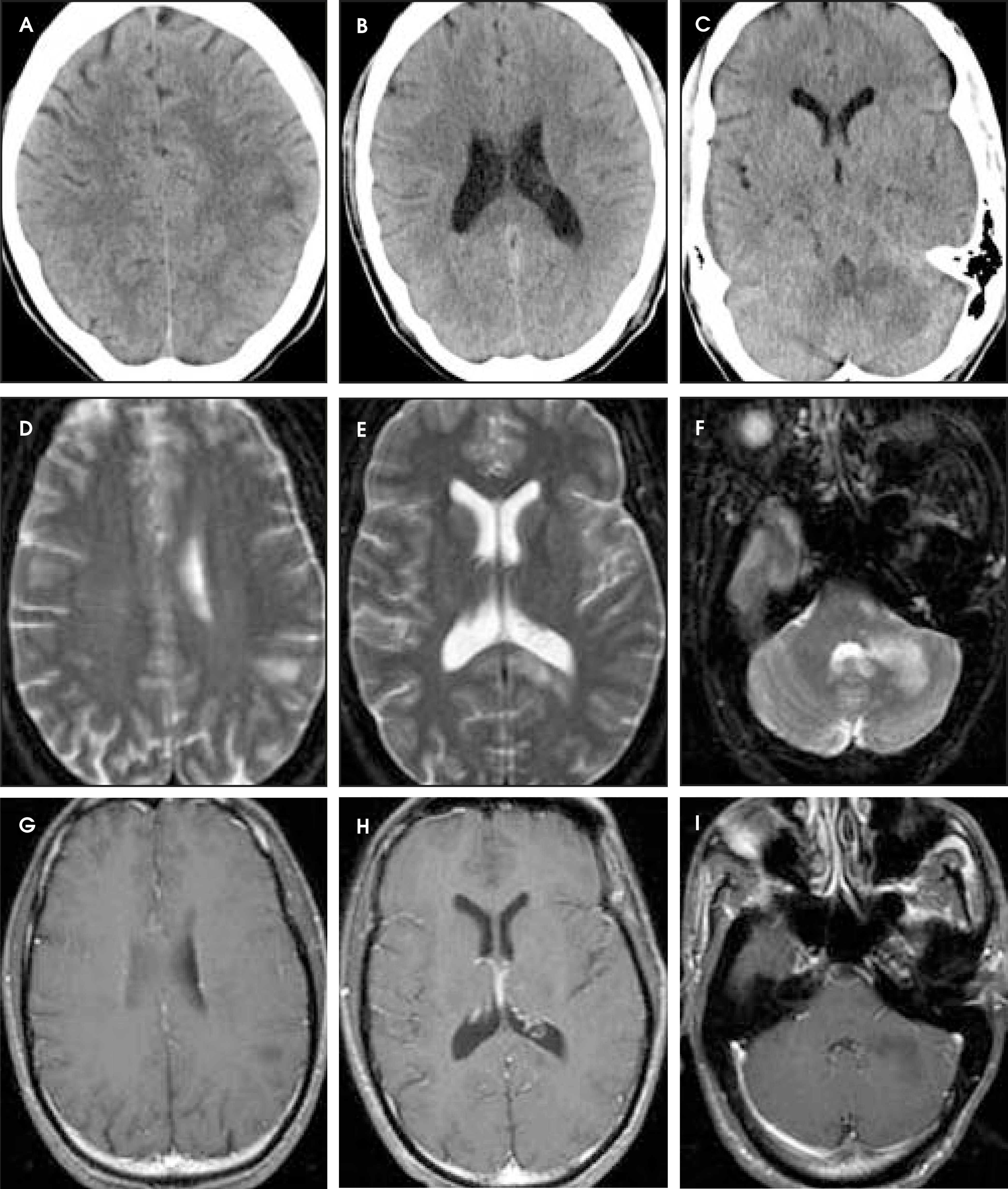
Berger et al15 reported that PML affects approximately 4% of all AIDS patients. The symptoms associated with PML include altered mental status and speech, motor, and visual disturbances.7 Classic CT imaging findings include single or multiple low-attenuation white matter lesions without edema or mass effect (Figures 3A, B, and C). MRI findings show hyperintensity of the white matter on T2-weighted and FLAIR images (Figures 3D, E, and F) with corresponding T1 hypointensity. Typically, there is no enhancement (Figures 3G, H, and I)16,17 Donovan-Post et all8 quantified the pretreatment MRI findings in their study of 48 patients with white matter lesions as bilateral in 91.7%, confluent in 93.8%, and discrete in 66.7%. The lesions were predominantly supratentorial (frontoparietal more than temporo-occipital) within the periventricular region, centrum semiovale, and subcortical white matter. Although patients with HIV encephalopathy exhibit atrophy on neuroimaging, up to 68.8% of patients with PML had mild cortical atrophy and 50% of them were found to have ventricular dilatation. Gray matter region lesions, typically in the thalamus and basal ganglia, are also common, likely representing involvement of the traversing white matter tracts. A definitive diagnosis of PML may require biopsy. However, in many cases, confirmatory clinical symptoms, CSF polymerase chain reaction (PCR) studies that are positive for the JC virus, and appropriate imaging findings may be sufficient for making a treatment decision, and biopsy can be avoided.18
Progressive multifocal leukoencephalopathy is a progressive disease, typically causing death within 2 to 5 months after diagnosis.18,19 The standard use of HAART in the United States since 1996 has led to the recent suggestion that AIDS patients with PML may live longer, although it has yet to be extensively documented.18 Most cases of PML are refractory to treatment. Some studies have attempted to predict prognosis from imaging analysis; however, this has not been analyzed in a large cohort of patients. The presence of mild mass effect has been shown to be a prognostic indicator of decreased survival.18 However, this finding was contradicted by Thurnher et al,20 who stated that the development of mass effect and temporary enhancement on MR images in the early phase of treatment might represent positive predictive factors for prolonged survival. Currently, no MRI findings have significantly correlated with patient survival. The imaging findings of PML that are seen in patients with AIDS significantly differ compared with other immunocompromised individuals, in that AIDS patients may present with a solitary discrete lesion and/or lesions that are not confined to the white matter. Thus, PML should not be excluded if it is unifocal and/or appears to involve deep cortical gray matter.4
Mass lesion
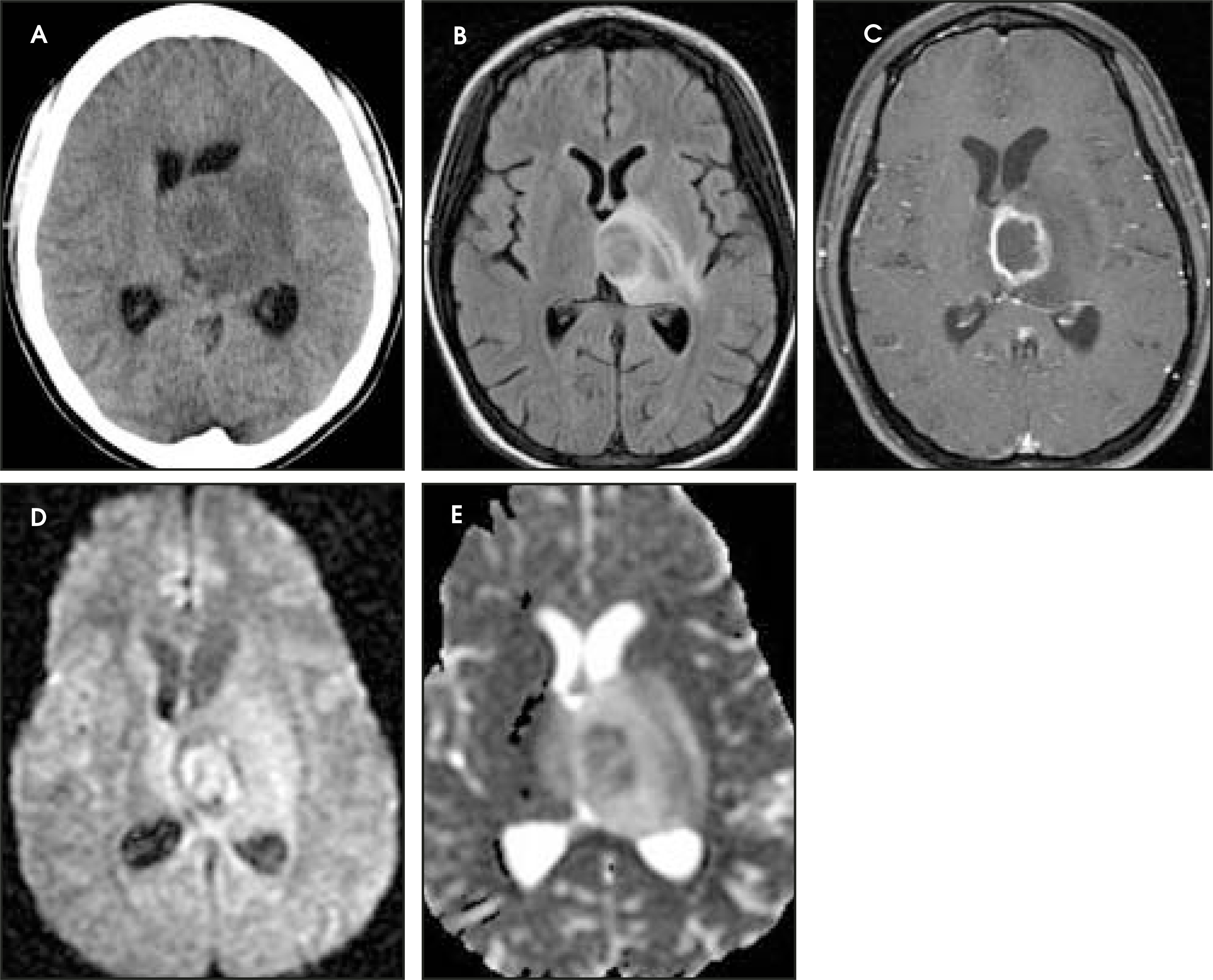
Toxoplasmosis—Toxoplasmosis is the most common cerebral mass lesion in patients with AIDS.7 The infection is caused by an obligate intracellular parasite, Toxoplasma gondii, with reservoirs in the house cat (feces) and uncooked meat (eg, pork and free-range chicken). Approximately 20% to 70% of the normal adult population in the United States is seropositive for antibodies to toxoplasmosis.2The incidence ranges between 3% and 40% of patients with CNS complications of AIDS.1,7,21 The high frequency of multicentric lesions and evidence of choroid plexus infection with toxoplasmosis suggests hematogenous dissemination of parasites.7Imaging findings are suggestive, but nonspecific. Head CT typically shows multiple, small (<2 cm), low-attenuation masses with ring enhancement. While the lesions are typically multiple, isolated lesions are also possible (Figure 4A). MR images show decreased T1 and increased T2/FLAIR (Figure 4B) signal intensity lesions with edema and ring enhancement (Figure 4C). Most lesions are within the cerebral hemisphere white matter; however, involvement in any area is possible. The lesions show restricted diffusion on diffusion-weighted imaging (DWI) (Figure 4D) and corresponding apparent diffusion coefficient maps (Figure 4E), similar to other abscesses.
One of the most significant clinical challenges in AIDS is the differentiation between toxoplasmosis and lymphoma, which can have similar clinical and imaging findings. Since toxoplasmosis occurs 2 to 3 times more frequently than lymphoma in AIDS patients in many geographic areas,2 empiric chemotherapy for toxoplasmosis (usually pyrimethamine and sulfadiazine for 3 weeks) has been advocated, with close clinical follow-up. Porter et al22 reported clinical improvement with treatment in 90% to 95% of patients by day 14. This clinical response to antitoxoplasma therapy has been the main criterion for diagnosis given the low specificity for serologic and PCR tests.23 If response is limited, treatment fails, or there is rapid deterioration, then imaging and biopsy should be performed.
Imaging and biopsy should be performed prior to any steroid treatment. Steroids may cause regression of lymphoma and may falsely give the impression that there was improvement secondary to antitoxoplasmosis medication, delaying diagnosis and further treatment for lymphoma. It is important to recognize that lymphoma can progress rapidly. This may be a clinically confusing sign that may suggest an infectious process. A fulminant illness should prompt immediate biopsy and early follow-up studies during empirical therapy for toxoplasmosis.5
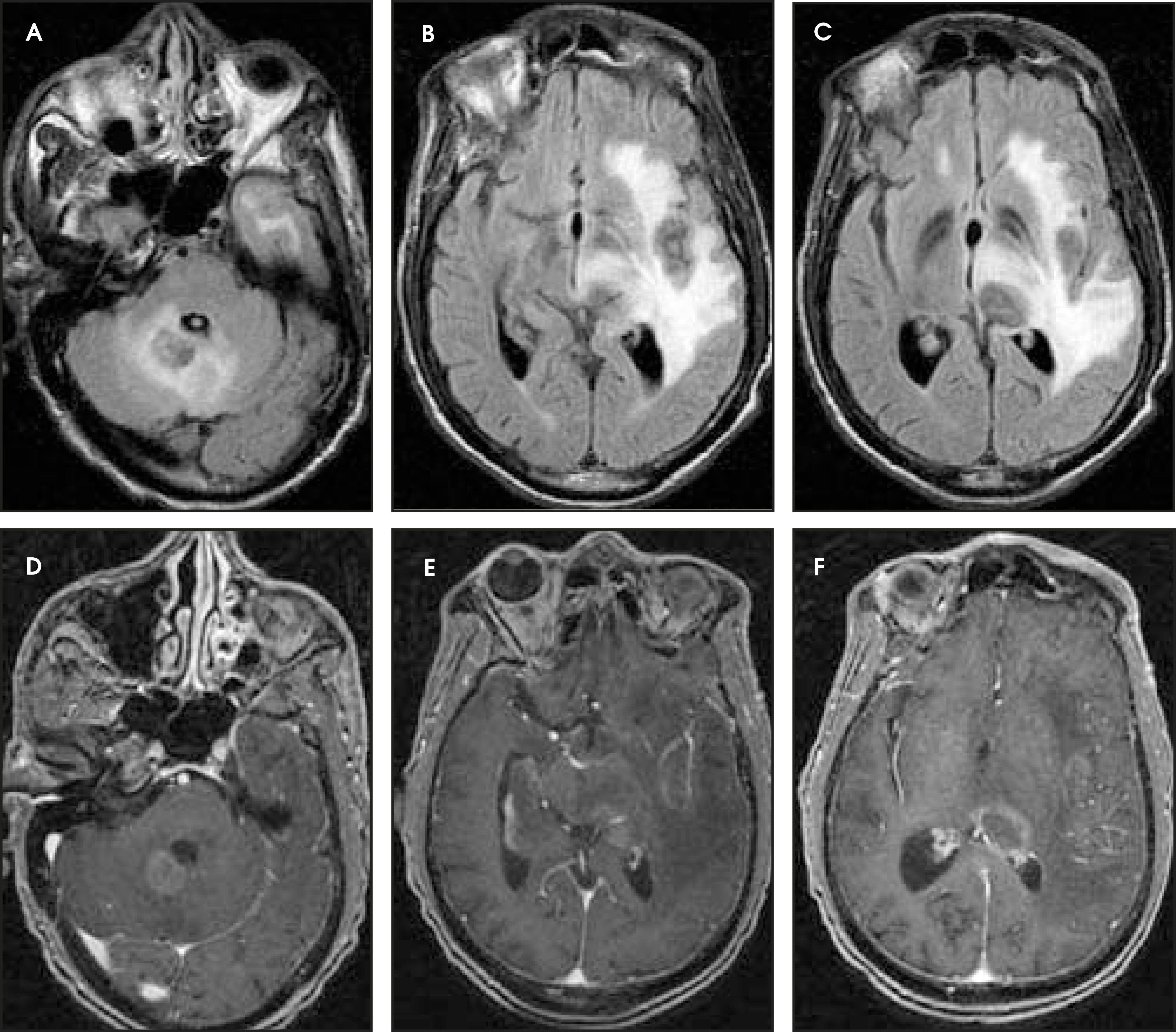
Lymphoma—Primary CNS lymphoma is a high-grade B-cell non-Hodgkin’s lymphoma with a strong association with Epstein-Barr virus (EBV) infection.24 Primary lymphoma accounts for 2% to 10% of brain lesions in AIDS patients.1,22,25,26 It is the second most common cause of a CNS mass lesion in AIDS patients.27 Histopathologically, this is a small, round blue-cell tumor that is often associated with increased attenuation on noncontrast head CT, because of a high nuclear-to-cytoplasmic ratio. However, low- attenuation white matter lesions may occur. Lymphoma may be unifocal or multifocal (Figure 5) with variable mass effect. Typically, there is a paucity of edema relative to the tumor size. On MRI, these lesions may be isointense or hypointense on T1-weighted images and may be of variable intensity on T2-weighted or FLAIR images. However, because of the highly cellular infiltrate of small, round blue cells, the lesions may show T2/FLAIR hypointensity (Figures 5A, B, and C). Most lesions exhibit homogenous enhancement; but ring enhancement is frequently seen in AIDS patients (Figures 5D, E, and F). Steroids may inhibit the visibility of contrast enhancement. Lymphoma is most often periventricular in location but has been described throughout the parenchyma.
Radiologic findings may be helpful in distinguishing between toxoplasmosis and lymphoma. While autopsy studies have shown primary brain lymphoma to be multifocal in 80% to 100% of AIDS patients, when confronted with a solitary mass lesion, it is important to recognize that lymphoma is more probable than toxoplasmosis and that empirical treatment is not likely to be effective.28 Biopsy should be performed as soon as possible for a nondiagnostic unifocal lesion.4 Lymphoma is most often periventricular in location and may encase the ventricles by subependymal spread, so-called rimphoma. This pattern is highly suggestive of primary CNS lymphoma (PCNSL) and is unusual for other mass lesions. Lymphoma often involves the corpus callosum, whereas edema from infection will not cross this tight white matter tract.
Initial data also suggest that diffusion coefficients may be helpful, with toxoplasmosis lesions showing slightly greater diffusion than lymphoma lesions, although further studies must be done to support this finding.29 Thallium (thallium-201 [201Tl] thallous chloride) radiopharmaceutical imaging is often useful, as lymphoma is thallium-avid and toxoplasmosis is not. Thallium behaves biologically like potassium. It is taken up by anabolic pumps, a process accentuated in actively growing and dividing cells in lymphoma. The sensitivity and specificity of 201TI–single-photon-emission CT (SPECT) in depicting lymphoma has been reported to be as high as 100% and 93%, respectively.30 Diagnostic accuracy may be improved when serum toxoplasma immunoglobulin-G (IgG) findings are included in the decision tree.31 While these radiologic findings may be suggestive of lymphoma, ideally, brain biopsy is required for diagnosis.
It is important to note that there are imaging differences between the non-AIDS population and the AIDS population. In the non-AIDS population, primary brain lymphoma shows a ho-mogeneous pattern of contrast enhancement on CT and MRI with ventricular encasement. In patients with AIDS, lymphoma is aggressive, is often multicentric (Figures 5A, B, and C), and can grow rapidly, more than doubling in size within 2 weeks.23,25,32 The AIDS-related lymphomas can look like rings or targets on CT and MR images, and are likely caused by internal necrosis (Figures 5D, E, and F).
Central nervous system lymphoma is sensitive to radiation therapy but is prone to recurrence. Survival is usually <1 year, despite therapy.23,32 Whole-brain irradiation of 4 to 5 cGY over a period of 3 weeks is standard treatment.4 There is a 4-fold increase in survival rates for patients who receive treatment compared with those who do not receive appropriate therapy.30 For this reason, treatment delay or inappropriate diagnosis after chemotherapy for toxoplasmosis and/or after treatment with steroids prompts more frequent monitoring during therapy, and biopsy for rapidly progressing lesions or for those failing treatment.32
Cystic mass lesion—Cryptococcus is the most common cause of CNS fungal infection in patients with AIDS in the United States.33 The overall incidence is approximately 5% of all patients with AIDS.1 Clinical findings are nonspecific and can be subtle. Headache, malaise, fever, nausea, and vomiting are common findings.7 The Cryptococcus neoformans organism is a common soil fungus that infects the lungs and then spreads hematogenously to the CSF, causing choroid plexitis, meningitis, and encephalitis. The gelatinous polysaccharide coat of the fungus hinders phagocytosis and impairs leukocyte migration.34 The CSF is the preferred site because of the lack of anticryptococcal factors in the CSF, which are usually present in the serum.35 This lack of defense mechanism in the CSF, in addition to the lack of exotoxin from the organism, mitigates the neurologic inflammatory re-sponse.34 CT imaging is frequently normal; however, when cystic-appearing lesions are present within the basal ganglia region, the imaging findings of cryptococcal infection can be quite specific. Cryptococci extend into the parenchyma through the Virchow-Robin spaces and expand these perivascular spaces, filling them with budding cryptococci. These pseudocystic perivascular lesions are characteristic of this disease.34The signal characteristics follow fluid signal with low T1 signal intensity and high T2 signal intensity on MRI (Figures 6A and B), typically without enhancement. Leptomeningeal enhancement as well as enhancement extending into the perivascular spaces may also be seen (Figures 6C, D, E, and F).
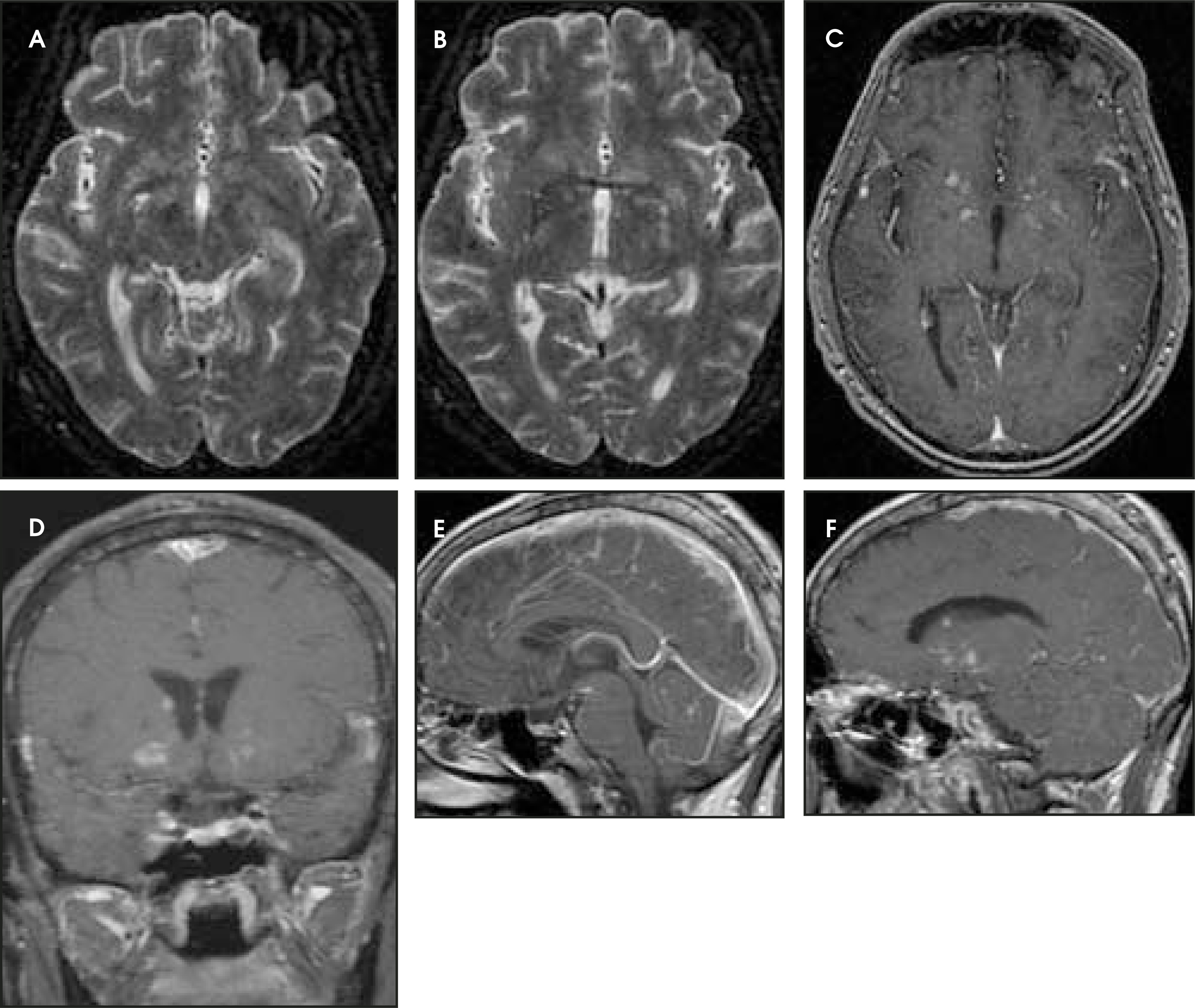
A cryptococcoma is a collection of organisms, inflammatory cells, and mucoid material in the brain parenchyma. Cryptococcomas can develop from extension of organisms from the perivascular spaces into the parenchyma or from direct invasion from other meningeal or ependymal surfaces. Choroid plexitis can result, with enlargement and enhancement of the choroid plexus associated with leptomeningeal enhancement.36 Unilateral cystic dilatation of the temporal horn of the lateral ventricle has been described secondary to CSF entrapment by an inflamed choroid plexus. The diagnosis of infection with Cryptococcus is made by detection of cryptococcal antigen in CSF or by identification of the cryptococci by an india-ink preparation of the CSF.34 Treatment involves antifungal therapy, which may have significant adverse effects. Despite appropriate treatment, recurrence is common, and the mortality rates can reach 40% in immuno-compromised individuals, with a mean survival of 2 to 3 months.1
Conclusion
Imaging studies performed for neurologic illness in AIDS play an important role in diagnosis and management. The authors described a simple algorithmic approach based on the imaging findings to guide differential diagnosis for the clinician and radiologist. Four patterns should be recognized: 1) normal; 2) atrophy; 3) intra-axial lesion without mass or mass effect; and 4) mass lesions. Each pattern provides information that is helpful in the differential diagnosis of the AIDS patient. Normal studies do not exclude pathology. Frequently, cryptococcal and other infectious processes have normal studies in the initial presentation. Normal studies require close clinical follow-up with repeat imaging, as necessary, to assist in the recognition of occult processes in patients with neurologic symptoms. Findings of atrophy may be related to normal aging or to pathologic processes. Pathologic processes, such as HIV encephalopathy and AIDS dementia complex (ADC), should be considered when atrophy is identified. Atrophy can also be seen in progressive multifocal leukoencephalopathy; however, underlying parenchymal lesions are also present. When encountering a parenchymal lesion without mass or mass effect, PML must be considered. Intra-axial mass lesions represent an ongoing debate, since 80% of brain masses in patients with AIDS are caused by toxoplasmosis or lymphoma.4-6 Prompt clinical diagnosis is necessary for proper treatment, so a mass lesion that is suggestive of toxoplasmosis should be treated empirically, with nonresponding patients undergoing biopsy for further clarification of diagnosis. Periventricular masses with thick subependymal spread are highly characteristic of lymphoma. Cystic masses in the region of the basal ganglia are pathognomonic for cryptococcal involvement.
References
- Levy R, Bredesen D, Rosenblum M, Levy J. AIDS Pathogenesis and Treatment. 1988:371-401.
- Levy R, Bredesen D, Rosenblum M. Neurological manifestations of the acquired immunodeficiency syndrome (AIDS): Experience at UCSF and review of the literature. J Neurosurg. 1985;62:475-495.
- Chinn R, Wilkinson I, Hall-Craggs M. Toxoplasmosis and primary central nervous system lymphoma in HIV infection: Diagnosis with MR spectroscopy. Radiology. 1995;197:649-654.
- Ciricillo S, Rosenblum M. Use of CT and MR imaging to distinguish intracranial lesions and to define the need for biopsy in AIDS patients. J Neurosurg. 1990;73:720-724.
- Chiang F, Miller B, Chang L. Fulminant cerebral lymphoma in AIDS. AJNR Am J Neuroradiol. 1996;17:157-160.
- Rosenblum M, Levy R, Bredesen D. Neurosurgical implications of the acquired immunodeficiency syndrome (AIDS). Clin Neurosurg. 1988;34:419-445.
- Simpson D, Tagliati M. Neurologic manifestations of HIV infection. Ann Intern Med Erratum in: Ann Intern Med.
- Navia B, Jordan B, Price R. The AIDS dementia complex: I. Clinical features. Ann Neurol. 1986;19:517-524.
- Post M, Tate L, Quencer R. CT, MR, and pathology in HIV encephalitis and meningitis. AJR Am J Roentgenol. 1988;151:373-380.
- Thurnher M, Schindler E, Thurnher S. Highly active antiretroviral therapy for patients with AIDS dementia complex: Effect on MR imaging findings and clinical course. AJNR Am J Neuroradiol. 2000;21:670-678.
- Filippi C, Sze G, Farber S. Regression of HIV encephalopathy and basal ganglia signal intensity abnormality at MR imaging in patients with AIDS after the initiation of protease inhibitor therapy. Radiology. 1998;206:491-498.
- Padgett B, Walker D, ZuRhein G. Cultivation of papova-like virus from human brain with progressive multifocal leucoencephalopathy. Lancet. 1971;1:1257-1260.
- Love S, Wiley C, Graham D, Lantos P. Greenfield’s Neuropathology. 2002:1-105.
- Tornatore C, Berger J, Houff S. Detection of JC virus DNA in peripheral lymphocytes from patients with and without progressive multi-focal leukoencephalopathy. Ann Neurol. 1992;31:454-462.
- Berger J, Kaszovitz B, Post M, Dickinson G. Progressive multifocal leukoencephalopathy associated with human immunodeficiency virus infection. A review of the literature with a report of sixteen cases. Ann Intern Med. 1987;107:78-87.
- Whiteman M, Post M, Berger J. Progressive multifocal leukoencephalopathy in 47 HIV-seropositive patients: Neuroimaging with clinical and pathologic correlation. Radiology. 1993;187:233-240.
- Mark A, Atlas S. Progressive multifocal leuko-encephalopathy in patients with AIDS: Appearance on MR Images. Radiology. 1989;173:517-520.
- Post M, Yiannoutsos C, Simpson D. Progressive multifocal leukoencephalopathy in AIDS: Are there any MR findings useful to patient management and predictive of patient survival? AIDS Clinical Trials Group, 243 Team. AJNR Am J Neuroradiol. 1999;20:1896-1906.
- Holman R, Janssen R, Buehler J. Epidemiology of progressive multifocal leukoencephalopathy in the United States: Analysis of national mortality and AIDS surveillance data. Neurology. 1991;41:1733-1736.
- Thurnher M, Post M, Rieger A. Initial and follow-up MR imaging findings in AIDS-related progressive multifocal leukoencephalopathy treated with highly active antiretroviral therapy. AJNR Am J Neuroradiol. 2001;22:977-984.
- Anders K, Guerra W, Tomiyasu U. The neuropathology of AIDS: UCLA experience and review. Am J Pathol. 1986;124:537-558.
- Porter S, Sande M. Toxoplasmosis of the central nervous system in the acquired immunodeficiency syndrome. N Engl J Med. 1992;327:1643-1648.
- Chang L, Cornford M, Chiang F. Radiologic-pathologic correlation. Cerebral toxoplasmosis and lymphoma in AIDS. AJNR Am J Neuroradiol. 1995;16:1653-1663.
- MacMahon E, Glass J, Hayward S. Epstein Barr Virus in AIDS-related primary central nervous system lymphoma. Lancet. 1991;338:969-973.
- So Y, Beckstead J, Davis R. Primary central nervous system lymphoma in acquired immune deficiency syndrome: A clinical and pathologic study. Ann Neurol. 1986;20:566-572.
- Rosenblum M, Levy R, Bredesen D. Primary central nervous system lymphomas in patients with AIDS. Ann Neurol. 1988;23(suppl):S13-S16.
- Lee V, Antonacci V, Tilak S. Intracranial mass lesions: Sequential thallium and gallium scintigraphy in patients with AIDS. Radiology. 1999;211:507-512.
- Morgello S, Petito C, Mouradian J. Central nervous system lymphoma in the acquired immunodeficiency syndrome. Clin Neuropathol. 1990;9:205-215.
- Camacho D, Smith J, Castillo M. Differentiation of toxoplasmosis and lymphoma in AIDS patients by using apparent diffusion coefficients. AJNR Am J Neuroradiol. 2003;24:633-637.
- Kessler L, Ruiz A, Post J. Thallium-201 brain SPECT of lymphoma in AIDS patients: Pitfalls and technique optimization. AJNR Am J Neuroradiol. 1998;19:1105-1109.
- Skiest D, Erdman W, Chang W. SPECT thallium-201 combined with toxoplasma serology for the presumptive diagnosis of focal central nervous system mass lesions in patients with AIDS. J Infect. 2000;40:274-281.
- Cordoliani Y, Derosier C, Pharaboz C. Primary cerebral lymphoma in patients with AIDS: MR findings in 17 cases. AJR Am J Roentgenol. 1992;159:841-847.
- Dismukes W. Cryptococcal meningitis in patients with AIDS. J Infect Dis. 1988;157:624-628.
- Kovoor J, Mahadevan A, Narayan J. Cryptococcal choroid plexitis as a mass lesion: MR imaging and histopathologic correlation. AJNR Am J Neuroradiol. 2002;23:273-276.
- Igel H, Bolande R. Humoral defense mechanisms in cryptococcosis: Substances in normal human serum, saliva, and cerebrospinal fluid affecting the growth of cryptococcus neoformans. J Infect Dis. 1966;116:75-83.
- Andreula C, Burdi N, Carella A. CNS cryptococcosis in AIDS: Spectrum of MR findings. J Comput Assist Tomogr. 1993;17:438-441.
Citation
. An algorithmic approach to neuroimaging in AIDS. Applied Radiology. 2005;34(12):18-32. doi:10.37549/AR1391.