Angiography and embolization in lower gastrointestinal bleeding
Applied Radiology — Vol. 33 , Issue 12 , pp. 9 -18
DOI: 10.37549/AR1302
Published: December 1, 2004
Categories
Acute arterial bleeding from the lower gastrointestinal tract has traditionally been treated with vasopressin infusion, but, more and more frequently, embolization is being selected as primary therapy. While favored proximal to the ligament of Treitz, embolization historically garnered little enthusiasm in the lower tract due to the less-developed collateral arterial network and early reports of intestinal infarctions and strictures.1-5 Accordingly, embolization was largely reserved for salvage therapy in patients who failed vasopressin infusion or were poor candidates for vasopressin and surgery.
Since the development of microcatheters and new embolic materials, embolization has been chosen more frequently as primary therapy, with fewer complications reported. Microcatheters allow more selective catheterization, often enabling embolization of the offending artery at the bowel wall. Though more technically challenging for the angiographer, successful superselective embolization results in prompt cessation of bleeding and minimizes the amount of bowel at risk. Furthermore, unlike vasopressin infusion, it allows immediate catheter removal and, often, earlier discharge from the intensive care unit.
Background
Most bleeding from the lower gastrointestinal tract will stop with conservative management and recur.6 However, some patients require therapy to halt life-threatening or prolonged bleeding. In addition to transcatheter therapies, endoscopy and surgery are options.
While useful for evaluation of slow or chronic bleeding, endoscopy has limited utility during acute arterial bleeding. Intraluminal blood and retained stool impair visibility at colonoscopy and limit the ability to reach the right colon, a common site of bleeding.7 Furthermore, the lower gastrointestinal tract includes all of the small intestine distal to the ligament of Treitz, a region that cannot be fully evaluated endoscopically.
While surgery does not share these limitations, emergent surgery for active bleeding carries a mortality of 20% to 30% with reports as high as 47%. 6,8,9
Further, when bleeding cannot be localized, empiric resection may be necessary, and, frequently, a subtotal colectomy is chosen.6,10 Nevertheless, as active bleeding can rarely be identified intraoperatively, the resected bowel may not include the bleeding site, and hemorrhage may recur.11-13
Angiography to diagnose gastrointestinal bleeding was first described in 1963, and, shortly thereafter, therapeutic vasoconstrictor drug infusions were reported.14,15 The most popular of these drugs, vasopressin, constricts smooth muscle in the arterial and bowel walls, causing vasospasm, reducing pulse pressure and promoting hemostasis. Though vasopressin controls hemorrhage in up to 90% of cases, 30% to 50% will have recurrent bleeding.16,17 In addition, complications from systemic vasoconstriction and prolonged arterial catheterization are not infrequent, and some patients, particularly those with known cardiac disease, are not candidates for vasopressin infusion.
The high failure and complication rates of available therapies contributed to investigation for an alternative treatment for lower gastrointestinal bleeding.
Pre-procedure evaluation
For maximal benefit, angiography should be performed during active bleeding. Therefore, the history and physical exam should seek to determine the location, the cause, and the rate of bleeding. These parameters give an indication of the likelihood of detection with angiography and of effective control with embolization.
Signs and symptoms of lower tract bleeding are typically nonspecific with the exception of profuse hematochezia or melena. Symptoms and signs often relate to hypovolemia and/or anemia and include lightheadedness, dizziness, syncope, angina, diaphoresis, tachycardia, hypotension, orthostasis, and cold distal extremities. However, patients with brisk bleeding may present with severe hemodynamic compromise or cardiovascular collapse. Tachycardia and hypotension are the best indicators of active bleeding. In fact, Nicholson et al18 noted a perfect correlation between contrast extravasation and a systolic blood pressure less than 100 mm Hg at the time of angiography. Laboratory values—including hematocrit, platelet count, coagulation parameters, and creatinine—are important to establish. Nevertheless, declining hematocrit values are not helpful in establishing active bleeding, though stable values usually exclude significant blood loss.
Review of the past medical history may reveal predisposing conditions or suggest etiologies (Table 1) that may alter the approach to or the success of therapy. For example, vasopressin is often favored for anastomotic bleeding or diffuse bowel processes. Further, knowledge of cardiovascular risk factors is important if embolization is not possible and vasopressin must be used.
Pertinent imaging should be reviewed. Endoscopy results may reveal potential bleeding sites. A Tc-99m red blood scan is helpful in documenting active bleeding, estimating bleeding rate, and localizing bleeding to a vascular distribution. Delayed positive images should be reviewed with caution, as extravasated radiotracer may have transited far from the bleeding focus. Further, though scintigraphy is estimated to detect bleeding rates as low as 0.1 mL/min, angiography, at best, detects bleeding of 0.5 to 1.0 mL/min.14
Contraindications
There are no absolute contraindications to angiography and embolization. However, several relative contraindications are worth mentioning. Coagulopathy is a relative contraindication because transcatheter therapies rely on effective native hemostasis. Embolization agents, though they partially block flow, primarily function as a thrombogenic matrix. Further, Encarnacion et al19 noted that coagulopathy was associated with a significantly higher volume of red blood cells administered, a significantly lower rate of success with embolization, and a significantly higher mortality due to bleeding. Therefore, a coagulopathy is best corrected prior to angiography. Nevertheless, a correctable coagulopathy should not delay evaluation and intervention. For patients with uncorrectable coagulopathy, transcatheter therapy may be attempted, but the patient and clinical team should be apprised of the lower success rate and higher complication rate.
Other relative contraindications include congestive heart failure, recent myocardial infarction, pregnancy, renal insufficiency and allergy to iodinated contrast media. Typically, the risk of a complication is acceptable when the procedure is potentially life-saving and the surgical alternative is more risky. For patients with renal insufficiency or a contrast allergy, risk may be minimized with the use of nonionic contrast media or an alternative contrast agent. One alternative agent, carbon dioxide, has had mixed results in the evaluation of gastrointestinal bleeding.20,21 Side effects are mild and include nausea, vomiting, and abdominal discomfort.
Procedure
Intensive nursing care is required during these procedures. The patient’s heart rate, blood pressure, oxygen saturation, and sedation level must be monitored. Typically, conscious sedation with a combination of midazolam and fentanyl or their equivalents is sufficient. Fluid resuscitation should be initiated prior to and be continued throughout the procedure, and deficient coagulation factors, platelets, and red blood cells should be replaced. If necessary, pressor support should be instituted and titrated to an acceptable blood pressure. A bladder catheter is useful for monitoring urinary output and for draining excreted contrast that may obscure pelvic bowel loops. Some practitioners may choose to administer antibiotics to protect against intestinal and/or skin flora.
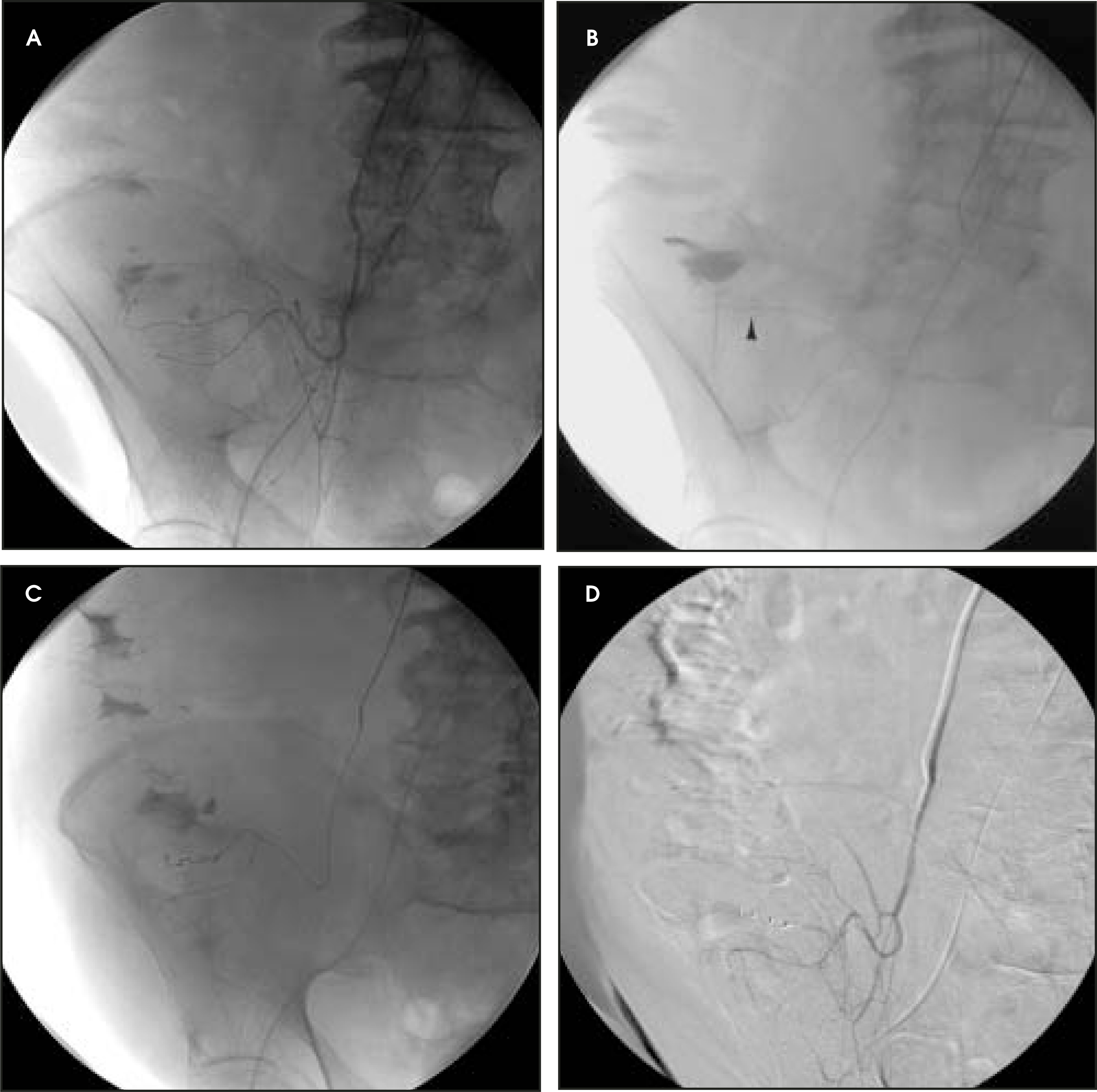
Typically, the right common femoral artery is catheterized. An arterial sheath facilitates catheter exchanges and is required for embolization. If the bleeding site is known, the artery to that site is selected first. Alternately, if the study is performed without a suggested site and if the inferior mesenteric artery (Figures 1A and 1B) is patent, it is selected before excreted contrast in the bladder obscures pelvic bowel loops. Negative evaluation of the inferior mesenteric artery should be followed by selective catheterization of the superior mesenteric artery (Figure 1C). Finally, the celiac artery should be evaluated. Typically not a source of blood supply to the lower tract, the celiac artery rarely gives origin to the middle colic artery. Furthermore, upper tract bleeding occasionally simulates lower tract bleeding clinically. Finally, if bleeding arises from the rectum, it may be necessary to evaluate the internal iliac arteries to exclude contribution from the hemorrhoidal arteries (Figure 2).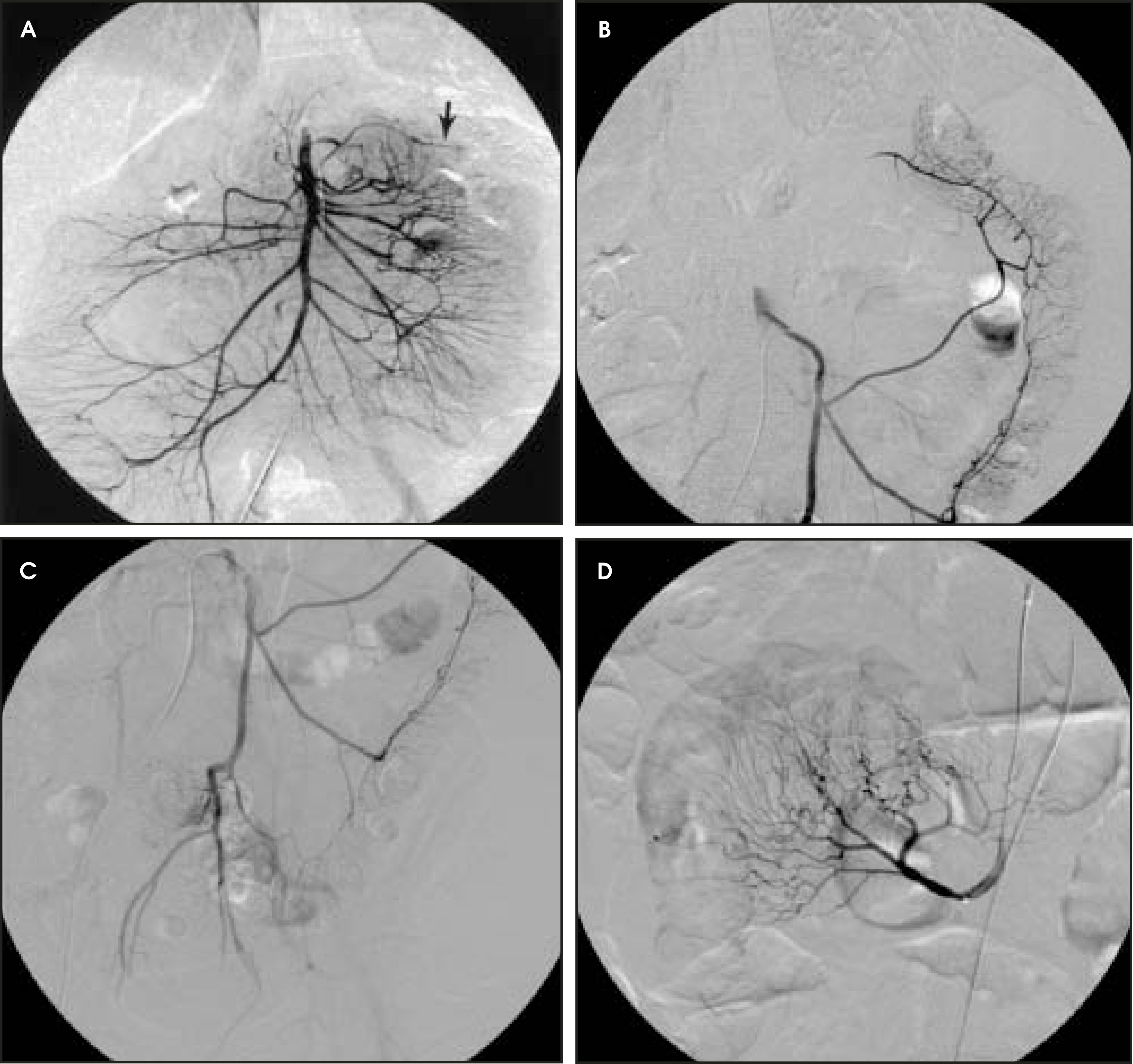
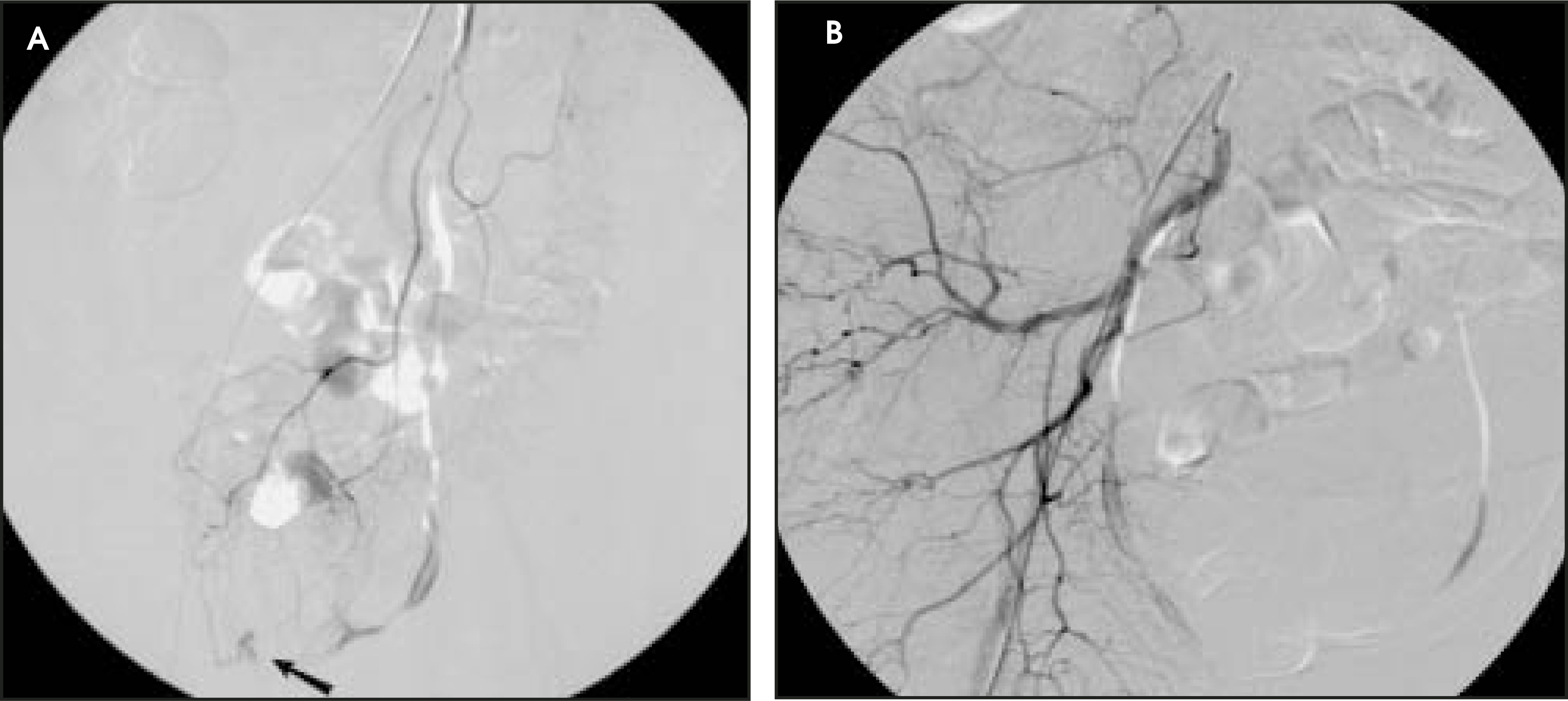
Subselective catheterization and/or magnification views may be necessary (Figure 1D). Injection rates should be adequate to opacify an artery’s complete distribution, and filming should be at least 2 frames per second with continuation into the venous phase. Digital subtraction angiography images must be reviewed in subtracted and unsubtracted viewing modes to distinguish respiratory and bowel motion from extravasation. Glucagon (1 mg) can be administered intravenously to reduce bowel peristalsis.
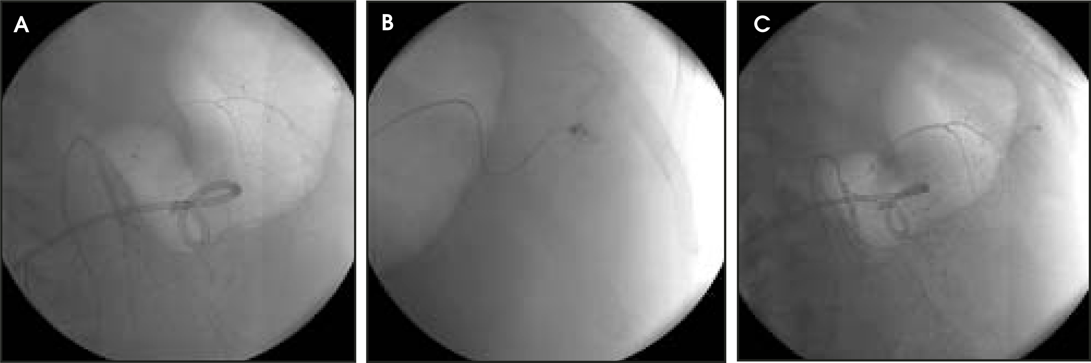
When extravasation is confirmed, superselective arterial catheterization is performed (Figure 3). First, the catheter is advanced to or into the truncal vessel supplying the bleeding site. Next, a microcatheter and wire combination are advanced coaxially through the catheter as distally as possible, often to the distal arcade or into the bleeding vasa recta (Figures 4 and 5).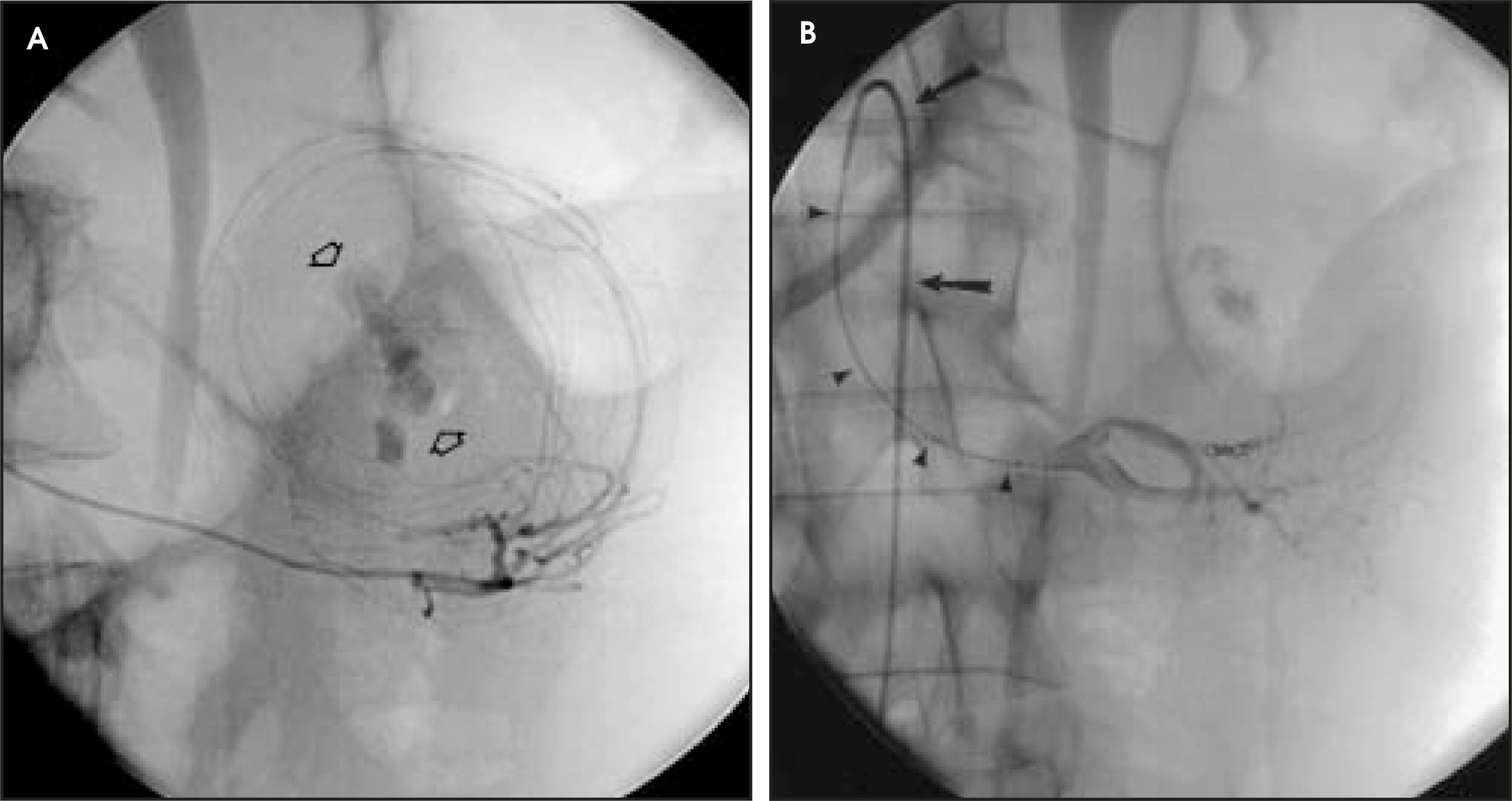
Superselective catheterization can be the most difficult portion of the procedure. Vascular tortuosity can be problematic, and satisfactory catheter position may not be achievable. Further, vessel spasm from wire and catheter manipulations occurs frequently. Intra-arterial nitroglycerin in 50- to 200-mg doses can be administered if the patient can tolerate the hemodynamic effects of the drug. Notably, catheter-induced spasm by itself has produced long-term hemostasis in some patients.22
Prior to embolization, contrast injection should confirm extravasation and define the territory that will be affected (Figure 5B). If embolization will affect too large a region, alternate therapies, including vasopressin infusion, and surgery should be considered.
Both temporary and permanent agents have been successfully used to treat lower tract bleeding. Autogenous clot, sometimes modified to enhance thrombogenicity, and Gelfoam pledgets (Pharmacia & Upjohn Co., Kalamazoo, MI) were used in early embolizations.1,2,23 However, both agents have limitations. They require added contrast for visibility and, due to their temporary nature, the artery can recanalize. In fact, use of autogenous clot may lead to recanalization within 24 hours, sometimes resulting in rebleeding.24 Polyvinyl alcohol (350 to 500 μm or larger) and metallic microcoils became favored because they could pass through microcatheters and were permanent. Microcoils in particular were commonly chosen because they could be precisely deployed and were easily visible—features that reduce the risk of nontarget embolization (Figures 4, 5, and 6). Agents that reach the capillary level, such as Gelfoam powder, polyvinyl alcohol particles smaller than 250 μm, alcohol, and Ethibloc (Johnson & Johnson Co., Brussels, Belgium) should be avoided because they can cause infarction.25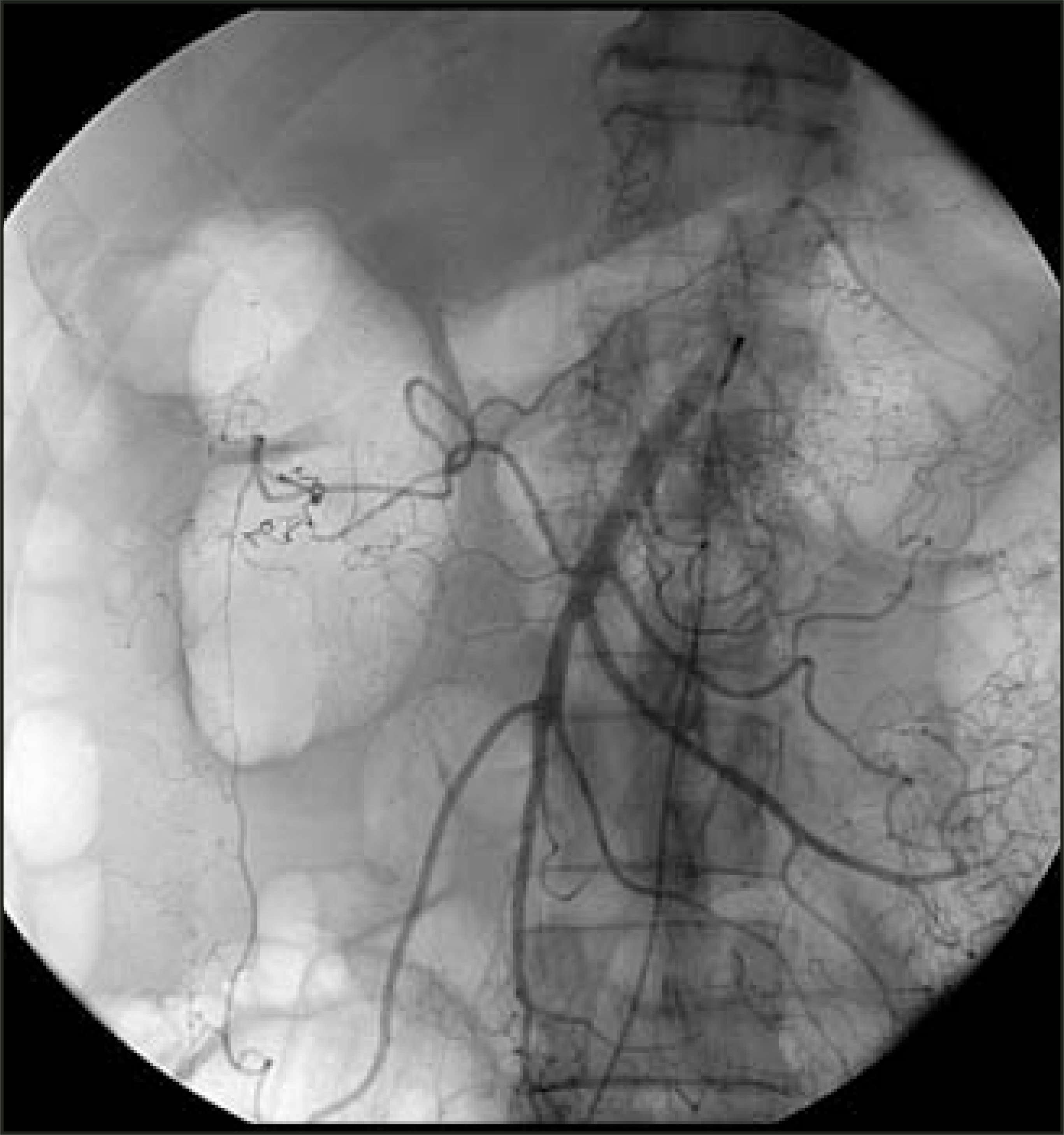
Typically, only small amounts of the embolized agent are necessary to stop bleeding. Therefore, arteriography should be performed periodically during embolization to determine whether extravasation has ceased (Figure 5C). Larger amounts risk infarction or nontarget embolization without added benefit.26
Final arteriography is performed to exclude residual extravasation and detect potential complications (Figure 6). Failure to stop bleeding can result from incomplete embolization, collateral arterial filling to the site, and coagulopathy. If extravasation persists and if further embolization risks bowel injury, endoscopy or surgery should be considered. Vasopressin is not advocated after unsuccessful embolization due to the risk of inducing ischemia. If surgery is selected for small-bowel bleeding, the catheter can facilitate localization via methylene blue injection at open laparotomy, or coils can be deployed in the feeding artery for identification by palpation or radiography.27-29
Catheters and sheaths should be removed as soon as coagulation parameters are acceptable. Arterial puncture site closure devices may allow sheath removal despite coagulopathy.
Follow-up
Following cessation of bleeding, the patient should be monitored in an intensive care unit. Some patients will require additional transfusions as they “catch up” from the large volumes lost. Though declining hematocrit values or inappropriate rises can indicate continued bleeding, they may reflect ongoing fluid shifts, particularly after large volume fluid resuscitation. Bloody stools may take days to resolve, particularly if bleeding arose from the small intestine. Strict bowel rest is advised until the exam and laboratory profile normalize. This reduces confusing clinical information and decreases morbidity should additional invasive procedures be necessary.
Abdominal pain, sometimes with fever and leukocytosis, is not uncommon after embolization.2 This pain may reflect the underlying bowel pathology, a postembolization syndrome, mild ischemia, or developing infarction. Therefore, periodic abdominal exams are necessary to evaluate evolution of pain over time and to detect early signs of ischemia and infarction.
Additional signs and symptoms of infarction include absent bowel sounds, abdominal distension, a palpable mass, and peritoneal signs. Laboratory parameters may reveal a leukocytosis and anion gap, and, if measured, the lactic acid level will likely be elevated. For sedated or obtunded patients, abdominal rigidity or an abnormal laboratory profile may be the first indicators of infarction. Prompt surgery is necessary to reduce morbidity and mortality.
In contrast, mild ischemia may be managed conservatively, and for some patients, will resolve or be undetectable. Guy et al30 reported 2 asymptomatic patients with endoscopic evidence of mucosal ischemia. Similarly, Nicholson et al18 described the uneventful recovery of a patient with symptoms of ischemic colitis following embolization who had mucosal necrosis at colonoscopy. Broad-spectrum antibiotics may be useful if ischemia is suspected.
Despite successful embolization, some patients will experience continued or recurrent bleeding. If bleeding is suspected, scintigraphy or arteriography may be necessary for confirmation. Repeat arteriography may reveal recanalization of the embolized vessel, bleeding from collaterals, bleeding from a new site, or no abnormality. Embolization may be repeated if extravasation is confirmed and bowel injury is unlikely.
Arteriography is rarely diagnostic in establishing a cause of bleeding. Thus, most patients require evaluation through endoscopy or radiologic studies to determine the origin of bleeding and to assess the need for additional therapy. This evaluation can often be delayed until the patient is fully recovered.
Results
Arteriography is positive in only 40% to 70% of patients with clinical evidence of active bleeding.17,31 Unfortunately, many patients undergo multiple exams due to continued or recurrent bleeds. As a result, heparin and thrombolytic agents have been used to provoke blood loss in patients with bleeding of obscure origin. Koval et al32 reported improvement in detection from 32% to 65% using intravenous heparin, an intra-arterial vasodilator, and thrombolytic administration. Subsequent studies have confirmed improved yield without any complications.33-36
Since the first report of embolization for lower gastrointestinal bleeding in 1974, the procedure has been reported in more than 40 publications.1 Technical success is defined as the cessation of extravasation after embolization. In a recent review, Darcy37 noted technical success rates ranging from 73% to 100%. He ascribed an overall decline in technical success between old studies (average 97%) and new studies using microcatheters (average 88%) to more stringent criteria for claiming success as operators seek more selective sites of embolization.
Clinical success, defined as prolonged cessation of bleeding, is lower than the technical success in most series due to recurrence of bleeding. Darcy37 noted clinical success rates ranging from 60% to 100% with an average of 83% in recent series. Overall clinical success is superior in the colon (range 86% to 100%). Otherwise, clinical success has not been convincingly related to the site of embolization, the embolization agent used, or the cause of bleeding.
Complications
Bowel infarction is the most feared complication of embolization. Contributing factors include overembolization and insufficient collateral flow, but often the cause is unclear. Alterations in wound healing from hemorrhage or other factors may contribute.38 Bowel infarction following embolization has occurred in conjunction with dissection of the artery and when embolization was performed immediately following vasopressin infusion.5,39 Though unproven, these reports suggest a threshold of inflow reduction that may contribute to infarction. Similarly, though anastomotic bleeding has been successfully treated with embolization, infarction has been reported.4,40,41 Embolization is thought to be risky due to surgical changes that limit collateral flow. Therefore, vasopressin is preferred for anastomotic bleeds.
Bowel strictures have been described on follow-up endoscopy and barium studies.3 First reported by Mitty3 in 1979, this complication has been corroborated in humans and dogs.18,24 Since strictures may be asymptomatic, the frequency of this complication is unknown, and the significance remains unclear.
Though the technical and clinical success rates have remained similar over time, Darcy37 described a decline in complication rates. Prior to 1990, he noted minor complication rates of 20.2% and major complication rates of 11.1%. However, newer studies, using microcatheters and superselective embolization, reveal a reduction in the minor complication rate to 15.3% and the major complication rate to 1.3%. Thus, the rate of infarction, though not zero, has declined with changes in practice.
Some authors propose that the risk of intestinal infarction is justifiable for life-threatening bleeding. They support their argument by noting the high rate of success with embolization, the low rate of intestinal injury, and the high rate of surgical morbidity.3,5,26 Moreover, successful embolization may delay surgery to a time when the patient can better tolerate the procedure.42
Additional complications include allergic reactions, renal failure, vascular damage and nontarget embolization. The severity of the latter will be determined by the tissue affected.
Conclusion
Superselective embolization is increasingly replacing vasopressin infusion as the transcatheter therapy of choice for treatment of lower gastrointestinal tract bleeding. Though technically more challenging, the procedure allows more rapid cessation of bleeding with lower rebleeding and complication rates.
References
- Bookstein J, Chlosta E, Foley D, Walter J. Transcatheter hemostasis of gastrointestinal bleeding using modified autogenous clot. Radiology. 1974;113:277-285.
- Chuang V, Wallace S, Zornoza J, Davis L. Transcatheter arterial occlusion in the management of rectosigmoidal bleeding. Radiology. 1979;133:605-609.
- Mitty H, Efremidis S, Keller R. Colonic stricture after transcatheter embolization for diverticular bleeding. AJR Am J Roentgenol. 1979;133:519-521.
- Shenoy S, Satchidanand S, Wesp E. Colonic ischemic necrosis following therapeutic embolization. Gastrointest Radiol. 1981;6:235-237.
- Rosenkrantz H, Bookstein J, Rosen R. Postembolic colonic infarction. Radiology. 1982;142:47-51.
- Rigg B, Ewing M. Current attitudes on diverticulitis with particular reference to colonic bleeding. Arch Surg. 1966;92:321-332.
- Casarella W, Kanter I, Seaman W. Right-sided colonic diverticula as a cause of acute rectal hemorrhage. N Engl J Med. 1972;286:450-453.
- Taylor F, Epstein L. Treatment of massive diverticular hemorrhage. Arch Surg. 1969;98:505-508.
- Pennoyer W, Vignati P, Cohen J. Management of angiogram positive lower gastrointestinal hemorrhage: Long term follow-up of non-operative treatments. Int J Colorect Dis. 1996;11:279-282.
- Olsen W. Hemorrhage from diverticular disease of the colon. The role of emergency subtotal colectomy. Am J Surg. 1968;115:247-263.
- Giffin J, Butcher H, Ackerman L. Surgical management of colonic diverticulitis. Arch Surg. 1967;94:619-626.
- Klein R, Gallagher D. Massive colonic bleeding from diverticular disease. Am J Surg. 1969;118:553-557.
- Sebrechts C, Bookstein J. Embolization in the management of lower-gastrointestinal hemorrhage. Semin Intervent Radiol. 1988;5:39-48.
- Nusbaum M, Baum S. Radiographic demonstration of unknown sites of gastrointestinal bleeding. Surg Forum. 1963;14:374-375.
- Baum S, Nusbaum M. The control of gastrointestinal hemorrhage by selective mesenteric arterial infusion of vasopressin. Radiology. 1971;98:497-505.
- Athanasoulis C, Baum S, Rosch J. Mesenteric arterial infusions of vasopressin for hemorrhage from colonic diverticulosis. Am J Surg. 1975;129:212-216.
- Browder W, Cerise E, Litwin M. Impact of emergency angiography in massive lower gastrointestinal bleeding. Ann Surg. 1986;204:530-536.
- Nicholson A, Ettles D, Hartley J. Transcatheter coil embolotherapy: A safe and effective option for major colonic haemorrhage. Gut. 1998;43:79-84.
- Encarnacion C, Kadir S, Beam C, Payne C. Gastrointestinal bleeding: Treatment with gastroin-testinal arterial embolization. Radiology. 1992;183:505-508.
- Sandhu C, Buckenham T, Belli A. Using CO2-enhanced arteriography to investigate acute gastrointestinal hemorrhage. AJR Am J Roentgenol. 1999;173:1399-1401.
- Hawkins I, Caridi J. Carbon dioxide digital subtraction angiography: 26 year experience at the University of Florida. Eur Radiol. 1998;8:391-402.
- Cynamon J, Atar E, Steiner A. Catheter-induced vasospasm in the treatment of acute lower gastrointestinal bleeding. J Vasc Interv Radiol. 2003;14:211-216.
- Goldberger L, Bookstein J. Transcatheter embolization for treatment of diverticular hemorrhage. Radiology. 1977;122:613-617.
- Cho K, Schmidt R, Lenz J. Effects of experimental embolization of superior mesenteric artery branch on the intestine. Invest Radiol. 1979;14:207-212.
- Kusano S, Murata K, Ohuchi H. Low-dose particulate polyvinylalcohol embolization in massive small artery intestinal hemorrhage. Experimental and clinical results. Invest Radiol. 1987;22:388-392.
- Uflacker R. Transcatheter embolization for treatment of acute lower gastrointestinal bleeding. Acta Radiologica. 1987;28:425-430.
- Zuckerman D, Gaz R. Catheter-guided intraoperative localization of a jejunal angiodysplasia using the tracker-18 coaxial catheter system: case report. Cardiovasc Intervent Radiol. 1991;14:358-359.
- Schmidt S, Boskind J, Smith D, Catalano R. Angiographic localization of small bowel angiodysplasia with use of platinum coils. J Vasc Interv Radiol. 1993;4:737-739.
- Athanasoulis C, Moncure A, Greenfield A. Intraoperative localization of small bowel bleeding sites with combined use of angiographic methods and methylene blue injection. Surgery. 1980;87:77-84.
- Guy G, Shetty P, Sharma R. Acute lower gastrointestinal hemorrhage: Treatment by superselective embolization with polyvinyl alcohol particles. AJR Am J Roentgenol. 1992;159:521-526.
- Colacchio T, Forde K, Patsos T, Nunez D. Impact of modern diagnostic methods on the management of active rectal bleeding. Am J Surg. 1982;143:607-610.
- Koval G, Benner K, Rosch J, Kozak B. Aggressive angiographic diagnosis in acute lower gastrointestinal hemorrhage. Dig Dis Sci. 1987;32:248-253.
- Glickerman D, Kowdley K, Rosch J. Urokinase in gastrointestinal tract bleeding. Radiology. 1988;168:375-376.
- Malden E, Hicks M, Royal H. Recurrent gastrointestinal bleeding: Use of thrombolysis with anticoagulation in diagnosis. Radiology. 1998;207:147-151.
- Mernagh J, O’Donovan N, Somers S. Use of heparin in the investigation of obscure gastrointestinal bleeding. Can Assoc Radiol J. 2001;52:232-235.
- Ryan J, Key S, Dumbleton S, Smith T. Nonlocalized lower gastrointestinal bleeding: provocative bleeding studies with intraarterial tPA, heparin, and tolazoline. J Vasc Interv Radiol. 2001;12:1273-1277.
- Darcy M. Treatment of lower gastrointestinal bleeding: Vasopressin infusion versus embolization. J Vasc Interv Radiol. 2003;14:535-543.
- McGinn F. Effects of haemorrhage upon surgical operations. Br J Surg. 1976;63:742-746.
- Walker W, Goldin A, Shaff M, Allibone G. Per catheter control of haemorrhage from the superior and inferior mesenteric arteries. Clin Radiol. 1980;31:71-80.
- Lawler G, Bircher M, Spencer J. Embolisation in colonic bleeding. Br J Radiol. 1985;58:83-84.
- DuBois J, Ostrow L, Smith G, Welling D. Transcatheter embolization of small bowel anastomosis: a case report and review of the literature. Milit Med. 1989;154:505-507.
- Rahn N, Tishler J, Han S, Russinovich N. Diagnostic and interventional angiography in acute gastrointestinal hemorrhage. Radiology. 1982;143:361-366.
Citation
. Angiography and embolization in lower gastrointestinal bleeding. Applied Radiology. 2004;33(12):9-18. doi:10.37549/AR1302.