Artery Embolization in the Treatment of Refractory Internal Hemorrhoids
Applied Radiology — Vol. 55 , Issue 2
Published: November 26, 2025
1 Joint & Vascular Institute, Libertyville, Illinois
2 Department of Interventional Radiology, UCLA Health, Los Angeles, California
* Corresponding author: Layth Alkhani (lalkhani2002@gmail.com)
Abstract
Hemorrhoids are naturally occurring vascular cushions within the anal canal, but today have become synonymous with the pathology of symptomatic hemorrhoid disease. Internal hemorrhoids are classified using the Goligher system, and management traditionally begins with conservative and office-based therapies. Many patients, however, experience recurrent or refractory symptoms, and surgical hemorrhoidectomy carries risks of pain, complications, and prolonged recovery. Hemorrhoid artery embolization (HAE) has emerged as a minimally invasive, nonsurgical option for patients with bleeding internal hemorrhoids. Appropriate patient selection is crucial, with ideal candidates experiencing significant bleeding refractory to conservative medical therapy. A comprehensive evaluation, including colonoscopy and bleeding/prolapse grading, is required. HAE involves selective embolization of hypertrophied superior rectal artery branches supplying the corpus cavernosum recti, using microspheres, coils, or both. The endpoint is decreased hemorrhoidal blush without complete stasis, followed by durable occlusion with coils. Data from nearly 400 patients demonstrate technical success rates of 93-100% and clinical success rates of 63-94%. Outcomes include significant reductions in bleeding scores, hemorrhoid size, prolapse, and pain. Patient satisfaction is high, with rates up to 94%. Early randomized controlled trial data show efficacy comparable to hemorrhoidectomy, but with shorter recovery, reduced pain, and fewer complications. HAE is a safe and effective treatment for internal hemorrhoids, particularly in patients with refractory bleeding who are not ideal surgical candidates or wish to avoid surgery. Future studies should refine patient selection, standardize procedural technique, and establish long-term comparative outcomes.
Categories
Introduction
Hemorrhoids are naturally occurring clusters of vascular tissue within the anal canal consisting of smooth muscle, connective tissue, and arteriovenous connections. 1,2 The hemorrhoidal cushions are naturally present and provide bulk to the anal canal that is believed to aid in stool continence. Classified by their location, either proximal or distal to the dentate line, hemorrhoids have become synonymous with symptomatic hemorrhoid disease. 3
Proximal hemorrhoids, also known as internal hemorrhoids, are most commonly characterized by rectal bleeding, particularly after defecation. 4,5 They are graded based on the degree of prolapse according to the Goligher classification system: grade I hemorrhoids bleed without prolapse, grade II hemorrhoids prolapse but spontaneously reduce, grade III hemorrhoids prolapse and require manual reduction, and grade IV hemorrhoids prolapse irreducibly. 6 Owing to the asymptomatic nature of up to 40% of cases, hemorrhoidal prevalence is difficult to quantify. 6 However, estimates of prevalence range from 4% to 40% globally; US studies report hemorrhoids to be the third-most common outpatient gastrointestinal diagnosis, with an estimated 3-4 million office and emergency department visits annually. 3,6-10
First-line treatment for symptomatic internal hemorrhoids typically consists of conservative medical management, including dietary changes to increase fiber and water intake, topical treatments such as corticosteroids or anti-inflammatory drugs, and other over-the-counter medications to relieve acute symptoms. 4,6,11,12 For patients with grade I-III internal hemorrhoids and symptoms refractory to conservative treatments, office-based therapies such as rubber band ligation, sclerotherapy, and infrared photocoagulation are often offered. These procedures, however, can be painful and have recurrence rates of up to 50%. 12-15 For grade III-IV internal hemorrhoids or recurrent hemorrhoids refractory to office-based treatments, surgical intervention is often required. 1,3,4 However, surgery is invasive and painful, and carries a risk of complications, with a median recovery time of approximately 6 weeks. 16,17
Here, we review the potential of hemorrhoid artery embolization (HAE) as a minimally invasive option for patients with bleeding internal hemorrhoids but who otherwise are either not ideal candidates for, or do not wish to pursue, traditional surgical treatment.
Patient Selection
Appropriate patient selection is crucial in ensuring a successful clinical response to HAE. While many patients with internal hemorrhoids report pain, HAE is most successful in those experiencing bleeding. In those without bleeding, the superior rectal arteries that supply the corpus cavernosum recti may not be hypertrophied, thus limiting the efficacy of embolization. Ideal candidates for HAE include those whose quality of life has been severely impacted by bleeding and who either want to avoid surgery or are nonsurgical candidates. Notably, conservative management continues to be an important adjunct to HAE, as with any treatment method, including diet modifications, lifestyle changes, sitz baths, and topical creams.
A comprehensive physical and medical history should be taken during the initial consultation with an interventional radiologist. Patients with a history of radiation to the pelvis should not be offered embolization, owing to potential compromise of collateral vasculature to the rectum, which increases the risk of ischemic complications. Patients must also undergo a colonoscopy to confirm the hemorrhoid grade and location, as well as ensure the absence of anorectal cancer. In addition to the degree of prolapse assessed using the Goligher classification (grade I-IV), the severity of bleeding should be measured using the French bleeding score, which ranges from 0 to 9. Both results should be documented during the initial visit. 18,19
As with any angiography-based procedure, patients with allergies to iodinated contrast media or renal failure have relative contraindications for HAE.
Procedural Technique
HAE seeks to reduce blood flow to the arteriovenous network within the internal hemorrhoidal cushions, known as the corpus cavernosum recti, via selective treatment of the corresponding terminal branches of the superior rectal arteries.
The interventional radiologist begins by obtaining femoral or radial arterial access using US guidance, after which they will place a vascular sheath and perform an aortogram to identify the origin of the inferior mesenteric artery (IMA). The IMA is then catheterized and a microcatheter is advanced into the superior rectal artery (SRA). Contrast is injected to identify the hypertrophied branches of the SRA supplying an area of contrast blush representing the corpus cavernosum recti within the hemorrhoidal cushions. The terminal branches of the SRA are then selectively catheterized, and angiography is used to confirm hemorrhoidal blush within the corpus cavernosum recti.
Embolization can be performed with microcoils, microspheres, or a combination of both. Microspheres are often diluted with iodinated contrast to a 1:20 ratio and are used sparingly to occlude distal arterial anastomoses within the middle and/or inferior rectal arteries. Microspheres range from 300 to 1200 µm in size; notably, larger microspheres showed better long-term efficacy and no minor ischemic findings compared with smaller sizes in one study. 20 Rather than complete stasis, the desired endpoint of microsphere embolization is decreased blush within the corpus cavernosum recti, which reduces the risk of ischemic complications. Microcoils are then used to embolize the distal SRAs to stasis and ensure durable occlusion.
Following embolization, the microcatheter is carefully withdrawn and angiography is performed to confirm major vessel patency and the absence of residual hemorrhoidal tissue blush below the pubic symphysis. The internal iliac arteries may also be catheterized to check for the presence of significant contributory middle rectal arteries. These may also be embolized using a technique similar to the one described. Some practices choose to perform middle rectal artery embolization only if they are visualized during SRA catheterization and/or if traditional superior rectal embolization fails to control bleeding.
Patients are observed for 2-6 hours and may be discharged the same day. Mild postprocedure discomfort can be treated with nonsteroidal anti-inflammatory drugs as needed. Follow-up is conducted 1-3 months post procedure.
Case Example
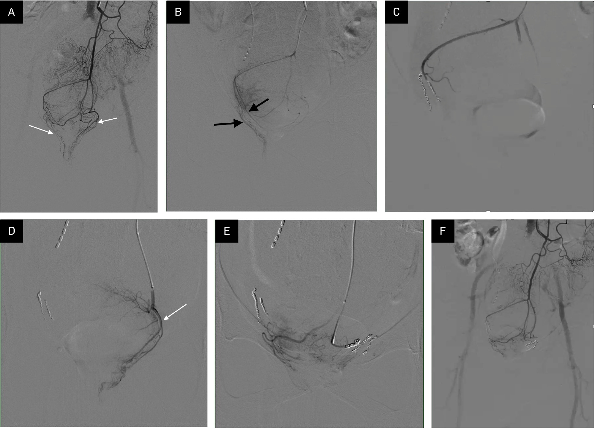
An adult patient presented with complaints of bright red bleeding upon defecation despite undergoing sclerotherapy and banding procedures for prolapsing internal hemorrhoids (Goligher grade II) 2 months prior. An arteriogram of the IMA (Figure 1) demonstrated dominant hypertrophied SRA branches supplying the corpus cavernosum recti below the pubic symphysis. These branches were embolized using a combination of a small amount of 500-700 μm microspheres and microcoils. A second arteriogram of the IMA demonstrated absence of blush in corpus cavernosum recti. Anoscopy performed 1 month post procedure demonstrated shrinkage of the internal hemorrhoids to Goligher grade I, with no evidence of anorectal complications. The procedure resulted in complete resolution of bleeding.
Clinical Efficacy
Literature, including nearly 400 patients, describes the feasibility and efficacy of treating hemorrhoids with HAE. 21,22 Rates of technical success, defined as embolization of at least one artery supplying the corpus cavernosum recti, range from 93% to 100% across all studies. Clinical success, often defined as reduction in French bleeding scores, is lower, ranging from 63% to 94%. 23-27 The leading cause of clinical failure in these studies was rebleeding; however, repeat embolization procedures have led to clinical success in some patients. 23 Meta-analyses have found statistically significant reductions in French bleeding scores (averaging ~2.7). 21,22 However, studies have also reported improvements in pain, prolapse scores, and prolapse size of >40%. 25,27 The variability in clinical success rates can be attributed to patient selection (most failures occurred in advanced cases) and technique.
Patient satisfaction rates correlate with high clinical success; studies report satisfaction rates as high as 94%. 25,28,29 A 2021 meta-analysis reported lower rebleeding rates for HAE using microspheres and coils. 22
Hemorrhoid Artery Embolization vs Surgery
Emerging evidence suggests efficacy rates for HAE comparable to surgical hemorrhoidectomy. A 2023 randomized control trial of 33 patients found no significant difference in hemorrhoidal symptom control between the embolization group and the surgical Ferguson hemorrhoidectomy group at 1 year post procedure. 30 By 12 months post treatment, the rates of bleeding, prolapse, and pruritus were similar between both groups. Along with similar clinical outcomes, the HAE group demonstrated a shorter recovery time. By 6 months post procedure, satisfaction rates between the 2 groups were also similar. The embolization group, however, reported lower pain and significantly lower medication usage. Patients undergoing hemorrhoidectomy report an average visual analog pain score of 6.6/10 during their first bowel movement compared with 0/10 in the embolization group. 30 These findings support the use of HAE as a clinically viable procedure that offers reduced pain and recovery time compared with hemorrhoidectomy.
Safety
Since 2015, major complications of HAE have been exceptionally rare, with only one case of rectosigmoidal ischemia reported. 31,32 In this instance, the patient developed severe sigmoidal stenosis and was treated with 3 endoscopic dilations using a balloon dilator. 32 This risk can be mitigated by careful embolic selection, placement, and coil use. Small superficial ulcerations have been reported in upward of 45% of patients. 20 However, these lesions are often asymptomatic and heal without intervention. The risk of ulceration may be reduced through the use of larger beads (>900 μm), according to one study. Notwithstanding these complications, HAE maintains a favorable safety profile. Some patients report mild transient symptoms such as pelvic or anal aching. This often resolves within a few days after conservative management. In contrast to surgery, severe pain following HAE is rare. Owing to the nature of the procedure, HAE does not cause anal sphincter damage or mucosal resection. The minimally invasive nature of the procedure also eliminates the risks of wound infection or delayed healing.
Conclusion
Data from nearly 400 patients suggest that HAE is a safe and effective treatment option for patients with internal hemorrhoids, particularly those with refractory bleeding and those seeking less invasive hemorrhoid treatment. Early evidence has shown outcomes comparable to hemorrhoidectomy, along with shorter recovery time, reduced pain, and fewer complications. Future studies should focus on the identification of ideal patient demographics, standardization of procedural techniques such as access and embolic, and compare long-term outcomes against traditional management to establish HAE’s role in hemorrhoid treatment.
References
- Fontem R, Eyvazzadeh D. In: StatPearls.
- Sandler R, Peery A. Rethinking what we know about hemorrhoids. Clin Gastroenterol Hepatol. 2019;17(1):8-15. doi:10.1016/j.cgh.2018.03.020.
- Sun Z, Migaly J. Review of hemorrhoid disease: presentation and management. Clin Colon Rectal Surg. 2016;29(1):22-29. doi:10.1055/s-0035-1568144.
- Mott T, Latimer K, Edwards C. Hemorrhoids: diagnosis and treatment options. Am Fam Physician. 2018;97(3):172-179.
- Sheikh P, Régnier C, Goron F, Salmat G. The prevalence, characteristics and treatment of hemorrhoidal disease: results of an international web-based survey. J Comp Eff Res. 2020;9(17):1219-1232. doi:10.2217/cer-2020-0159.
- Riss S, Weiser F, Schwameis K. The prevalence of hemorrhoids in adults. Int J Colorectal Dis. 2012;27(2):215-220. doi:10.1007/s00384-011-1316-3.
- Johanson J, Sonnenberg A. The prevalence of hemorrhoids and chronic constipation. An epidemiologic study. Gastroenterology. 1990;98(2):380-386. doi:10.1016/0016-5085(90)90828-o.
- Elbatea H, Enaba M, Elkassas G, El-Kalla F, Elfert A. Indications and outcome of colonoscopy in the middle of Nile delta of Egypt. Dig Dis Sci. 2011;56(7):2120-2123. doi:10.1007/s10620-010-1538-1.
- Lee J, Kim H, Kang J, Shin J, Song Y. Factors associated with hemorrhoids in Korean adults: Korean national health and nutrition examination survey. Korean J Fam Med. 2014;35(5):227. doi:10.4082/kjfm.2014.35.5.227.
- Peery A, Crockett S, Barritt A. Burden of gastrointestinal, liver, and pancreatic diseases in the United States. Gastroenterology. 2015;149(7):1731-1741. doi:10.1053/j.gastro.2015.08.045.
- Acheson A, Scholefield J. Management of haemorrhoids. BMJ. 2008;336(7640):380-383. doi:10.1136/bmj.39465.674745.80.
- MacRae H, McLeod R. Comparison of hemorrhoidal treatment modalities. A meta-analysis. Dis Colon Rectum. 1995;38(7):687-694. doi:10.1007/BF02048023.
- Pastor Peinado P, Ocaña J, Abadía Barno P. Quality of life and outcomes after rubber band ligation for haemorrhoidal disease. Langenbecks Arch Surg. 2023;408(1):243. doi:10.1007/s00423-023-02990-6.
- Brown S, Tiernan J, Watson A. Haemorrhoidal artery ligation versus rubber band ligation for the management of symptomatic second-degree and third-degree haemorrhoids (HubBLe): a multicentre, open-label, randomised controlled trial. Lancet. 2016;388(10042):356-364. doi:10.1016/S0140-6736(16)30584-0.
- Albuquerque A. Rubber band ligation of hemorrhoids: a guide for complications. World J Gastrointest Surg. 2016;8(9):614-620. doi:10.4240/wjgs.v8.i9.614.
- Cristea C, Lewis C. In: StatPearls.
- Bouchard D, Abramowitz L, Castinel A. One-year outcome of haemorrhoidectomy: a prospective multicentre french study. Colorectal Dis. 2013;15(6):719-726. doi:10.1111/codi.12090.
- Fathallah N, Beaussier H, Chatellier G. Proposal for a new score: hemorrhoidal bleeding score. Ann Coloproctol. 2021;37(5):311-317. doi:10.3393/ac.2020.08.19.
- Goligher J. Surgery of the Anus, Rectum, and Colon. 1980.
- Küçükay M, Küçükay F. Superior rectal artery embolization with tris-acryl gelatin microspheres: a randomized comparison of particle size. J Vasc Interv Radiol. 2021;32(6):819-825. doi:10.1016/j.jvir.2021.02.011.
- Nguyenhuy M, Xu Y, Kok H. Clinical outcomes following rectal artery embolisation for the treatment of internal haemorrhoids: a systematic review and meta-analysis. Cardiovasc Intervent Radiol. 2022;45(9):1351-1361. doi:10.1007/s00270-022-03154-7.
- Makris G, Thulasidasan N, Malietzis G. Catheter-directed hemorrhoidal dearterialization technique for the management of hemorrhoids: a meta-analysis of the clinical evidence. J Vasc Interv Radiol. 2021;32(8):1119-1127. doi:10.1016/j.jvir.2021.03.548.
- Vidal V, Sapoval M, Sielezneff Y. Emborrhoid: a new concept for the treatment of hemorrhoids with arterial embolization: the first 14 cases. Cardiovasc Intervent Radiol. 2015;38(1):72-78. doi:10.1007/s00270-014-1017-8.
- Sun X, Xu J, Zhang J, Jin Y, Chen Q. Management of rectal bleeding due to internal haemorrhoids with arterial embolisation: a single-centre experience and protocol. Clin Radiol. 2018;73(11):985. doi:10.1016/j.crad.2018.07.105.
- Zakharchenko A, Kaitoukov Y, Vinnik Y. Safety and efficacy of superior rectal artery embolization with particles and metallic coils for the treatment of hemorrhoids (Emborrhoid technique). Diagn Interv Imaging. 2016;97(11):1079-1084. doi:10.1016/j.diii.2016.08.002.
- Moussa N, Bonnet B, Pereira H. Mid-term results of superior rectal artery and coils for hemorrhoidal embolization with particles bleeding. Cardiovasc Intervent Radiol. 2020;43(7):1062-1069. doi:10.1007/s00270-020-02441-5.
- Bagla S, Pavidapha A, Lerner J. Outcomes of hemorrhoidal artery embolization from a multidisciplinary outpatient interventional center. J Vasc Interv Radiol. 2023;34(5):745-749. doi:10.1016/j.jvir.2023.01.023.
- Tradi F, Louis G, Giorgi R. Embolization of the superior rectal arteries for hemorrhoidal disease: prospective results in 25 patients. J Vasc Interv Radiol. 2018;29(6):884-892. doi:10.1016/j.jvir.2018.01.778.
- Panneau J, Mege D, Di Bisceglie M. Hemorrhoidal disease: what role can rectal artery embolization play?. Front Surg. 2024;11. doi:10.3389/fsurg.2024.1474799.
- Falsarella P, Nasser F, Affonso B. Embolization of the superior rectal arteries versus closed hemorrhoidectomy (Ferguson technique) in the treatment of hemorrhoidal disease: a randomized clinical trial. J Vasc Interv Radiol. 2023;34(5):736-744. doi:10.1016/j.jvir.2023.01.022.
- Talaie R, Torkian P, Moghadam A. Hemorrhoid embolization: a review of current evidences. Diagn Interv Imaging. 2022;103(1):3-11. doi:10.1016/j.diii.2021.07.001.
- Eberspacher C, Ficuccilli F, Tessieri L. Annoyed with haemorrhoids? Risks of the emborrhoid technique. Dig Dis Sci. 2021;66(11):3725-3729. doi:10.1007/s10620-021-07208-7.
Disclosures
The authors have no conflicts of interest to disclose. None of the authors received outside funding for the production of this original manuscript and no part of this article has been previously published elsewhere.
Citation
. Artery Embolization in the Treatment of Refractory Internal Hemorrhoids. Applied Radiology. 2025;55(2). doi:10.37549/AR-D-25-0134.