Ascarisin the gallbladder lumbricoides
Applied Radiology — Vol. 38 , Issue 1 , pp. 40A -40B
DOI: 10.37549/AR1676
Published: January 1, 2009
Categories
CASE SUMMARY
A 34-year-old woman was admitted to the hospital with increased temperature, general weakness and exhaustion, chills from fever, loss of appetite, vomiting, pain in the abdomen, a salty bitter taste in her mouth, and dysuria. The discomfort started suddenly, 2 days before hospitalization, and did not stop after taking painkillers (metamizol).
Until this episode, the patient had been healthy, with no similar problems in her family. Her drinking water source is a well, which is treated with the addition of chlorine from time to time.
On admission, she was febrile (38.6°C), with normal blood pressure and moderate tachycardia (120 beats per min). The color of the patient’s skin was pale yellow, and the mucoses of the mouth and conjunctives were pale. The sclera of each eye was yellow, and the conjunctiva of each was pale. In addition, she had dental caries, pain in her abdomen below the right rib, and swollen ankles. The physical examination of other systems was normal.
The patient was anemic when she was admitted to the hospital (hemoglobin: 3.6 mmol/L; erythrocytes: 2.01× 10¹²/L; reticulocytes:40%; leukocytes: 5.1 × 10⁸/L). Eosinophyls were 0.06% (0.02 to 0.04); the level of the proteins was low (total: 58 mmol/L; albumin:36 mmol/L; globulin: 22 mmol/L). The bilirubin values were normal (total bil-irubins: 6.5 μmol/L). Enzymes of the liver were in thenormal range: aspartate transaminase 151 μkat/L (normal to 567 μkat/L), ALT: 428 μkat/L (normal to 750 μkat/L). The level of the fibrinogen was high: 15.88 g/L (normal to 11.0 g/L). Eggs of Ascaris lumbricoides were found in the patient’s stool. Chest radiography and abdominal ultrasonography was ordered.
IMAGING FINDINGS
Chest radiography of the patient showed minimal hilar shadows with no other lung changes, but the heart shadow was increased and pericarditis was suspected.
Ultrasonography showed a tubular, nonshadowing structure with highly echogenous walls and a less echogenous center in the lumen of the gallbladder. The mobile, tubular structure was approximately 15 cm long, with a diameter of approximately 0.5 mm (Figure 1).
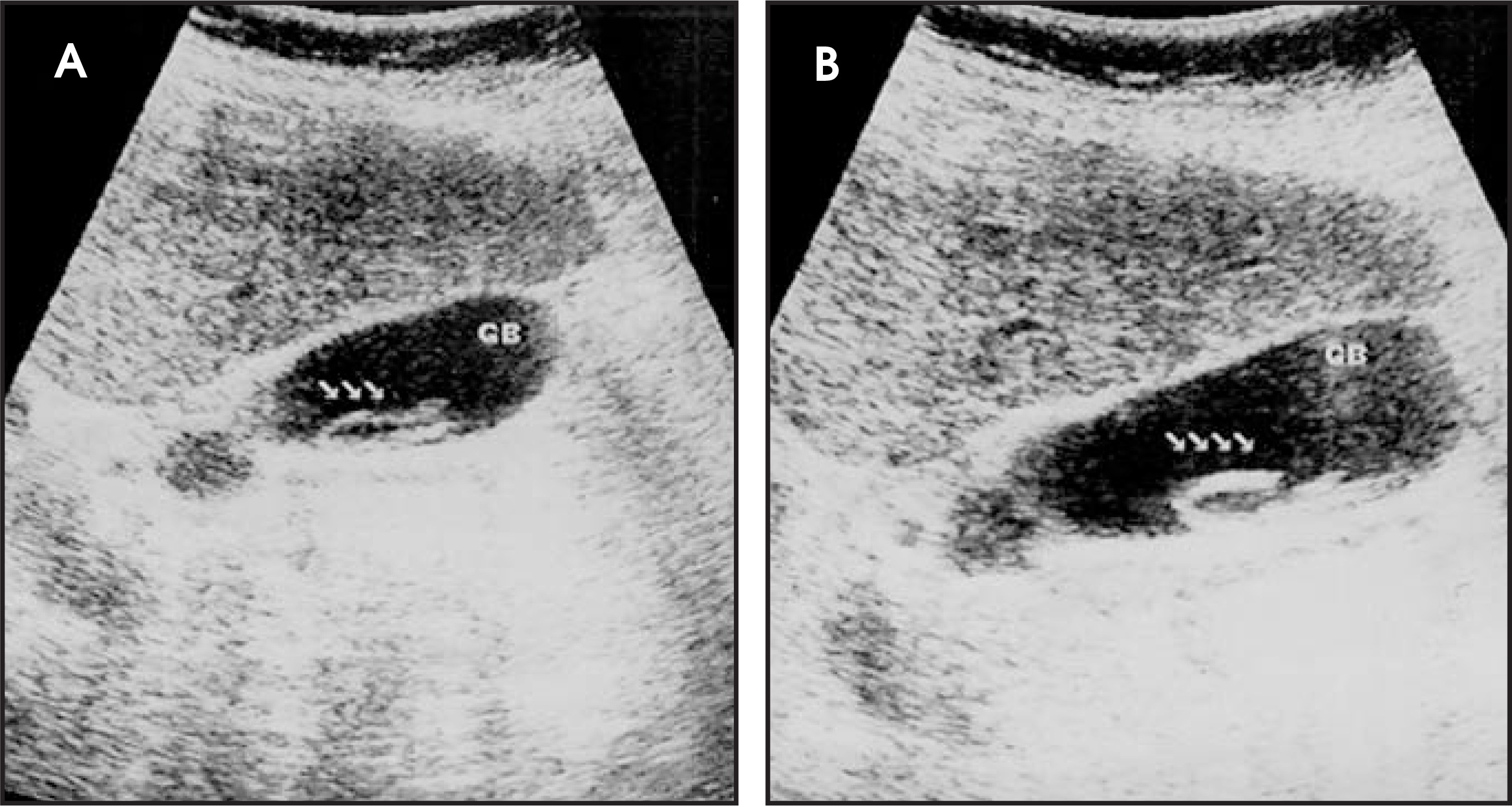
DIAGNOSIS
Ascaris lumbricoides in gallbladder
DISCUSSION
The intestinal roundwormAscaris lumbricoides is a cylindrical nonsegmented worm measuring 15 to 40 cm long.1 Roughly 25% of the world’s population is believed to be infected by this worm.2 Infection is primarly spread through poor sanitation and bad hygienic conditions.2 The adult form lives primarily in the small intestine (99%), duodenum, stomach, gallbladder, and ductus pancreaticus.3
Each worm can produce approximately 200,000 eggs daily. Roughly 2 weeks after the eggs move from the bowel, the larvae hatch. Adult worms can penetrate the small intestine and spread to the heart and lungs through the circulation. Larvae that penetrate the lungs can cause Löffler’s syndrome. The movement of adult worms can be asymptomatic and therefore go unrecognized.2 In children, however, adult forms of the worms can produce abdominal pain.4 Massive infection with Ascaris lumbricoides can cause a bowel obstruction.5 Aberrant migration of the adult forms can produce obstructive cholangitis and cholecystitis,6 abscesses in the liver, pancreatitis, appendicitis,7 or peritonitis. A fever caused by another disease or the use of some medication, ie tetrachloretilen, can provoke such migration.3
The infection with Ascaris lumbricoides is diagnosed by identifying the characteristic eggs in the stool of the infected person. Filling effects that are 15 to 35 cm long or small circles (from bended worms) can be seen on radiography. They can be seen on ultrasonography, mostly by accident. Combined with a history of the patient’s symptoms and signs of the disease, imaging findings should be confirmed with the pathologic analysis of the stool.7
When found in the gallbladder, an Ascaris lumbricoides infection most often requires a cholecystectomy.8 Once the patient reported here began antihelmintic therapy with mebendazol and metronidazol, there was spontaneous elimination of the ascaris from the gallbladder and the intestines. But the infection was not cleared from the small intestine. One month later, the anemia, hypoproteinemia, eosinophilia, and pericarditis were reduced significantly.
CONCLUSION
The infection of the gallbladder with Ascaris lumbricoides is not frequent, but ultrasonography screening can be the key diagnostic tool in the diagnosis and evaluation of response to treatment.
References
- Laing F, Rumac C, Wilson S, Chabonau J, Johnson J. Diagnostic Ultrasound. 1998.
- Liu L, Weller P, Fauci A, Braunwald E, Isselbacher K. Intestinal nematodes Harrison’s Principles of Internal Medicine. 1998:1208-1209.
- Khuroo M, Zargar S, Yattoo G. Sonographic findings in gallbladder ascariasis. J Clin Ultrasound. 1992;20:587-591.
- Ozmen M, Oguzkurt L, Ahmet B. Ultrasonographic diagnosis of intestinal ascariasis. Pediatr Radiol. 1995;25(suppl 1):S171-S172.
- Coskun A, Ozcan N, Durak A. Intestinal ascariasis as a cause of bowel obstruction in two patients: Sonographic diagnosis. J Clin Ultrasound. 1996;24:326-328.
- Bude R, Bowerman R. Biliary ascariasis. Radiology. 2000;214:844-847.
- Misra S, Dwivedi M, Misra V. Preoperative sonographic diagnosis of acute appendicitis caused by Ascaris lumbricoides. J Clin Ultrasound. 1999;27:96-97.
- Javid G, Wani N, Gulzar G. Gallbladder ascaris: Presentation and management. Br J Surg. 1999;86:1526-1527.
Citation
. Ascarisin the gallbladder lumbricoides. Applied Radiology. 2009;38(1):40A-40B. doi:10.37549/AR1676.