Beyond Reconstruction: The Next Frontiers in AI Imaging Acquisition
Applied Radiology — Vol. 55 , Issue 4
Published: June 1, 2026
1 Subtle Medical, Menlo Park, California
2 San Fernando Valley Interventional Radiology, RadNet, Inc, Los Angeles, California
3 DRT Consulting, Riverside, Connecticut
* Corresponding author: Thomas Campbell Arnold (campbell@radaccess.com)
Categories
Deep learning reconstruction (DLR) is a transformative technology that is rapidly becoming the standard of care across clinical practices. Faster scans, lower noise, and sharper images are no longer a competitive edge, but a baseline expectation. The question now is no longer whether artificial intelligence (AI) can accelerate acquisition, but whether it can make acquisition itself smarter.
The next frontier in acquisition AI is emerging across 3 principles: Acquire less. Acquire smarter. Acquire anywhere. Together, these principles reflect a broadening scope, from AI as a tool for image enhancement to AI as a system for workflow orchestration. As AI becomes more deeply embedded within imaging systems, scanners are evolving from passive hardware into intelligent, adaptive platforms.
The most efficient scan is the one that never needs to be acquired. DLR has already substantially shortened MRI and PET exam times, but there are limits to how much faster conventional acquisition can become. Another path to efficiency is not simply to accelerate scanning, but extracting greater value from the data already collected. This is the promise of image synthesis. By leveraging information from a limited range of already acquired sequences, AI can infer tissue contrast characteristics to synthetically generate images, making additional acquisitions unnecessary (Figure 1).1
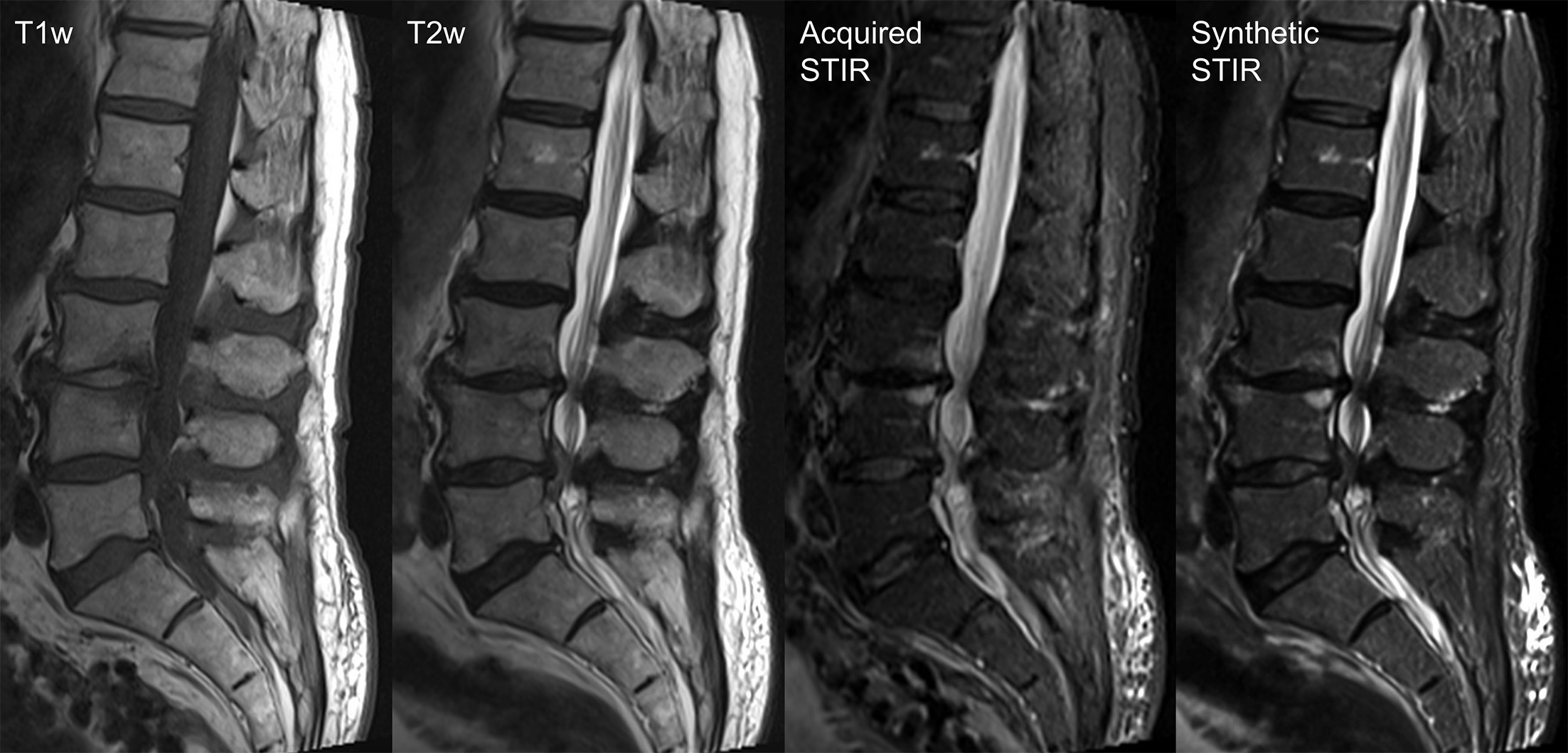
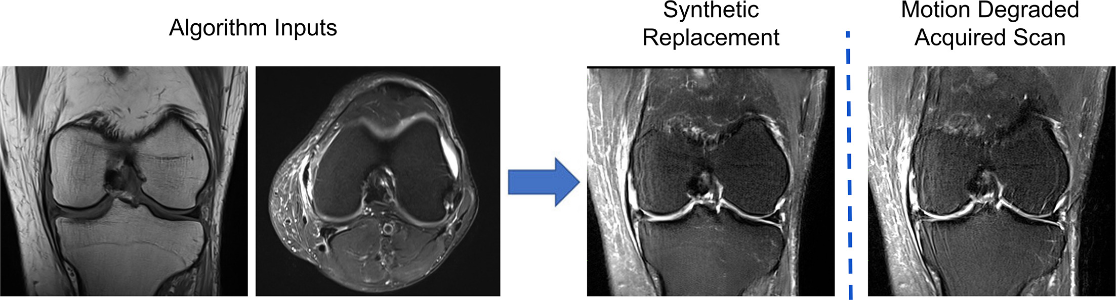
In practice, this approach can substantially shorten total exam times by reducing the number of series that must be routinely acquired on the scanner. By synthesizing missing information, these tools can also reduce the need for patients to return for additional scanning due to incomplete or motion-degraded studies (Figure 2).
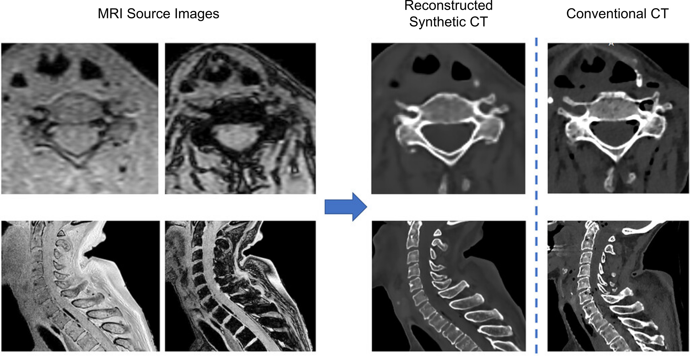
AI tools can also extract information that was previously impractical or undesirable to acquire. DLR is widely used to reduce radiation dose in CT and PET. Additionally, now deep learning can synthesize a CT-like image from a spine MRI (Figure 3), and for many use cases avoid an additional scan and radiation exposure, which is particularly beneficial for pediatric patients.2 This technology has high fracture sensitivity, which is useful as MRI reports often miss spondylolysis,3 resulting in significant delays in specialist evaluation.
With image synthesis, the goal is not simply faster scanning, but more efficient use of the information already available. AI tools capable of deriving monochromatic, monoenergetic spectral information from a conventional polychromatic CT study are also becoming increasingly prevalent. These techniques can routinely boost the visibility of contrast-enhancing lesions and, in some cases, salvage suboptimal vascular studies by increasing iodine conspicuity.
Synthetic acquisition represents only one component of the AI-enabled opportunity. Traditionally, imaging studies consist of sequences prescribed in advance. Fixed protocols are one of MRI’s hidden inefficiencies, consuming valuable scanner time acquiring sequences that are ultimately unnecessary, or omitting needed sequences and resulting in patient recalls.
Ideally, AI can make acquisition more responsive to real-time context by suggesting subsequent imaging steps based on what it has already identified. Rather than relying solely on protocols derived from limited clinical history, AI can detect relevant findings while the patient is still on the table and recommend protocol modifications in real time (Figure 4). For example, a routine brain MRI could be automatically redirected toward a dedicated internal auditory canal or pituitary protocol, thereby increasing diagnostic yield and avoiding the need for a callback.
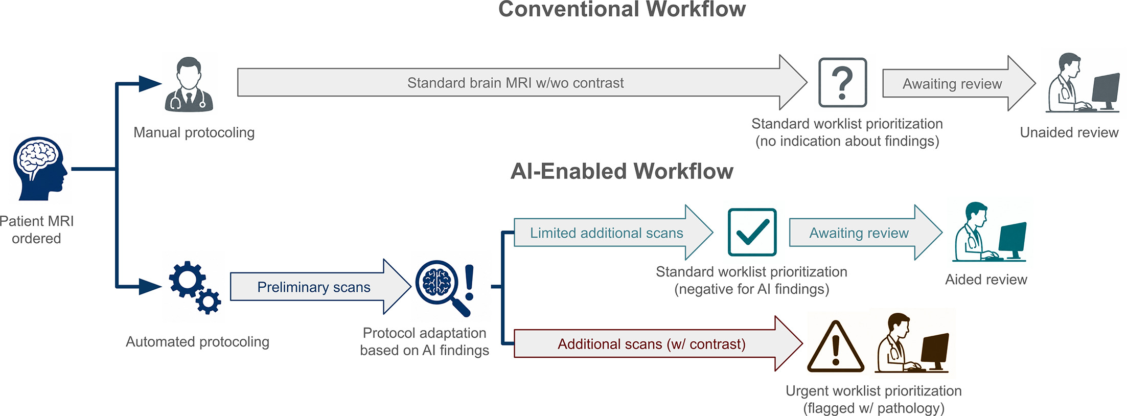
In some cases, AI could determine that no further imaging is required because the sequences already acquired indicated a low likelihood of clinically significant pathology. Conversely, AI could alert imaging teams about the potential value of adding contrast injection when unexpected findings are detected, while reducing overreliance on routine contrast when it’s unlikely to provide additional diagnostic value.
AI can also detect urgent findings in real-time and prioritize workflows through integration with PACS. In practical terms, fewer recalls, fewer repeat scans, and better technologist leverage translate directly into improved throughput and greater scanner efficiency. Additionally, AI can communicate urgent findings and key images to the care team through Health Insurance Portability and Accountability Act-compliant mobile applications before formal radiologist interpretation. In acute ischemic stroke, these technologies have been associated with reductions of up to 87 minutes from emergency department arrival to thrombectomy,4 with resultant shorter hospital stays and significant improvements in patient outcomes.
Even optimized protocols still depend on technologists to execute them. As imaging volumes rise, the availability of skilled personnel remains one of radiology’s most significant operational challenges. Companies are increasingly applying AI to automate complex acquisitions, such as cardiac MRI, enabling sites to offer advanced studies that might otherwise require specialized technologist expertise and distributing enterprise capabilities to better match scanner capacity and patient needs.
In addition, it is becoming increasingly challenging for even experienced technologists to tailor examinations as acquisition speeds continue to increase, especially with rising demands from simultaneous, remote scanning. This is where AI-enabled operational tools provide substantial value. Real-time scan monitoring, technologist decision support, and automated quality control can reduce the number of expert decisions required per exam while also reducing sequence repeats and patient call-backs.
MRI has been the clearest example of AI-driven transformation in acquisition, largely because its historically longer scan times made inefficiencies easier to identify. However, every imaging modality stands to benefit as AI expands beyond reconstruction and into workflow orchestration and operational decision-making. The long-term implication is not merely more efficient imaging, but progressively more autonomous imaging systems, with scanners increasingly capable of adapting examinations with minimal human intervention.
The next competitive advantage in radiology will not be faster scans; it will be smarter examinations.
References
- Tanenbaum L, Bash S, Zaharchuk G. Deep learning-generated synthetic MR imaging STIR spine images are superior in image quality and diagnostically equivalent to conventional STIR: a multicenter, multireader trial. AJNR Am J Neuroradiol. 2023;44(8):987-993. doi:10.3174/ajnr.A7920.
- Lafranca P, Rommelspacher Y, Muijs S. Safety and accuracy of cervical pedicle screw navigation using artificial intelligence–generated, MRI-based synthetic CT versus conventional CT. J Neurosurg Spine. 2025;44(2):235-241. doi:10.3171/2025.6.SPINE25213.
- Yamaguchi K, Skaggs D, Acevedo D. Spondylolysis is frequently missed by MRI in adolescents with back pain. J Child Orthop. 2012;6(3):237-240. doi:10.1007/s11832-012-0409-z.
- Hassan A. Artificial intelligence–parallel stroke workflow tool improves reperfusion rates and door‐in to puncture interval. Stroke Vasc Interv Neurol. 2022;2(5). doi:10.1161/SVIN.121.000224.
Disclosures
T. Arnold is an employee of Subtle Medical and the managing editor of radaccess.com. S. Shankaranarayanan is an employee of Subtle Medical. S. Bash is a consultant for Cortechs.ai, Subtle Medical, Darmiyan, GE Healthcare, Philips, Siemens, FujiFilm, Viz.ai, Eli Lilly, Eisai, and Positrigo. L. Tanenbaum is a consultant for Subtle Medical, MRIguidance, Neurophet, GE Healthcare, and Siemens. None of the authors received outside funding for the production of this original manuscript and no part of this article has been previously published elsewhere.
Citation
. Beyond Reconstruction: The Next Frontiers in AI Imaging Acquisition. Applied Radiology. 2026;55(4). doi:10.37549/AR-D-26-0044.