Bilateral endometriomas
Applied Radiology — Vol. 48 , Issue 5 , pp. 46 -47
DOI: 10.37549/AR2606
Published: September 1, 2019
Categories
Case Summary
A 34-year-old female with history of abnormal uterine bleeding, iron deficiency anemia, and uterine polyps status post-polypectomy presented to the gynecology clinic with worsening dysmenorrhea. Her pain occurred only during menses. Due to a history of gastric bypass and omental adhesions, laparoscopic evaluation for endometriosis was not possible.
Imaging Findings
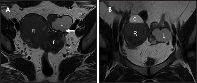
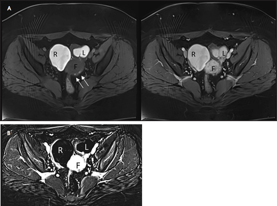
Transvaginal ultrasound demonstrated bilateral indeterminate ovarian cysts. Pelvic MRI with and without gadolinium contrast (Figures 1 and 2) was ordered for further characterization of the ovarian cysts. Imaging findings were compatible with bilateral endometriomas, which was later confirmed by pathology from bilateral cystectomies.
Diagnosis
Bilateral endometriomas
Discussion
Contrast-enhanced MRI of the pelvis can be useful to characterize indeterminate adnexal lesions. As demonstrated in this case, endometriomas have a characteristic appearance. Due to repeated cyclical hemorrhage and varying levels of deoxyhemoglobin and methemoglobin, endometriomas will appear hyperintense on T1-weighted sequences with varying levels of hypointensity on T2-weighted sequences. This has been described as the shading sign,1 and is characteristic of endometriomas, which more commonly show complete loss of signal intensity on T2-weighted images, or as hypointense layering fluid level.
T2 shading is particularly useful in distinguishing between endometriomas and hemorrhagic cysts. Hemorrhagic cysts also appear hyperintense on T1-weighted images, but typically do not have T2 shading. Since they do not have repeated cyclical hemorrhage, the concentration and viscosity of hemorrhagic cysts is lower, and they are typically bright on T2-weighted images.
Endometriomas can also be distinguished from hemorrhagic cysts by the presence of a T2 dark spot.2 Defined as a well-defined hypointense focus on T2-weighted images, the T2 dark spot is a marker of chronic hemorrhage and when present, highly specific for endometrioma.
Rarely, endometriomas may under- go malignant transformation. Contrast-enhanced MRI, particularly with digital subtraction, is useful for identifying enhancing mural nodules, the most important finding for identifying malignant transformation.3 Interval increase in size or loss of T2 shading can also be seen. Wall enhancement is a normal finding associated with endometriomas.
Other differential considerations include dermoid cysts, cystic neoplasm, and tubo-ovarian abscess. Dermoid cysts can be characterized by the presence of fat, manifesting as signal loss on fat-saturated sequences. Cystic neoplasms often have more soft tissue components with heterogeneous T1 signal that typically enhance.4 Abscess typically presents as a T1 hypointense lesion with T2 heterogeneous to hyperintense signal, with wall and surrounding soft tissue enhancement.5
Conclusion
Indeterminate adnexal lesions that cannot be diagnosed on ultrasound are often followed up with contrast-enhanced pelvic MRI for further characterization. T2 shading, presence of a T2 dark spot, persistent T1 hyperintensity on fat-saturated sequences, and lack of internal contrast enhancement can be used reliably distinguish endometriomas from hemorrhagic cysts, dermoid cysts, ovarian neoplasms, and tubo-ovarian abscess, as well as rule out malignant transformation.
References
- Glastonbury C. The shading sign.. Radiology. 2002;224(1):199-201.
- Corwin M, Gerscovich E, Lamba R. Differentiation of ovarian endometriomas from hemorrhagic cysts at MR imaging: utility of the T2 dark spot sign.. Radiology. 2014;271(1):126-132.
- Takeuchi M, Matsuzaki K, Uehara H. Malignant transformation of pelvic endometriosis: MR imaging findings and pathologic correlation.. Radiographics. 2006;26(2):407-417.
- Lee S. Radiological reasoning: imaging characterization of bilateral adnexal masses.. AJR Am J Roentgenol. 2006;187(3):S460-466.
- Kim M, Rha S, Oh S. MR Imaging findings of hydrosalpinx: a comprehensive review.. Radiographics. 2009;29(2):495-507.
Citation
. Bilateral endometriomas. Applied Radiology. 2019;48(5):46-47. doi:10.37549/AR2606.