Budd-Chiari syndrome
Applied Radiology — Vol. 37 , Issue 9 , pp. 40 -41
DOI: 10.37549/AR1645
Published: September 1, 2008
Categories
CASE SUMMARY
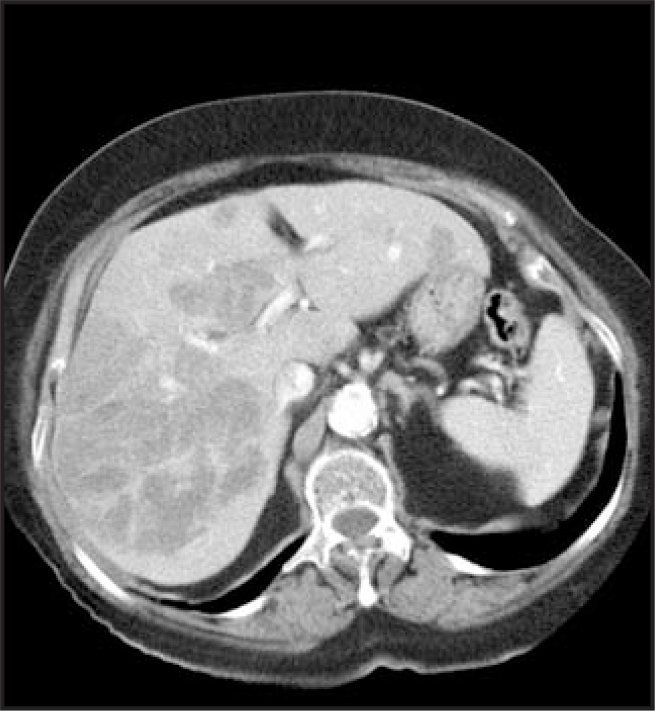
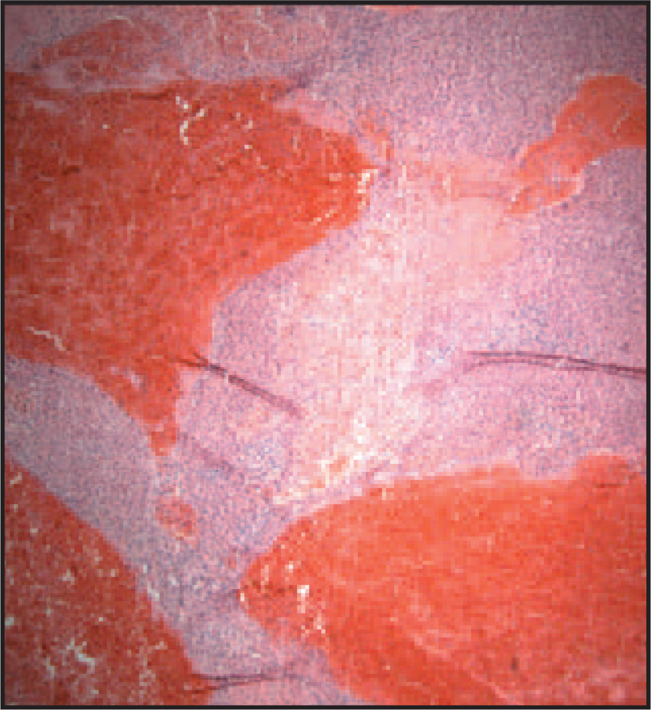
A 77-year-old woman presented with right upper quadrant pain. To evaluate the pain, she was examined with ultrasound, which revealed a mass in the right lobe of the liver. For further evaluation, contrast-enhanced computed tomography (CT) was performed, which showed a low-attenuation mass at the same location (Figure 1). Since the patient had a history of pulmonary embolism and was on chronic warfarin therapy, a transjugular liver biopsy of the low-attenuation mass in the right lobe of the liver was performed in the interventional radiology suite. Samples were sent to the pathology laboratory for analysis (Figure 2). The next morning, the patient became hypotensive. A noncontrast abdominal CT scan revealed a hematoma (not shown). After resuscitation, she was taken to the interventional radiology suite for emergent hepatic arterial embolization. She was then taken to the operating room, where the hematoma and the right hepatic lobe were removed. The intrahepatic tumor was confirmed by intraoperative ultrasound and by direct inspection of the liver. The right lobe was also sent to pathology for analysis.
IMAGING FINDINGS
Ultrasound examination showed a normal-appearing gallbladder without evidence of disease that would cause acute or chronic right upper quadrant pain. It also revealed a heterogeneous nodular-appearing right lobe of the liver, suggesting tumor infiltration. Contrast-enhanced CT showed a diffuse infiltration of the liver with areas of mass effect from the venous system, which is suggestive of a primary liver neoplasm (Figure 1).
Correlation of the CT findings with histologic examination was recommended. Pathologic examination of the transjugular biopsy specimen from the right lobe of the liver showed thrombosis of the central veins, zonal parenchymal necrosis, and early recanalization of smaller thombosed veins (Figure 2). The findings suggested an acute or subacute course of the thrombosis. The imaging and pathologic findings are consistent with Budd-Chiari syndrome. There was no evidence of either benign or malignant intrahepatic neoplasm.
Direct intraoperative inspection and intraoperative ultrasound confirmed a large, right hepatic lobe mass. Gross pathologic examination suggested a hamartoma, which was thought to be responsible for the imaging and operative findings; however, microscopic evaluation gave a completely different picture.
DIAGNOSIS
Budd-Chiari syndrome (BCS)
DISCUSSION
Budd-Chiari syndrome may be asymptomatic but commonly presents with abdominal pain, hepatomegaly, and ascites. Ultrasonography is generally the imaging modality of choice with a reported overall sensitivity and specificity of ≥85%.1,2 The accuracy of detecting hepatic venous thrombosis with CT scanning has been reported to be 50%, but magnetic resonance imaging (MRI) is believed to offer the best visualization and permits differentiation between the different forms of BCS.3,4
The preferred method of biopsy in an anticoagulated patient is transjugular liver biopsy. Indications for transjugular biopsy of the liver include coagulopathy and ascites. Complications include arterio venous fistula, cardiac arrhythmias, and intraperitoneal hemorrhage.5 The risk of intraperitoneal bleeding is 0% to 9%.5-7 Management of BCS may be medical or surgical. Medical therapy has a 1-year survival rate of 92% and is highly effective when BCS is diagnosed and the underlying hypercoaguable disorder is treated early.8 Transjugular intrahepatic portosystemic shunting procedures may be useful in patients who have failed to improve with thrombolytic therapy.9 Liver transplantation is the therapy of choice if clinical deterioration continues despite medical therapy.10
CONCLUSION
Since specific therapy for the benign but potentially devastating sequellae of BCS is available, prompt diagnosis is vital. Though a combination of imaging modalities can be used to diagnose BCS, proper diagnosis can be made only by histopathologic evaluation. Budd-Chiari syndrome should be considered in the differential diagnosis of a primary liver tumor.
References
- Chawla Y, Kumar S, Dhiman R. Duplex Doppler sonography in patients with Budd-Chiari syndrome. J Gastroenterol Hepatol. 1999;14:904-907.
- Bolondi L, Gaiani S, Li Bassi S. Diagnosis of Budd-Chiari Syndrome by pulsed Doppler ultrasound. Gastroenterology. 1991;100:1324-1331.
- Miller W, Federle M, Straub W, Davis P. Budd-Chiari syndrome: Imaging with pathologic correlation. Abdom Imaging. 1993;18:329-335.
- Noone T, Semelka R, Siegelman E. Budd-Chiari syndrome: Spectrum of appearances of acute, subacute, and chronic disease with magnetic resonance imaging. J Magn Reson Imaging. 2000;11:44-50.
- Bruzzi J, O’Connell M, Thakore H. Trans jugular liver biopsy: Assessment of safety and efficacy of the Quick-Core biopsy needle. Abdom Imaging. 2002;27:711-715.
- Maciel A, Marchiori E, de Barros S. Trans jugular liver biopsy: Histological diagnosis success comparing the trucut to the modified aspiration Ross needle. Arq Gastroenterol. 2003;4:80-84.
- Psooy B, Clark T, Beecroft J, Malatjalian D. Transjugular liver biopsy with use of the shark jaw needle: Diagnostic yield, complications, and cost-effectiveness. J Vasc Interv Radiol. 2001;12:61-65.
- Min A, Atillasoy E, Schwartz M. Reassessing the role of medical therapy in the management of hepatic vein thrombosis. Liver Transpl Surg Liver Transpl Surg.
- Putnam C, Porter K, Weil R. Liver transplantation for Budd-Chiari syndrome. JAMA. 1976;236:1142-1143.
- Menon K, Shah V, Kamath P. The Budd-Chiari syndrome. N Engl J Med N Engl J Med.
Citation
. Budd-Chiari syndrome. Applied Radiology. 2008;37(9):40-41. doi:10.37549/AR1645.