Cardiac imaging update; another PET/CT enters the market
Applied Radiology — Vol. 33 , Issue 12 , pp. 26 -28
DOI: 10.37549/AR1307
Published: December 1, 2004
Categories
The American Heart Association (AHA) Scientific Session 2004—held November 7 to 10 in New Orleans, LA—was the setting for the introduction of a variety of new cardiovascular imaging products.
Siemens highlights cardiovascular imaging line
Siemens Medical Solutions (Malvern, PA) featured several cardiovascular products at the meeting and introduced the company’s new Encompass Release for the Acuson Sequoia C512 and C256 echocardiography systems. This release features the new 8V3 transducer option for pediatric and fetal echocardiography examinations and an image optimization technology, called Native TEQ dynamic ultrasound technology, which is designed to improve workflow and ergonomics.
The Native TEQ technology enables the system’s computer to optimize images in real-time by reading the signals as they return from the patient and by automatically adjusting the parameters throughout the scan without the need for the operator to touch the keyboard. There are no gain or depth-gain-compensation (DGC) control adjustments required during scanning. The computer is programmed to perform advanced real-time motion analysis and to detect and differentiate noise and artifacts from soft tissue. The image gain is optimized automatically in the axial and lateral dimensions once the transducer touches the patient.
The new release also includes the Sequoia system’s new transducer, the 8V3, which is designed for pediatric cardiology and is based on proprietary Hanafy lens technology. According to the company, this transducer provides continuous focusing and image uniformity, broad bandwidth, and differentiation of near-field structures with high resolution.
The company also demonstrated new features on the Acuson CV70 cardiovascular system, including fourSight transesophageal (TEE) view for integrated 3-dimensional (3D) TEE imaging, and the new P9-4 transducer for pediatric cardiac imaging. The 3D TEE imaging allows for access to 3D/4-dimensional (4D) images through simple acquisition, review, manipulation, and dynamic display capabilities of gated 3D datasets using the V5Ms TEE transducer. The addition of the new P9-4 transducer for pediatric imaging expands the system’s transducer capabilities, allowing for a wide range of cardiovascular studies, including: stress echo-cardiography; adult, pediatric, and fetal echocardiograms; and epiaortic imaging.
Finally, Siemens also showcased the new Acuson AcuNav 8F ultrasound cath-eter for visualization of vascular and cardiac anatomy and physiology, measurement of blood flow, and visualization of other devices throughout the entire heart. The AcuNav 8F catheter is 33% smaller in cross-sectional area than is the current AcuNav 10F catheter, enabling access during closure of atrial septal defects on smaller patients, and left-heart applications for both interventional cardiology and electrophysiology.
Philips showcases array of cardiovascular imaging products
Royal Philips Electronics (Best, The Netherlands) featured a variety of cardiovascular imaging products at AHA, including the iE33 Intelligent Echocardiography system, the Allura Xper FD20 flat detector X-ray system, and the recently approved Allura 3D Coronary Angiography (3DCA) heart imaging tool.
iE33 Intelligent Echocardiography—The new iE33 Intelligent Echocardiography system features enhanced 2-dimensional (2D) image quality, live 3D echo imaging, 2D and 3D cardiac quantification, and enhanced ergonomics. Fully integrated 3D cardiac quantification software (QLAB 3DQ) permits measurement of the left ventricle volume and ejection fraction. The system is also able to analyze wall motion in 17 different segments of the heart simultaneously.
The system’s new Explora family of transducers includes the S5-1, with the company’s new PureWave crystal technology designed to deliver high-quality images of the beating heart. It also includes several enhancements in ergonomics, including hands-free voice command and one-button automated optimization controls.
Allura Xper FD20—According to the company, the Allura Xper FD20 flat detector X-ray system (Figure 1), powered by the proprietary 2k × 2k imaging chain, captures and processes information at a resolution up to 4 times greater than that of conventional systems. With a 30- × 38-cm field-of-view, this system was designed for mixed cardiac and vascular applications, and the ceiling-suspended C-arm allows for full-body coverage. It also includes the company’s proprietary interventional tool, StentBoost, to provide a view of stent deployment in coronary arteries, thereby allowing clinicians to take immediate corrective action, if necessary.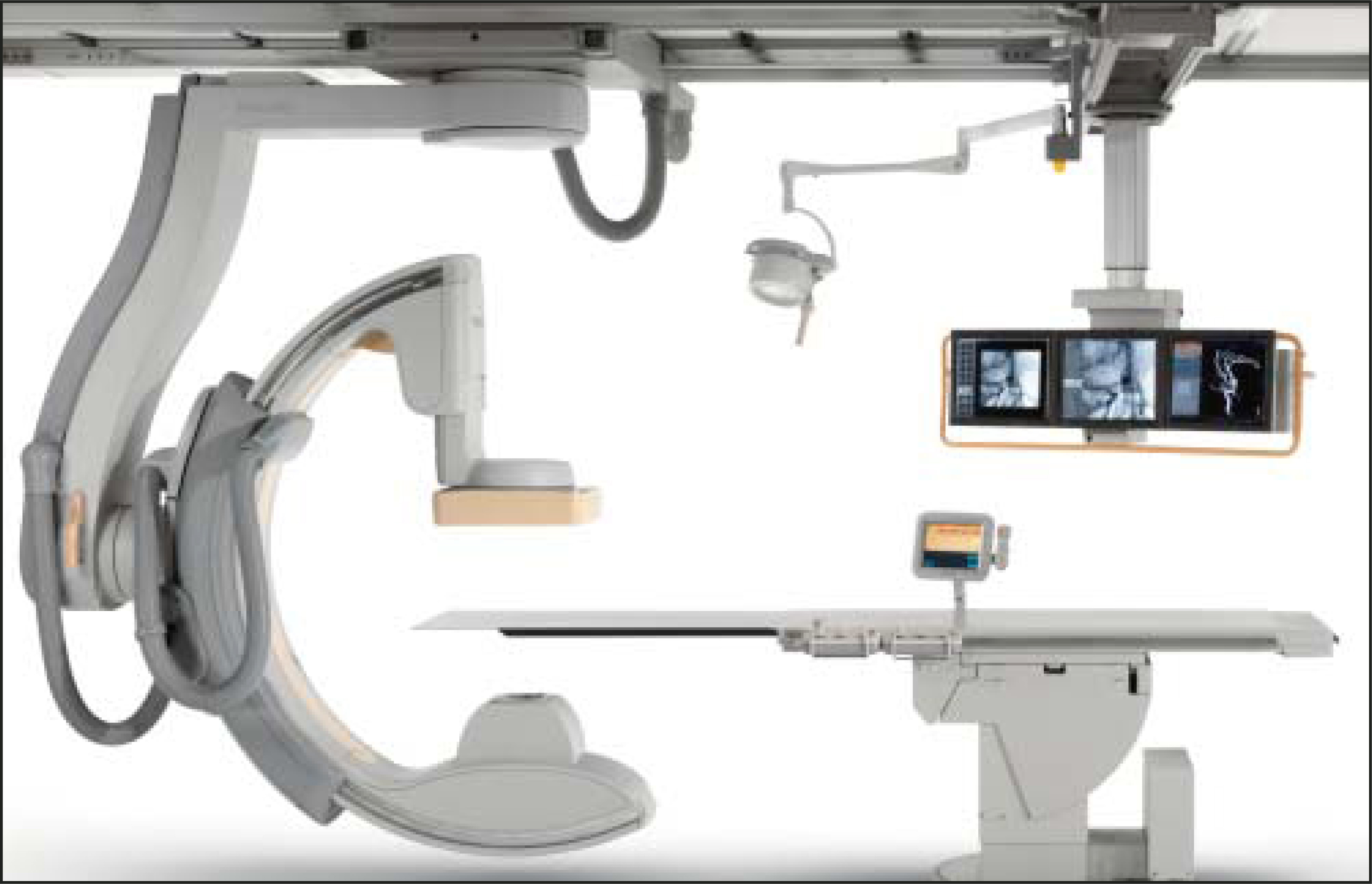
Allura 3D-CA—The company also highlighted its newly approved Allura 3D-CA heart imaging tool, which produces 3D images from multiple viewpoints and angles.
Other cardiovascular imaging products—In addition, Philips showcased its new Brilliance Computed Tomography (CT) Private Practice CV Configuration. This system, which was recently approved exclusively for the analysis of cardiac and peripheral vasculature, was designed for use by cardiologists. According to the company, it provides the clinician with rapid results (within 15 to 30 minutes) prior to requesting more extensive examinations.
“Our new specialty system gives cardiologists a fast and consistent CT unit to help determine within minutes if the correct treatment approach is medication, catheterization laboratory examinations, or surgery,” said Phil Prather, Director, Global Cardiology CT, at Philips. “With the potential time savings, cardiologists can increase patient volume, increase efficiency, and provide better, more precise patient care.”
In the area of magnetic resonance (MR) imaging, Philips showcased Easy Coronaries 3D. This whole-heart, free-breathing technique provides 3D representations of the heart and coronaries with one scan and does not require the use of contrast agents.
On the information management front, the company featured the Xcelera WebForum, a new Web-based results distribution solution designed to provide instant access to images and reports in Xcelera from any location, at any time. This new addition to the Xcelera information solutions portfolio allows the user to access all Xcelera images, from dynamic cardiac angiography runs and echocardiography to cardiac MR and CT studies, via any Internet connection. With this scalable system, images can also be incorporated into the electronic patient record. All data are 128-bit Secure Sockets Layer (SSL)-encrypted, and role-based permission functions define user rights for each user.
SonoSite unveils IMT software
Also at the AHA conference, SonoSite, Inc. of Bothell, WA, introduced new software designed to measure the intima-media thickness (IMT) of the carotid arteries and assess a patient’s risk of cardiovascular disease (Figure 2). According to the company, the SonoCalc IMT software can be used to detect thickening of the IMT of the carotid arteries, which is often an early sign of increased risk of cardiovascular disease.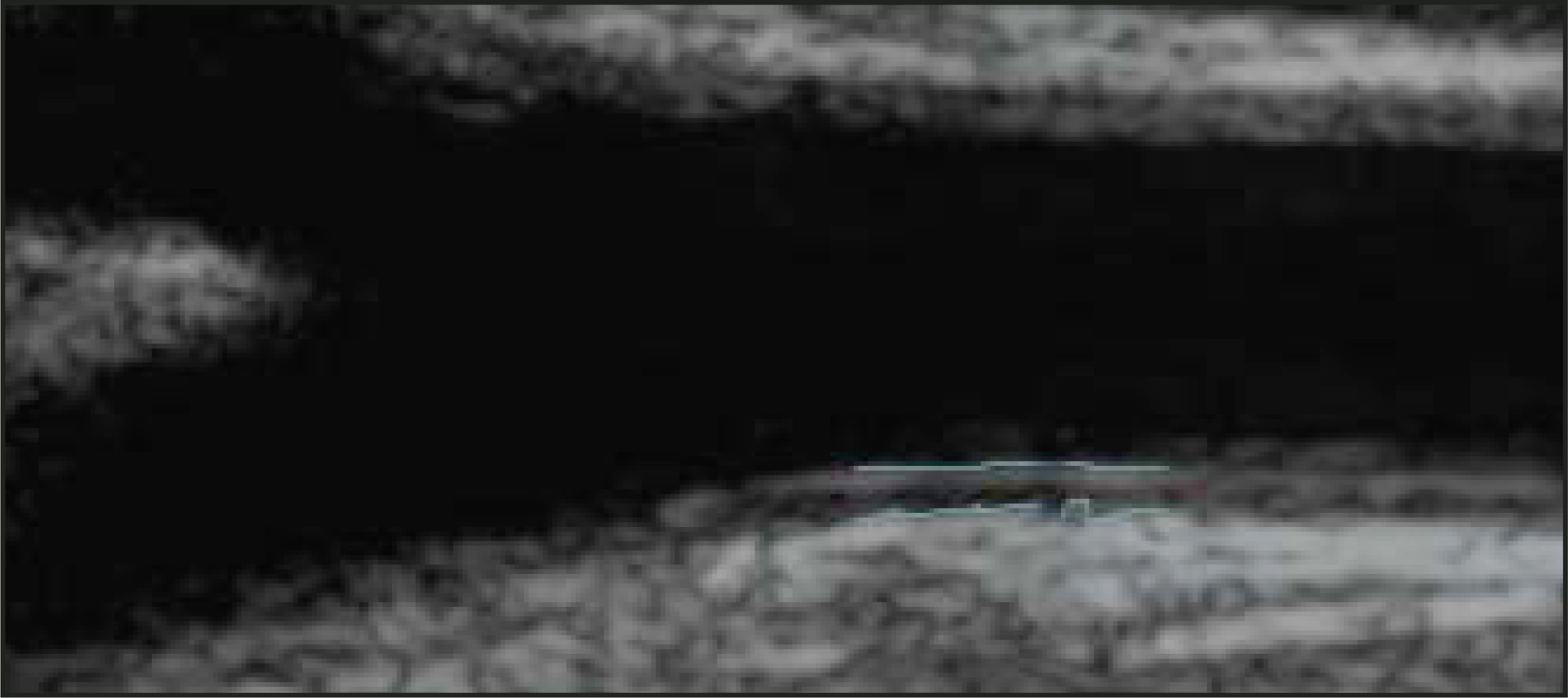
“I have found that patients with intermediate or limited risk factors, such as moderately elevated cholesterol, who would normally go untreated, may truly be at high risk for a cardiovascular event,” said Regina Drueding, MD, an internal medicine physician from Bountiful, UT, who has used the software. “An IMT scan, using ultrasound at the point of care, provides us with the right information to help prevent and better manage the disease. Currently, the most effective way of measuring a patient’s IMT is with ultrasound to easily and noninvasively capture images of the carotid artery wall for analysis.”
Used in conjunction with the company’s Titan high-resolution, handheld ultrasound system, the SonoCalc’s proprietary automated edge-detection technology provides physicians with the ability to analyze the IMT of the patient’s carotid artery and compare it with published population data to generate an individual cardiovascular health profile report for the patient. It also enables doctors to visualize disease progression to the patient by showing the patient the IMT scan.
SonoCalc features fully automated, single-click IMT measurement; semi-automated, user-guided IMT measurement for difficult images; statistical analysis of multiple IMT measurements; IMT measurements stored with corresponding patient information; and professional report generation summarizing one or more IMT measurements for a particular patient.
Hitachi introduces new PET/CT system
In other news, Hitachi Medical Systems America (Twinsburg, OH) recently received U.S. Food and Drug Administration (FDA) 510(k) clearance to market its new positron emission tomography/CT (PET/CT) system, the Sceptre P3.
According to the company, the Sceptre P3, which combines the company’s LSO PET with Pico Electronics and the Quad Slice CT, integrates the “Power of Three” suite of differentiating capabilities. Dual attenuation correction (DAC) technology allows the combination of both CT and sealed source attenuation correction to image patients with metal implants. The FDA-approved nonrigid Fusion7D algorithm provides registration by correcting for respiration differences between PET and CT acquisitions. The integration of the AVIA-PACS architecture enhances software communication and multimodal connectivity and supports RAID archiving capabilities.
According to the company, LSO (lutetium oxyorthosilicate) detector technology offers the best combination of physical and optical properties, providing improved count rate performance and reduced patient acquisition times. The CT component features a Fine-Slot multidetector with an ultrahigh-speed optical transmission system to deliver high-efficiency imaging. The DICOM-compliant AVIA-PACS architecture combines advanced connectivity with accelerating workflow solutions on a scaleable, multiconfiguration platform.
Citation
. Cardiac imaging update; another PET/CT enters the market. Applied Radiology. 2004;33(12):26-28. doi:10.37549/AR1307.