Characterization of coronary atherosclerotic plaques and the significance of vessel calcification
Applied Radiology — Vol. 30 , Issue 11 , pp. 40 -46
DOI: 10.37549/AR1055
Published: November 1, 2001
Categories
Plaque accumulation in coronary arteries eventually leads to the obstruction of blood flow and angiographic stenosis. Several angiographic studies have shown the prognostic value of stenosis severity.1 However, it has become clear that dynamic changes of coronary plaques residing in the vessel wall precede luminal stenosis and, therefore, better reflect the development of coronary artery disease. In fact, most plaques that eventually cause acute coronary syndromes (vulnerable plaques) are not severely stenotic before the acute event2-4 because early plaque development leads to expansion of the vessel wall (positive remodeling), thereby delaying luminal stenosis despite plaque accumulation.5 More recently, investigators have focused on the composition of coronary plaques as an important factor in the development of acute coronary events. The first evidence that morphologic characteristics of the vessel wall have prognostic value came from angiography, demonstrating that the extent of coronary calcification is correlated to plaque burden and predicts the likelihood of coronary events. The prognostic value of coronary calcifications has been further defined with electron-beam computed tomography (EBCT) and more recently with fast multidetector CT.
Other methods, particularly intravascular ultrasound (IVUS) and magnetic resonance imaging (MRI), attempt to characterize the morphology of individual lesions to assess plaque vulnerability. There is accumulating evidence that plaque characterization can be used in clinical practice. The overall plaque burden and morphology has prognostic value and can influence preventive treatment plans; serial observation of plaque burden allows recognition of disease progression or regression. In addition, the pre-interventional morphology of individual lesions affects outcome after coronary interventions and could be used to guide the interventional treatment.
Coronary plaque development and plaque vulnerability
Histologic and intravascular ultrasound studies have shown that coronary lesion development starts relatively early in life.6 Macrophages accumulate at future plaque sites and, by incorporating cholesterol, become “foam cells.” In advanced lesions, further cell accumulation leads to the formation of a lipid-rich core, which is separated from the lumen by a fibrous cap.6-8 Atheroma size is not well reflected by luminal stenosis because outward-directed growth of the plaque initially maintains lumen size despite increasing plaque burden. This compensatory vessel enlargement in response to plaque growth is termed positive arterial remodeling and was first described by Glagov et al5 in human coronary atherosclerosis. Coronary artery wall calcification is part of the development of atherosclerosis.9 Absent in the normal vessel wall, coronary artery calcification occurs in small amounts in early lesions and is found more frequently in advanced lesions.
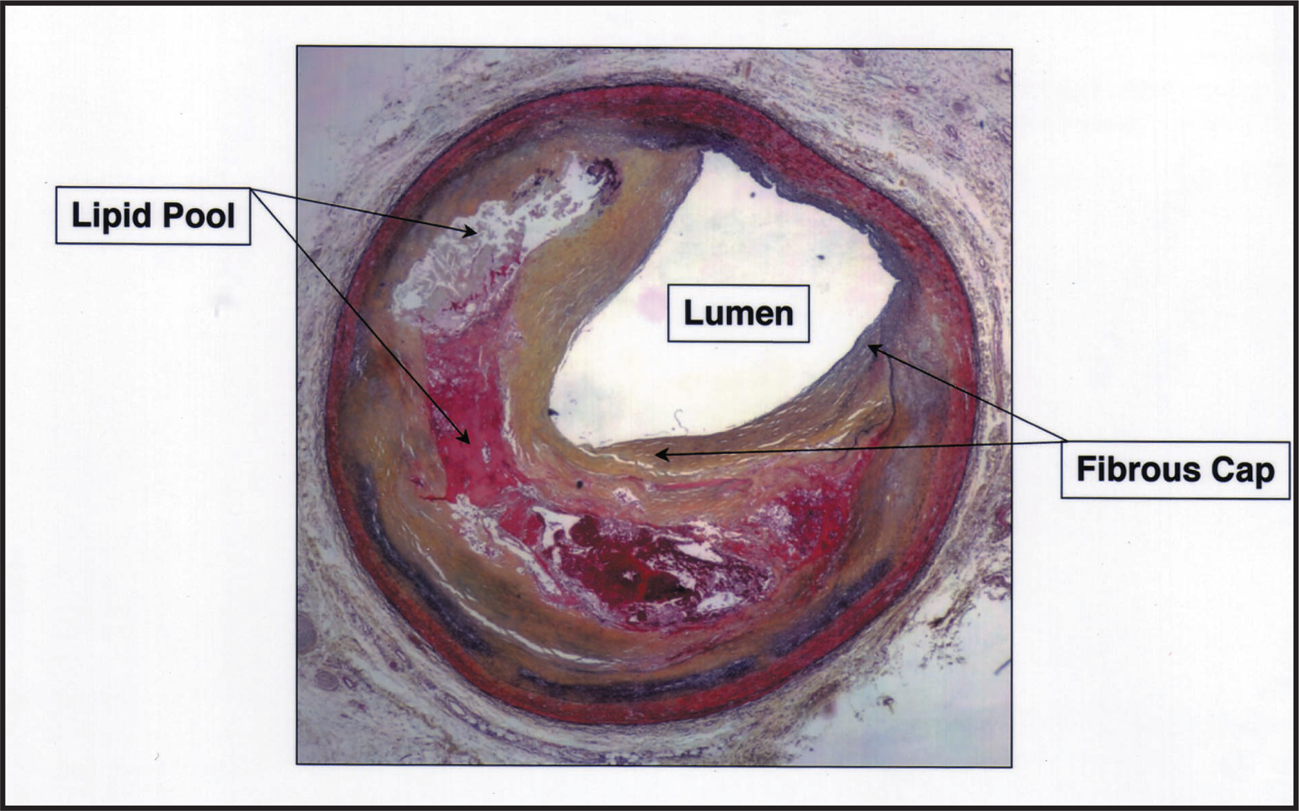
The size of the lipid pool within the atherosclerotic plaque and the thickness of the overlying fibrous “cap” are important characteristics predicting the stability of advanced plaques. Vulnerable plaques are characterized by a large lipid pool with a thin fibrous “cap.”10 The shoulder area at the margin of the plaque is a location of high stress, predisposed to rupture (figures 1 and 2).11,12 The accumulation of inflammatory cells and secretion of enzymes causes degradation of the fibrous “cap.”7,8,10 The relation of coronary calcifications to the probability of plaque rupture is unknown,9 and plaques vulnerable to fissure or erosion are frequently present in the absence of lesion calcification. The pathophysiological and clinical significance of lesions is related to plaque size and morphology, which in turn determine plaque stability. Chronic lesions with severe obstruction reduce blood flow and cause stable angina pectoris. However, most acute coronary syndromes are caused by sudden changes of mildly stenotic, vulnerable plaques. The initiating event is rupture or erosion of the fibrous cap, followed by luminal thrombus formation.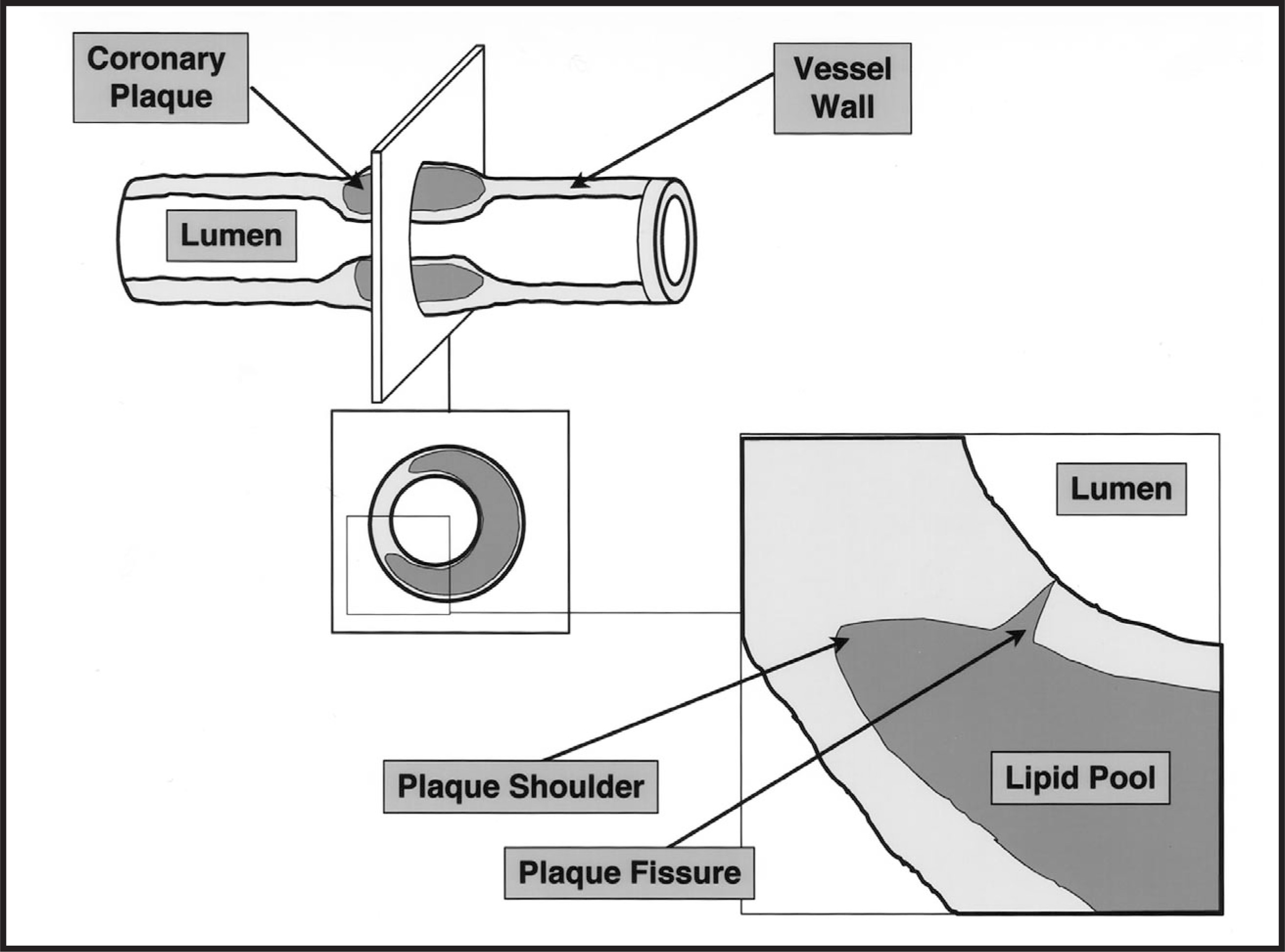
Diagnostic methods to characterize lesions
Coronary angiography
Imaging techniques visualizing coronary plaque morphology in vivo may allow identification of vulnerable lesions before rupture. A coronary angiogram shows the geometry of the vessel lumen, but not the vessel wall and plaque.13 However, it provides limited information about the surface of the plaque (smooth, irregular) and calcifications of the vessel wall. Several studies described that a majority of culprit lesions in patients with unstable angina had a characteristic angiographic appearance. These lesions have been termed Ambrose type II lesions.14,15 However, these characteristics describe plaques that have already ruptured, rather than those at risk to rupture.
The extent of fluoroscopic calcifications is a marker of the overall disease burden and, therefore, has prognostic value.16-18 However, angiography is not as sensitive a method to evaluate calcification as intravascular ultrasound, EBCT, or fast multidetector CT.19-23
Computed tomography
Computed tomography is very sensitive in detecting and quantitating coronary artery calcifications and can survey the entire coronary tree noninvasively. Epicardial coronary arteries are visualized easily by CT because the lower CT density of periarterial fat markedly contrasts to blood in the coronary arteries, and mural calcifications are identified because of their high CT density.
Electron-beam computer tomography allows the acquisition of serial transaxial images with a thickness of 3 to 6 mm and a temporal resolution of 100 ms. Thirty to 40 adjacent axial scans are obtained during 1 to 2 breath-holding periods. The short time window for acquiring each image virtually eliminates motion-related artifacts caused by cardiac and respiratory movements. Conventional CT, using fast multidetector technology, has been validated as an alternative technique to EBCT for coronary calcium detection and quantitation. Several recent reports describe the visualization of coronary calcifications with multidetector CT scanners, capable of acquiring 4 slices simultaneously and covering the entire heart in a single breath-hold with a slice thickness between 1.25 and 2.5 mm.23-25 Figure 3 shows a calcium scoring image obtained with fast multidetector CT. Calcium is clearly displayed in the left anterior descending coronary artery.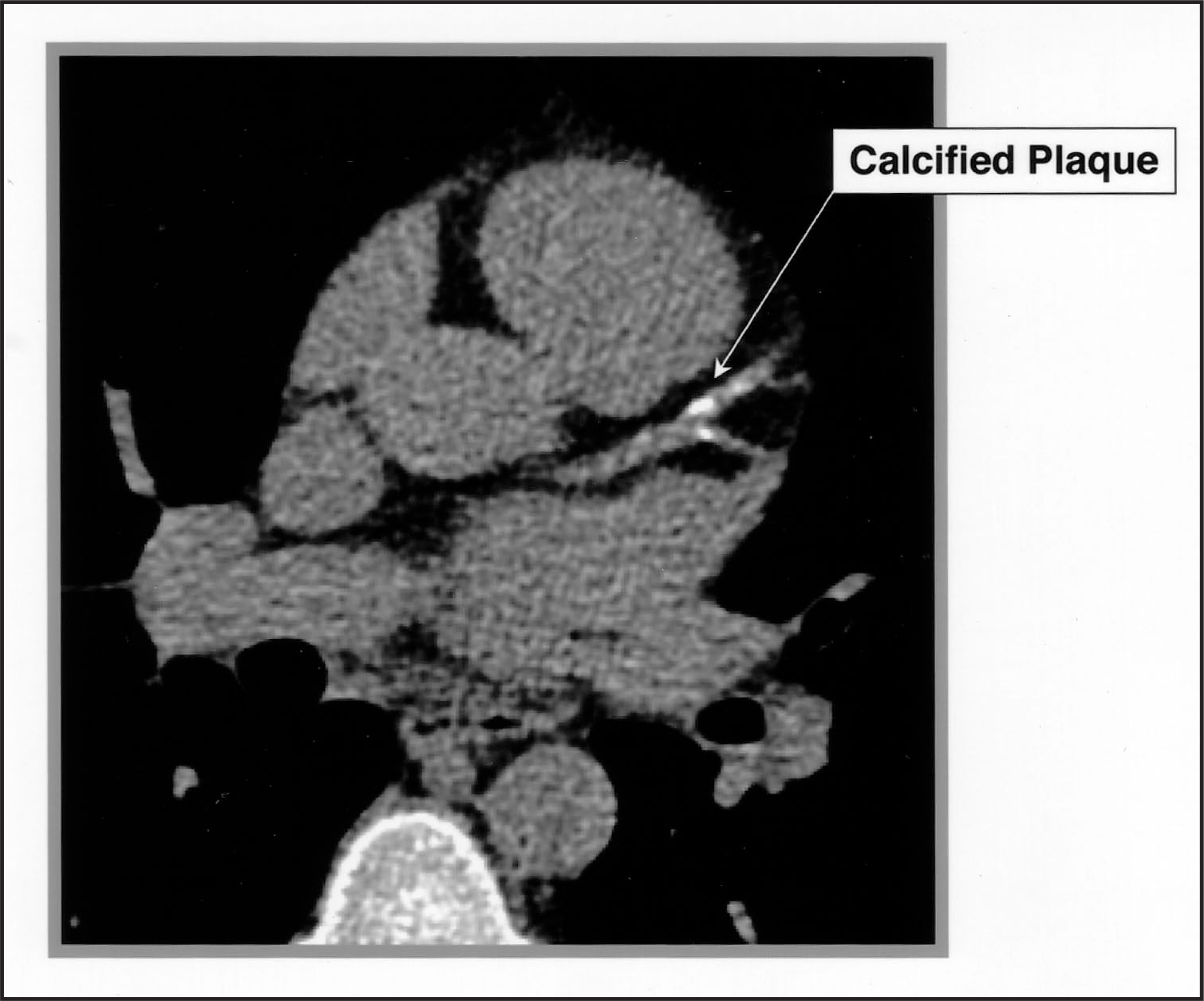
For the purposes of coronary calcium evaluation, CT examinations are performed without contrast agent administration and, therefore, display only calcified components of plaque. Usually, tissue densities ≥ 130 Hounsfield units are set as the attenuation level corresponding to calcified plaque. A calcium scoring system has been devised based on the maximum x-ray attenuation coefficient, or CT number measured in Hounsfield units, and the area of calcium deposits26 (traditional Agatston calcium score). Callister et al27 evaluated an alternative method of determining the calcium score by quantifying the actual volume of plaque analogous to that possible in prior histological studies (total calcium volume score, CVS).28 Both calcium scoring algorithms can be applied to either EBCT or fast multidetector CT images and provide a measure of total coronary plaque burden. The prognostic value of this information has been examined in several studies.29 Because compensatory vessel enlargement (positive remodeling) allows plaque accumulation without stenosis, the correlation of calcium area with luminal dimension is only moderate.30,31
Magnetic resonance imaging
Magnetic resonance imaging is a noninvasive two- or three-dimensional imaging technique that differentiates tissue structures on the basis of their proton magnetic properties. A wide range of image contrast can be obtained with different pulse sequences. Using clinical MRI scanners with customized software, these techniques are used to study atherosclerotic lesion morphology in animal models and human vessels.32-35 The extent of atherosclerotic plaque burden has been defined with high-resolution MRI in human popliteal arteries, demonstrating significant plaque burden in angiographically “normal” vessel segments.36 In recent studies, MRI has been shown to quantify vessel wall area accurately, allowing assessment of arterial remodeling.36,37 Shinnar et al38 describes the diagnostic accuracy of MRI for plaque characterization but also underlines the significant technical improvements in resolution and gating that are needed before this technique can be applied to the examination of coronary arteries in clinical settings. Figure 4 shows a three-dimensional T1-weighted gradient-echo MRI image from a human coronary artery segment from a post-mortem study.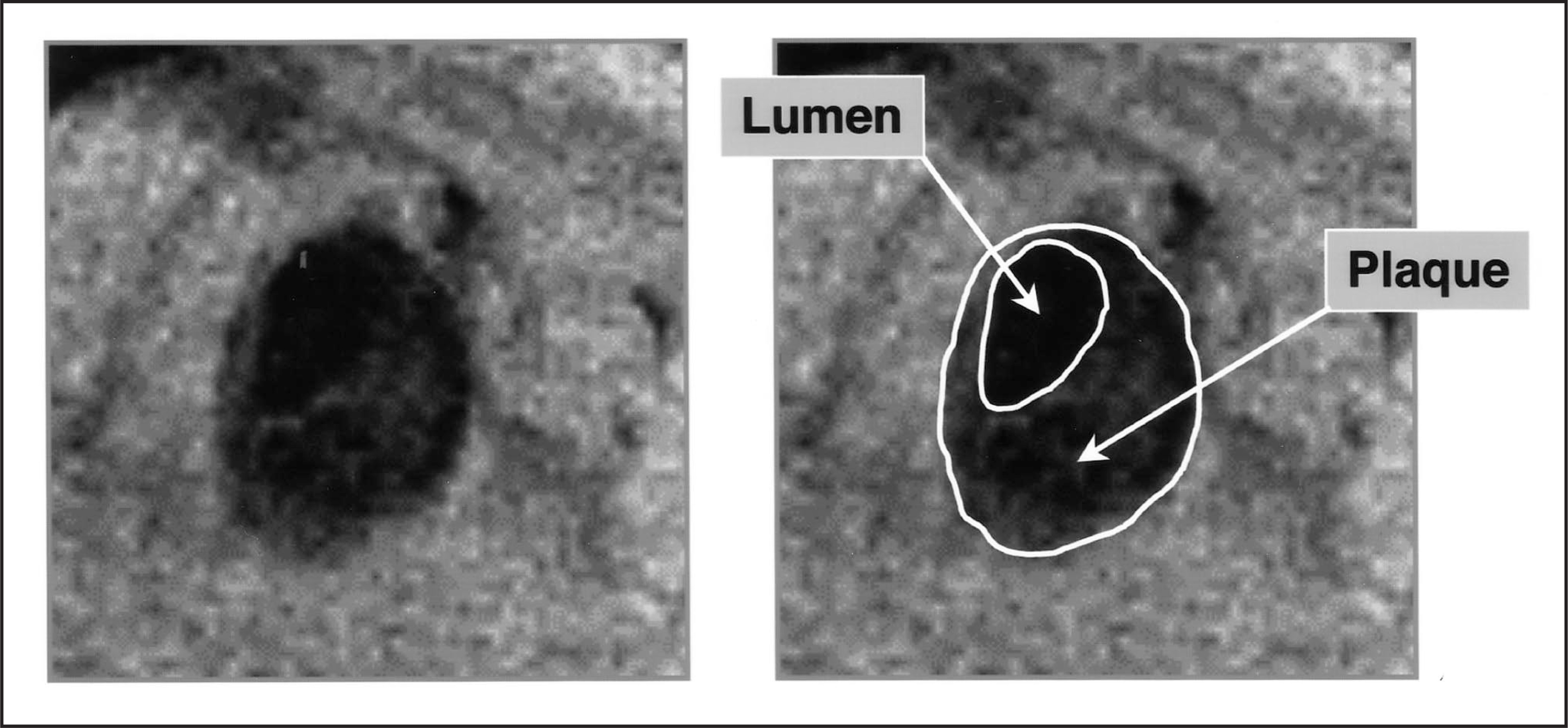
Intravascular ultrasound
Intravascular ultrasound provides tomographic images of the vessel wall including vessel size, plaque size, and plaque morphology (figure 5).39 During cardiac catheterization, a miniature ultrasound catheter is placed beyond the target lesion site. The ultrasound catheter is then withdrawn during continuous imaging, resulting in a series of cross-sections. The vessel wall of each cross-section can be described by its signal characteristics on a continuum from echodense (bright echo signal) to echolucent (faint echo signal). The measurement of lumen area, intima-media area, and external elastic membrane (EEM) area allows assessment of arterial remodeling and plaque burden. Several IVUS studies have demonstrated the reliability of ultrasound imaging in predicting the composition of the atherosclerotic plaque when compared with histology.40-42 Lipid-laden lesions appeared as hypoechoic, “soft” areas and fibrous or calcified tissues were recognized as bright echoes. In lipid-laden lesions with prominent overlying fibrous caps, a more reflective structure separating the soft echoes from the lumen was identified on the corresponding images.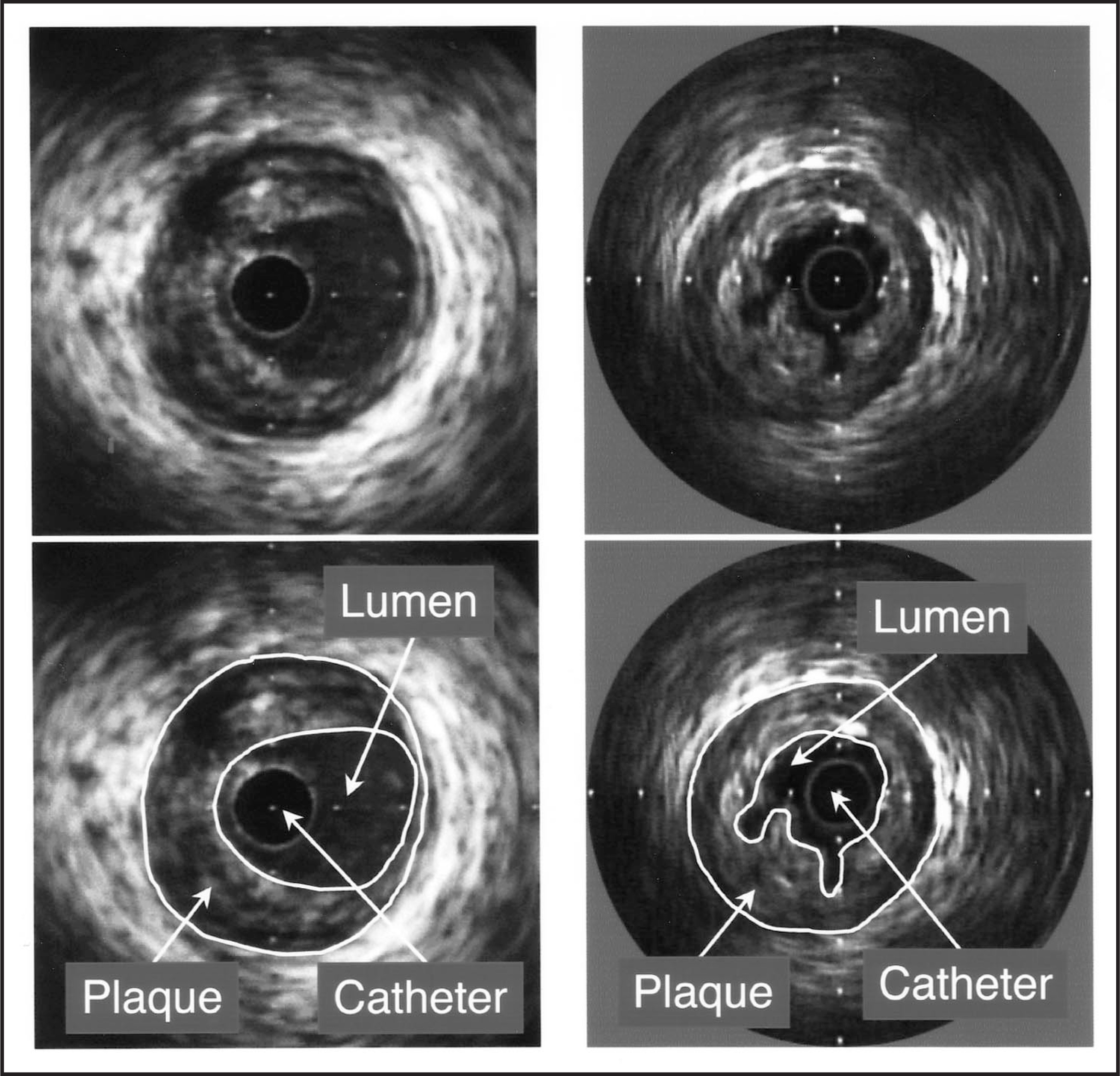
Advances in data analysis of the intravascular ultrasound information may allow a more detailed description of vessel wall and plaque morphology.43-45 In particular, radiofrequency analysis has been used for the characterization of plaque structures such as the lipid pool and fibrous cap.44-46
Other investigational catheter-based techniques, such as IVUS-elastography, optical-coherence-tomography (OCT) have been reviewed recently.47 An indirect approach to plaque characterization is the development of serum markers reflecting characteristics of coronary lesions. In particular, markers of inflammation like C-reactive protein (CRP) and high-sensitivity CRP may reflect lesion development and plaque vulnerability.48,49
Clinical applications
Coronary risk assessment in primary and secondary prevention
Total atherosclerotic plaque burden and its progression or regression reflects disease severity and activity. Quantification of coronary calcium with EBCT calcium scores has been shown to reflect the total atherosclerotic plaque burden.9,50,51 Therefore, CT can be used to predict future risk and offers the potential to follow disease progression, stabilization, and possible regression through serial imaging.52 In a serial study, the CVS was determined in 149 patients before and after 1 year of treatment with either HMG-CoA reductase inhibitors or diet alone. The average increase of the CVS during the follow-up period in the statin-treated and diet group was 5% ± 28% and 52% ± 36%, respectively (P < 0.001).53 However, according to a current AHA/ACC consensus statement, the incremental value of calcium scores over “traditional” multivariate risk-assessment models has not yet been established. These guidelines, therefore, do not recommend CT screening of coronary calcification for asymptomatic individuals, but conclude that it is justified in selected patient groups with intermediate risk.54
Serial studies have evaluated IVUS assessment of plaque burden. A small study examined the effect of 3-year treatment with pravastatin or diet in mildly diseased coronary arteries.55 During follow-up, plaque area increased by 41% in the control group but decreased by 7% in the treatment group. External elastic membrane area increased by 9% in the control group but did not change in the treatment group. Lumen area decreased by 9% in the control group but increased by 10% in the treatment group. The overall plaque burden demonstrated by IVUS or CT may direct preventive treatment plans. Theoretically, the progression or regression of plaque burden in serial studies may be used to assess the effectiveness of these treatments.
Assessment of plaque vulnerability
An important goal of plaque characterization is to correlate plaque structure to the subsequent risk of rupture leading to acute coronary syndromes. Currently, a reliable identification of these vulnerable plaques in not yet possible.56 However, using IVUS, several groups have found differences between stable and vulnerable coronary plaques. Plaque echolucency has been associated with the clinical presentation of unstable angina.41 Recent studies describe an association between acute coronary syndromes and positive remodeling.57-59 Our group has studied 85 patients with unstable and 46 patients with stable coronary syndromes using IVUS. Positive remodeling was significantly more frequent in unstable than in stable lesions (51.8% vs. 19.6%), while negative remodeling was more frequent in stable lesions (56.5% vs. 31.8%) (P = 0.001).57
A recent, prospective, serial IVUS study found that mildly stenotic lesions exhibiting positive remodeling and echolucent plaques at baseline more frequently caused acute coronary syndromes during follow-up than did echodense lesions with negative remodeling at baseline.60 The evidence for the ability of CT to identify vulnerable plaques remains limited. While a higher calcium-score may predict an overall greater number of vulnerable plaques, coronary calcifications of individual lesions are not a marker for vulnerability and several studies found vulnerable plaques without calcium.61 Recent reports describe the visualization of noncalcified coronary plaque with contrast-enhanced fast multidetector CT, but the experience is limited (figure 6).62,63 Plaque morphology has been studied with MRI in animal models and human peripheral arteries. The clinical role of MRI in the assessment of vulnerable lesions is not yet known.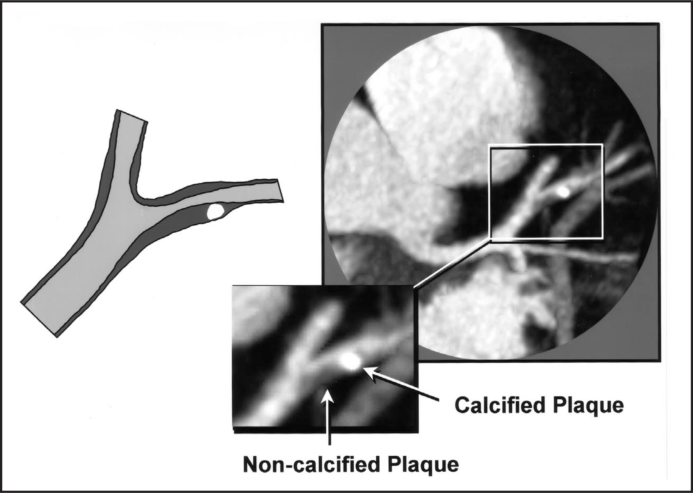
Role in interventional cardiology
Interventional catheter-based techniques can be used to evaluate the morphology of individual lesions before coronary interventions are performed. This baseline morphology predicts the response of the lesion to the intervention. The depth and circumferential extent of calcifications affect lesion expansion during balloon angioplasty, and dissections frequently occur at the junction between soft tissue and calcium.64 In recent IVUS studies, positive remodeling was associated with higher target-lesion revascularization rate after nonstent interventions. This effect was not seen in stented lesions.65-67 Therefore, not only the degree of luminal stenosis, but also the composition of the atherosclerotic plaque may determine the decision of which interventional strategy would be most suitable.
Conclusion
Plaque characterization describes imaging methods assessing morphologic characteristics of coronary plaques. Catheter-based imaging techniques provide characterization of individual lesions in the catheterization laboratory. Noninvasive imaging techniques evaluate characteristics of the entire coronary tree.
There is accumulating evidence that plaque characterization can be used in several ways in clinical practice. The overall plaque burden and plaque morphology has prognostic value and could influence preventive treatment plans. Serial observation of plaque burden allows the recognition of disease progression or regression and can be used to assess the effect of treatment. The pre-interventional morphology of individual lesions affects outcome after coronary interventions and can guide interventional treatment. AR
References
- Proudfit W, Bruschke A, MacMillan J. Fifteen year survival study of patients with obstructive coronary artery disease. Circulation.. 1983;68:986-997.
- Giroud D, Li J, Urban P. Relation of the site of acute myocardial infarction to the most severe coronary arterial stenosis prior to angiography. Am J Cardiol.. 1992;69:729-732.
- Ambrose J, Tannenbaum M, Alexopoulos D. Angiographic progression of coronary artery disease and the development of myocardial infarction. J Am Coll Cardiol.. 1988;12:56-62.
- Little W, Constantinescu M, Applegate R. Can coronary angiography predict the site of a subsequent myocardial infarction in patients with mild-to-moderate coronary artery disease?. Circulation.. 1988;78:1157-1166.
- Glagov S, Weisenberg E, Zarins C. Compensatory enlargement of human atherosclerotic coronary arteries. N Engl J Med.. 1987;316:1371-1375.
- Ross R. The pathogenesis of atherosclerosis: A perspective for the 1990s. Nature.. 1993;362:801-809.
- Moreno P, Falk E, Palacios I. Macrophage infiltration in acute coronary syndromes: Implications for plaque rupture. Circulation.. 1994;90:775-778.
- Shah P, Falk E, Badimon J. Human monocyte-derived macrophages induce collagen breakdown in fibrous caps of atherosclerotic plaques. Circulation.. 1995;92:1565-1569.
- Wexler L, Brundage B, Crouse J. Coronary artery calcification: Pathophysiology, epidemiology, imaging methods, and clinical implications. Circulation. 1996;94:1175-1192.
- Libby P. Molecular bases of the acute coronary syndromes. Circulation. 1995;91:2844-2850.
- Richardson P, Davies M, Born G. Lancet.. 1989;2:941-944.
- Loree H, Kamm R, Stringfellow R, Lee R. Circ Res.. 1992;71:850-858.
- Topol E, Nissen S. Our preoccupation with coronary luminology: The dissociation between clinical and angiographic findings in ischemic heart disease. Circulation.. 1995;92:2333-2342.
- Kalbfleisch S, McGillem M, Simon S. Automated quantification of indexes of coronary lesion complexity. Comparison between patients with stable and unstable angina. Circulation.. 1990;82:439-447.
- Ambrose J, Winters S, Arora R. Angiographic evolution of coronary artery morphology in unstable angina. J Am Coll Cardiol. 1986;7:472-478.
- Detrano R, Wong N, Tang W. Prognostic significance of cardiac cineflourocopy for coronary calcific deposits in asymptomatic high risk subject. J Am Coll Cardiol.. 1994;24:354-358.
- Detrano R, Hsiai T, Wang S, Puentes G. Prognostic value of coronary calcification and angiographic stenoses in patients undergoing coronary angiography. J Am Coll Cardiol.. 1996;27:285-290.
- Margolis J, Chen J, Kong Y. The diagnostic and prognostic significance of coronary artery calcification. Radiology.. 1980;137:609-616.
- Mintz G, Douek P, Pichard A. Target lesion calcification in coronary artery disease: An intravascular ultrasound study. J Am Coll Cardiol.. 1992;20:1149-1155.
- Tuzcu E, Berkalp B, De Franco A. The dilemma of diagnosing coronary calcification: Angiographic versus intravasular ultrasound. J Am Coll Cardiol.. 1996;27:832-838.
- Baumgart D, Schmermund A, Goerge G. Comparison of electron beam computed tomography with intracoronary ultrasound and coronary angiography for detection of coronary atherosclerosis. J Am Coll Cardiol.. 1997;30:57-64.
- Kajinami K, Seki H, Takekoshi N, Mabuchi H. Coronary calcification and coronary atherosclerosis: Site by site comparative morphologic study of electron beam computed tomography and coronary angiography. J Am Coll Cardiol.. 1997;29:1549-1556.
- Becker C, Jakobs T, Aydemir S. Helical and single-slice conventional CT versus electron beam CT for the quantification of coronary artery calcification. AJR Am J Roentgenol.. 2000;174:543-547.
- Becker C, Knez A, Leber A. Initial experience with multi-slice detector spiral CT in diagnosis of arteriosclerosis of coronary vessels [in German]. Radiologe.. 2000;40:118-122.
- Halliburton S, Kuzmiak S, Schvartzman P. Coronary artery calcium quantification: Impact of spiral CT acquisition on multi-slice analysis [abstract]. Radiology.. 2000;217(P):232.
- Agatston A, Janowitz W, Hildner F. Quantification of coronary artery calcium using ultrafast computed tomography. J Am Coll Cardiol.. 1990;15:827-832.
- Callister T, Cooil B, Raya S. Coronary artery disease: Improved reproducibility of calcium scoring with and electron-beam CT volumetric method. Radiology.. 1998;208:807-814.
- Callister T, Janowitz W, Raggi P. Sensitivity of two electron beam tomography protocols for the detection and quantification of coronary artery calcium. AJR Am J Roentgenol.. 2000;175:1743-1746.
- Secci A, Wong N, Tang W. Electron beam computed tomographic coronary calcium as a predictor of coronary events. Circulation.. 1997;96:1122-1129.
- Tanenbaum S, Kondos G, Veselik K. Detection of calcific deposits in coronary arteries by ultrafast computed tomography and correlation with angiography. Am J Cardiology.. 1989;63:870-872.
- Sangiorgi G, Rumberger J, Severson A. Arterial calcification and not lumen stenosis is highly correlated with atherosclerotic plaque burden in humans: A histologic study of 723 coronary artery segments using nondecalcifying methodology. J Am Coll Cardiol.. 1998;31:126-33.
- Worthley S, Helft G, Fuster V. Noninvasive in vivo magnetic resonance imaging of experimental coronary artery lesions in a porcine model. Circulation.. 2000;101:2956-2961.
- Toussaint J, LaMuraglia G, Southern J. Magnetic resonance images lipid, fibrous, calcified, hemorrhagic, and thrombotic components of human atherosclerosis in vivo. Circulation.. 1996;94:932-938.
- Rogers W, Prichard J, Hu Y. Characterization of signal properties in atherosclerotic plaque components by intravascular MRI. Arterioscler Thromb Vasc Biol.. 2000;20:1824-1830.
- Coulden R, Moss H, Graves M. High resolution magnetic resonance imaging of atherosclerosis and the response to balloon angioplasty. Heart.. 2000;83:188-191.
- Worthley S, Helft G, Fuster V. Serial in vivo MRI documents arterial remodeling in experimental atherosclerosis. Circulation.. 2000;101:586-589.
- Fayad Z, Fuster V, Fallon J. Noninvasive in vivo human coronary artery lumen and wall imaging using black-blood magnetic resonance imaging. Circulation.. 2000;102:506-510.
- Shinnar M, Fallon J, Wehrli S. The diagnostic accuracy of ex vivo MRI for human atherosclerotic plaque characterization. Arterioscler Thromb Vasc Biol.. 1999;19:2756-2761.
- Nissen S, Gurley J, Grines C. Intravascular ultrasound assessment of lumen size and wall morphology in normal subjects and patients with coronary artery disease. Circulation.. 1991;84:1087-1099.
- Gussenhoven E, Essed C, Lancee C. Arterial wall characteristics determined by intravascular ultrasound imaging: An in vitro study. J Am Coll Cardiol.. 1989;14:947-952.
- Hodgson J, Reddy K, Suneja R. Intracoronary ultrasound imaging: Correlation of plaque morphology with angiography, clinical syndrome and procedural results in patients undergoing coronary angioplasty. J Am Coll Cardiol.. 1993;21:35-44.
- Hiro T, Leung C, De Guzman S. Are soft echoes really soft? Intravascular ultrasound assessment of mechanical properties in human atherosclerotic tissue. Am Heart J.. 1997;133:1-7.
- Masseroli M, Cothren R, Meier D. Quantification of intramural calcification in coronary intravascular ultrasound images with automated image analysis. Am Heart J.. 1998;136:78-86.
- Moore M, Spencer T, Salter D. Characterization of coronary atherosclerotic morphology by spectral analysis of radiofrequency signal: In vitro intravascular ultrasound study with histological and radiological validation. Heart.. 1998;79:459-467.
- Komiyama N, Berry G, Kolz M. Tissue characterization of atherosclerotic plaques by intravascular ultrasound radiofrequency signal analysis: An in vitro study of human coronary arteries. Am Heart J.. 2000;140:565-574.
- Lee D, Sigel B, Swami V. Determination of carotid plaque risk by ultrasonic tissue characterization. Ultrasound Med Biol.. 1998;24:1291-1299.
- Pasterkamp G, Falk E, Woutman H, Borst C. Techniques characterizing the coronary atherosclerotic plaque: Influence on clinical decision making?. J Am Coll Cardiol.. 2000;36:13-21.
- Ridker P, Hennekens C, Buring J, Rifai N. C-reactive protein and other markers of inflammation in the prediction of cardiovascular disease in women. N Engl J Med.. 2000;342:836-843.
- Ridker P, Cushman M, Stampfer M. Inflammation, aspirin, and the risk of cardiovascular disease in apparently healthy men. N Engl J Med.. 1997;336:973-979.
- Simons D, Schwartz R, Edwards W. Noninvasive definition of anatomic coronary artery disease by ultrafast computed tomographic scanning: A quantitative pathologic comparison study. J Am Coll Cardiol. 1992;20:1118-1126.
- Rumberger J, Simons D, Fitzpatrick L. Coronary artery calcium area by electron-beam computed tomography and coronary atherosclerotic plaque area. Circulation.. 1995;92:2157-2162.
- Janowitz W, Agatston A, Viamonte M. Comparison of serial quantitative evaluation of calcified coronary artery plaque by ultrafast computed tomography in persons with and without coronary artery disease. Am J Cardiol.. 1991;68:1-6.
- Callister T, Raggi P, Cooil B. Effect of HMG-CoA reductase inhibitors on coronary artery disease by electron-beam computed tomography. N Engl J Med.. 1998;339:1972-1978.
- O'Rourke R, Brundage B, Froelicher V. American College of Cardiology/American Heart Association expert consensus document on electron-beam computed tomography for the diagnosis and prognosis of coronary artery disease. Circulation.. 2000;102:126-140.
- Takagi T, Yoshida K, Akasaka T. Intravascular ultrasound analysis of reduction in progression of coronary narrowing by treatment with pravastatin. Am J Cardiol. 1997;79:1673-1767.
- Schoenhagen P, Strauss McErlean E, Nissen S. The vulnerable coronary plaque. J Cardiovasc Nurs.. 2000;15:1-12.
- Schoenhagen P, Ziada K, Kapadia S. Extent and direction of arterial remodeling in stable and unstable coronary syndromes. Circulation.. 2000;101:598-603.
- Smits P, Pasterkamp G, de Jaegere P. Angioscopic complex lesions are predominantly compensatory enlarged: An angioscopic and intracoronary ultrasound study. Cardiovasc Res.. 1999;41:458-464.
- Nakamura M, Nishikawa H, Mukai S. Impact of coronary artery remodeling on clinical presentation of coronary artery disease: An intravascular ultrasound study. J Am Coll Cardiol.. 2001;37:63-69.
- Yamagishi M, Terashima M, Awano K. Morphology of vulnerable coronary plaque: Insights from follow-up of patients examined by intravascular ultrasound before and acute coronary syndrome. J Am Coll Cardiol.. 2000;35:106-111.
- Mintz G, Pichard A, Popma J. Determinants and correlates of target lesion calcium in coronary artery disease: A clinical, angiographic and intravascular ultrasound study. J Am Coll Cardiol.. 1997;29:268-274.
- Knez A, Becker C, Ohnesorge B. Noninvasive detection of coronary artery stenosis by multislice helical computed tomography. Circulation.. 2000;101.
- Becker C, Knez A, Ohnesorge B. Imaging of noncalcified coronary plaques using helical CT with retrospective ECG gating. AJR Am J Roentgenol.. 2000;175:423-424.
- Fitzgerald P, Ports T, Yock P. Contribution of localized calcium deposits to dissection after angioplasty in vivo assessed by intravascular ultrasound imaging. Circulation.. 1992;86:64-70.
- Dangas G, Mintz G, Mehran R. Stent implantation neutralizes the impact of preintervention arterial remodeling on subsequent target lesion revascularization. Am J Cardiol.. 2000;86:452-455.
- Dangas G, Mintz G, Mehran R. Stent implantation neutralizes the impact of preintervention arterial remodeling on subsequent target lesion revascularization. Am J Cardiol.. 2000;86:453-455.
- Wexberg P, Gyongyosi M, Sperker W. Pre-existing arterial remodeling is associated with in-hospital and late adverse cardiac events after coronary interventions in patients with stable angina pectoris. J Am Coll Cardiol.. 2000;36:1860-1869.
Citation
. Characterization of coronary atherosclerotic plaques and the significance of vessel calcification. Applied Radiology. 2001;30(11):40-46. doi:10.37549/AR1055.