CLINICAL Quiz
Applied Radiology — Vol. 31 , Issue 2 , pp. 36 -37
DOI: 10.37549/AR1072
Published: February 1, 2002
Categories
CASE SUMMARY
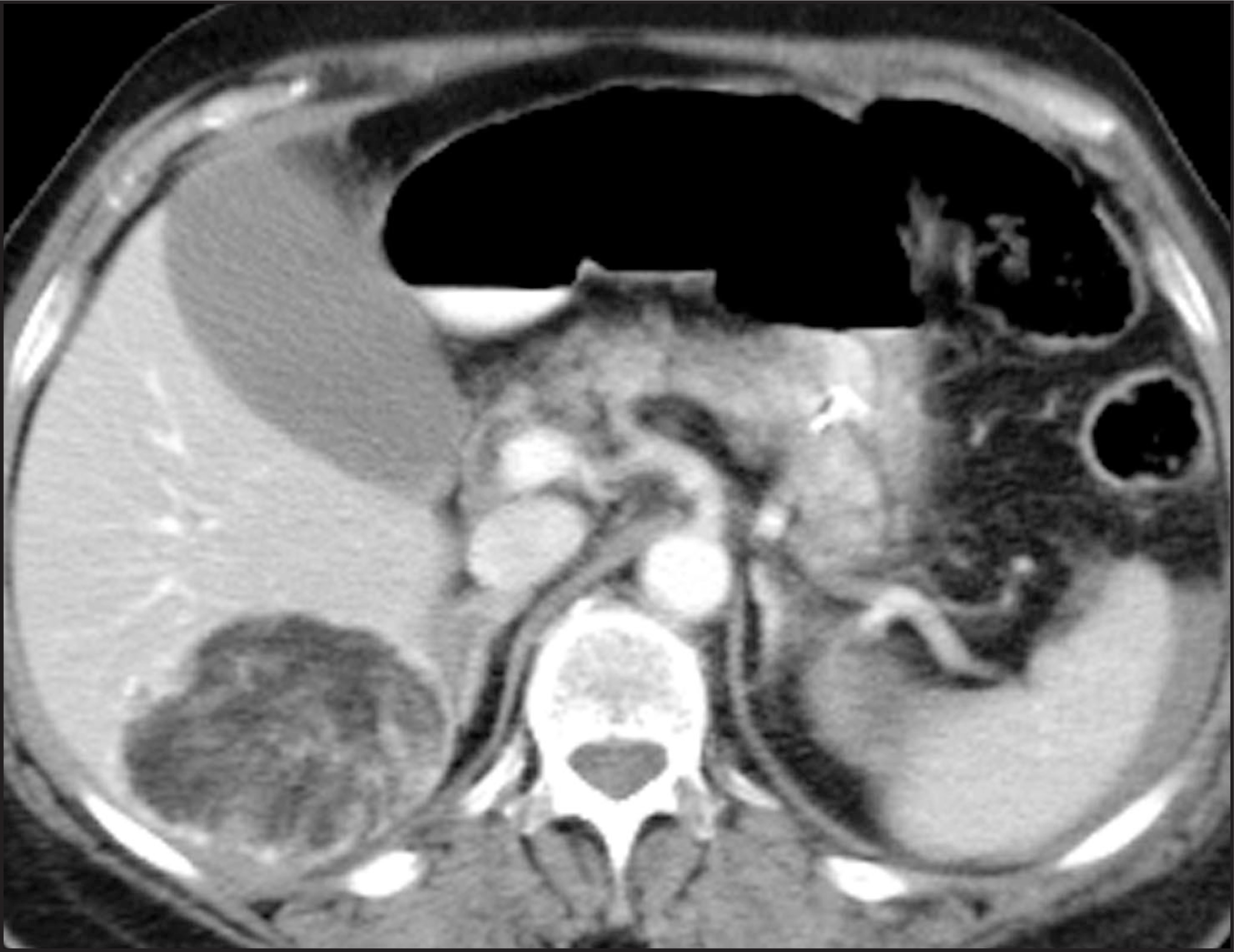
A 74-year-old woman presented to a Level-1 trauma center following a motor vehicle accident. A computed tomography (CT) of the abdomen was performed (figure 1).
DIAGNOSIS
Hepatic angiomyolipoma
IMAGING FINDINGS
A contrast-enhanced CT of the abdomen reveals a 5-cm mass in the posterior segment of the right hepatic lobe composed mostly of fatty tissue with some internal soft tissue stranding the hepatic lobe (figure 1).
DISCUSSION
Hepatic angiomyolipoma is a rare entity with only 70 cases reported in U.S./Japanese literature during the past 25 years.1 It is considered a benign mesenchymal tumor, often with a distinguishing fatty component.
Unlike the more common renal angiomyolipoma, which is associated with tuberous sclerosis in 40% to 50% of cases, hepatic angiomyolipoma is usually solitary. In addition, intratumoral hemorrhage, a common complication in renal angiomyolipoma, has not been reported in its hepatic counterpart.2
Angiomyolipoma, as its name implies, is composed of varying proportions of fat, blood vessel, and muscle, though the proportion of fat can be smaller in the hepatic form than in its renal analogue. In a study by Goodman and Ishak,3 fat occupied ≤ 10% of cut sections in 42% of hepatic angiomyolipomas.
Differential considerations include lipoma, adenoma, focal fatty infiltration, pseudolipoma, and metastatic neoplasms, such as malignant teretoma and liposarcoma. Among fat-containing hepatic lesions, however, the most important and common malignant diagnosis that must be distinguished from angiomyo-lipoma is hepatocellular carcinoma with fatty metamorphosis. Usually, hepatocellular carcinomas are well differentiated and therefore hypovascular; in contrast, an angiomyolipoma is hypervascular.4
Ahmadi et al5 attempted to differentiate angiomyolipoma from hepatocellular carcinoma based on dynamic contrast CT where angio-myolipoma showed early and prolonged enhancement (>4 min) with delayed peak enhancement at 40 to 80 seconds; as opposed to hepatocellular carcinoma that has peak enhancement at 10 seconds and absent or minimal delayed enhancement. Differentiation of angiomyolipoma from cavernous hemangioma can also be made based on the former’s earlier peak enhancement.
Similiarly, Sakamoto et al1 described MRI studies showing extensive enhancement on gadolinium-enhanced images. In addition, fat could be seen with great sensitivity on T1-weighted images as high-signal intensity. Differentiation from hepatocellular carcinoma with fatty metamorphosis can also be made based on the angiomyolipomas prolonged tumor enhancement (>6 min) relative to hepatocellular carcinoma.
CONCLUSION
Angiomyolipomas have a benign course, making preoperative diagnosis important prior to performing a major surgery, such as a hepatic wedge resection. In the recent past, differentiation of angiomyolipoma from hepato-cellular carcinoma with fatty metamorphosis was problematic, requiring biopsy or surgical resection. However, the introduction of improved diagnostic protocols with CT and MRI should permit accurate imaging diagnosis without the need for tissue sampling.
References
- Sakamoto Y, Inoue K, Ohtomo K. Magnetic resonance imaging of an angiomyolipoma of the liver. Abdom Imaging.. 1998;23:158-160.
- Grino P, Such J. Hepatic angiomyolipoma: A rarely described tumor. J Hepatol.. 2000;33:684.
- Goodman Z, Ishak K. Angiomyolipoma of the liver. Am J Surg Pathol.. 1984;8:745-750.
- Irie H, Honda H, Kuroiwa T. Hepatic angiomyolipoma: Report of changing size and internal composition on follow-up examination in two cases. J Comput Assist Tomogr. 1999;23:310-313.
- Ahmadi T, Itai Y, Takahashi M. Angiomyolipoma of the liver: Significance of CT and MR dynamic study. Abdom Imaging.. 1998;23:520-526.
Citation
. CLINICAL Quiz. Applied Radiology. 2002;31(2):36-37. doi:10.37549/AR1072.