CLINICAL QUIZ
Applied Radiology — Vol. 33 , Issue 1 , pp. 33 -39
DOI: 10.37549/AR1212
Published: January 1, 2004
Categories
CASE SUMMARY
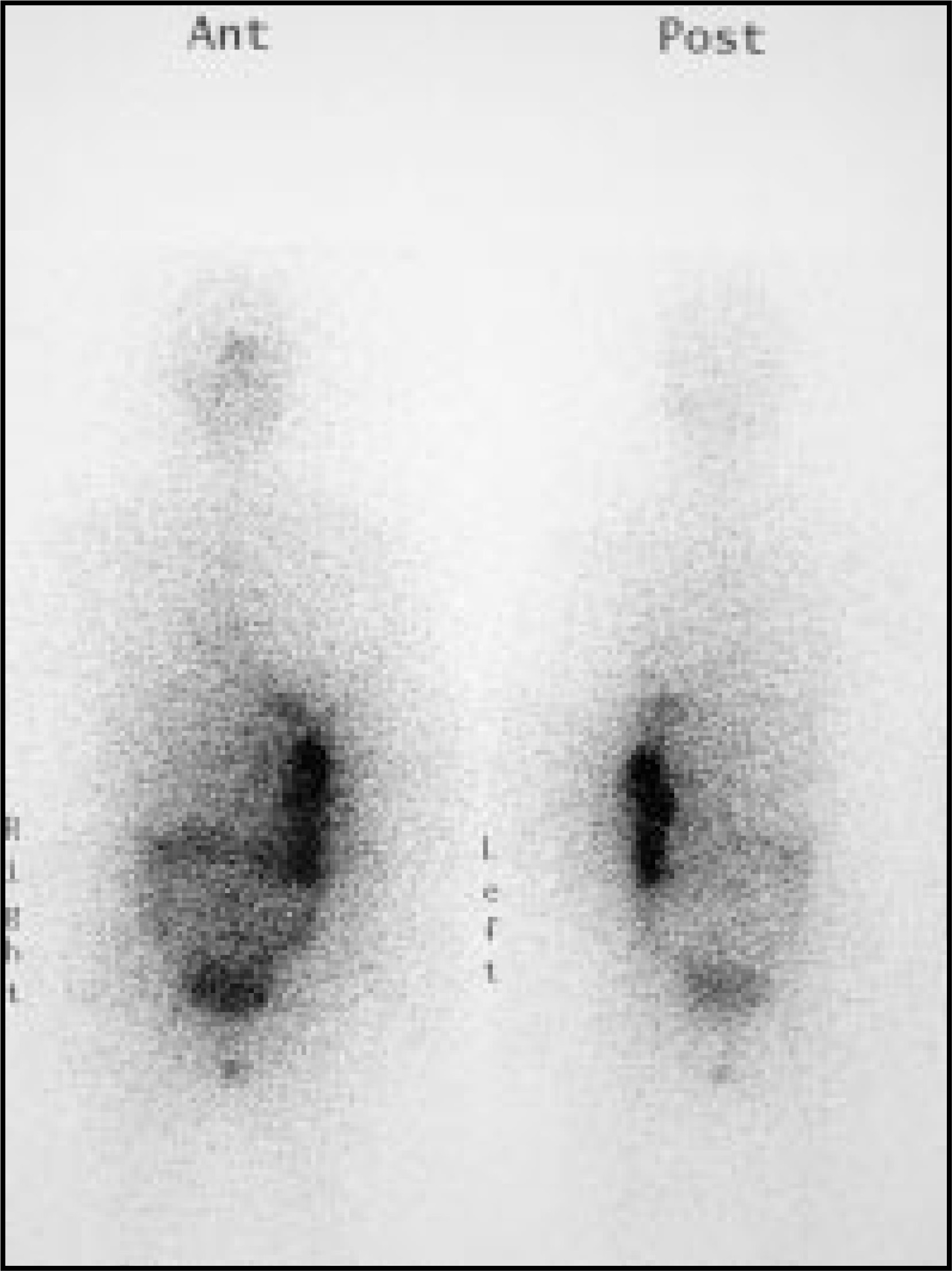
A 41-year-old woman with a history of multifocal papillary thyroid carcinoma had a total thyroidectomy performed 2 months prior to presentation. A diagnostic whole-body scan was obtained 48 hours after the administration of 185 MBq (5 mCi) of 131I (Figure 1). One day later, the patient was treated with 3700 MBq (100 mCi) of 131I. A post-therapy scan was performed 7 days later (Figure 2).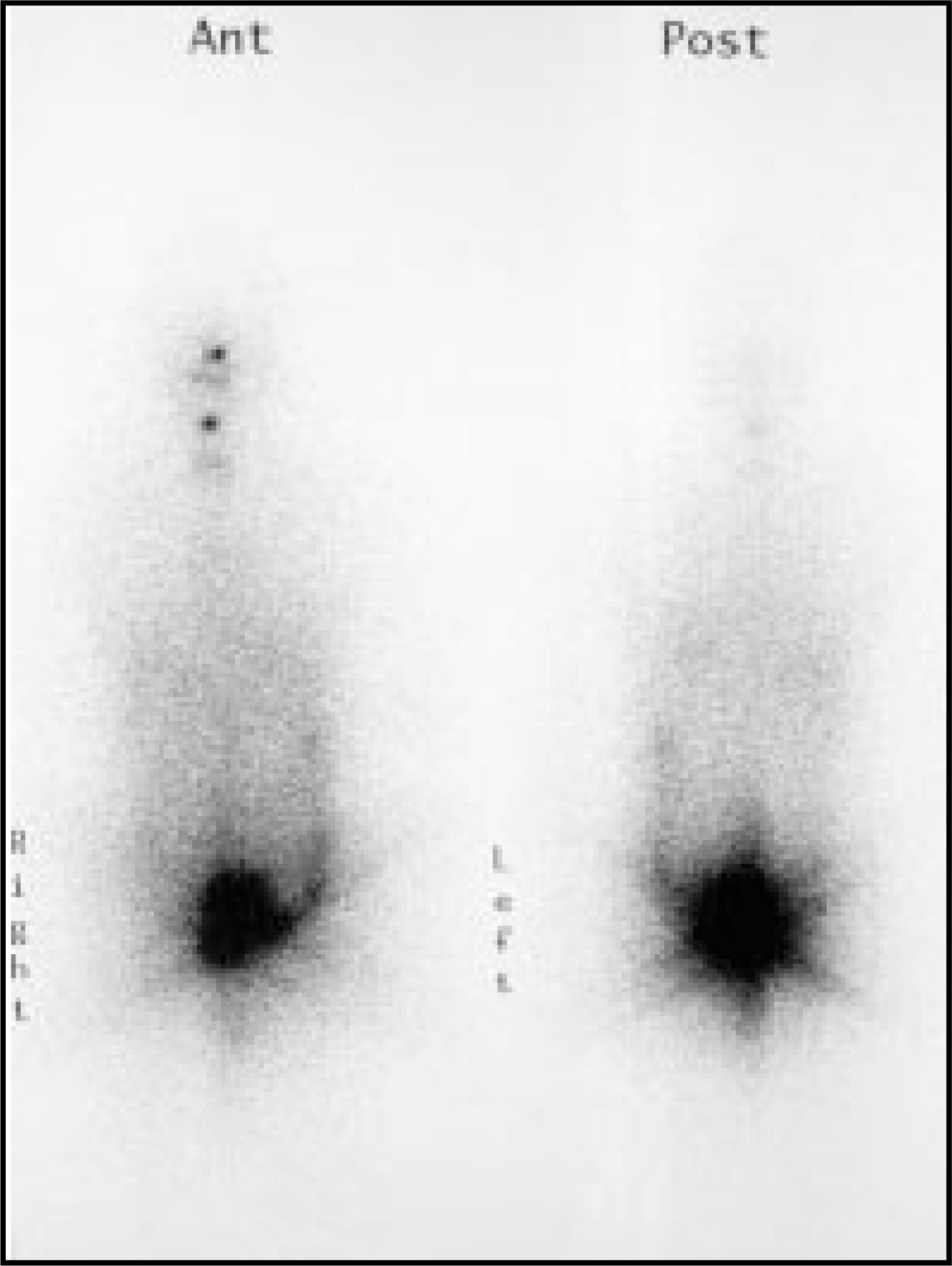
The diagnosis and discussion are presented beginning on page 38.
DIAGNOSIS
Thyroid stunning
IMAGING FINDINGS
DISCUSSION
Radioiodine plays an important role in the management of well-differentiated thyroid cancer. There are three principle nuclear medicine procedures performed on patients with thyroid cancer: 1) diagnostic scan, 2) 131I therapy, and 3) post-therapy scan. Given the wide availability and relatively low cost, most institutions use 131I for the diagnostic radiotracer, as opposed to 123I. Several days after the administration of the diagnostic dose, a diagnostic scan is obtained, which serves several purposes: 1) to show any postsurgical thyroid remnant, 2) to reveal any regional or distant metastases for staging, and 3) to demonstrate that the thyroid tissue takes up radioactive iodine and is therefore amenable to 131I therapy.
Once the diagnostic scan reveals functioning thyroid remnant or metastases, the administered activity of 131I therapy can then be prescribed based on histologic classification and stage of disease. There are several treatment strategies for thyroid cancer therapy, ranging from standardized fixed doses to individual dosimetry.1 A post-therapy scan is then obtained several days after 131I therapy. With the much higher dose of 131I therapy compared with the lower diagnostic 131I dose, it is not uncommon to detect functioning thyroid tissue that was not seen on the diagnostic scan. Therefore, the post-therapy scan can upstage patients’ disease.
Several authors have hypothesized that the diagnostic 131I dose impairs the thyroid cells such that the subsequent 131I therapy is less effective.2-6 This phenomenon has been termed thyroid “stunning.” The stunning effect may be observed when activity on a diagnostic scan appears less intense on the post-therapy scan. The existence of thyroid stunning remains controversial, since other authors have not been able to produce it.7,8 These authors argue that stunning may be merely a product of differences in protocol between the diagnostic and post-therapy scans. However, at a cellular level, researchers have shown a dose-dependent decrease in iodide transport 3 days following a 48-hour 131I irradiation of cultured porcine thyroid epithelial cells.9
Due to the potential for stunning, many physicians have altered their protocols for treating thyroid carcinoma. Since stunning may be dose-dependent, many have lowered the diagnostic 131I dose but at the expense of sensitivity.2,9-11 Due to the lack of reported stunning with 123I, some clinicians have exclusively used the more expensive 123I but again compromising sensitivity compared with 131I.2,4,5 As another alternative, some physicians have abandoned the diagnostic scan altogether.12,13 The two main drawbacks of this approach are: 1) the thyroid carcinoma may not be 131I-avid so 131I therapy may be inadvertently given to patients who would not benefit from it; and 2) the diagnostic scan may reveal unsuspected metastases requiring a larger therapy dose.13
Those authors who believe in thyroid stunning are still divided regarding the clinical effects of stunning. Some researchers have shown no significant difference in 131I therapy success rates between stunned and nonstunned patients.6,14 In addition, when comparing patients with 131I diagnostic scans with those without diagnostic scans, no significant difference in therapeutic efficacy was found.12 Contrary to these other reports, one group of investigators actually demonstrated higher ablation success rates in the stunned versus the nonstunned population.15
CONCLUSION
There has been no consensus achieved in the literature concerning 1) the existence of thyroid stunning; or 2) its effect on clinical outcome. Reports include myriad different protocols varying from institution to institution that are based on uncontrolled retrospective studies, making it difficult to reproduce results. Further investigation with prospective studies and large sample sizes should help clarify these issues.
References
- Mazzaferri E, Kloos R. Current approaches to primary therapy for papillary and follicular thyroid cancer. J Clin Endocrinol Metab. 2001;86:1447-1463.
- Park H, Perkins O, Edmundson J. Influence of diagnostic radioiodines on the uptake of ablative dose of 131iodine. Thyroid. 1994;4:49-54.
- Kao C, Yen T. Stunning effects after a diagnostic dose of iodine-131. Nuklearmedizin. 1998;37:23-25.
- Leger F, Izembart M, Dagousset F. Decreased uptake of therapeutic doses of 131iodine after 185-MBq 131iodine diagnostic imaging for thyroid remnants in differentiated thyroid carcinoma. Eur J Nucl Med. 1998;25:242-246.
- Park H, Park Y, Zhou X. Detection of remnant/metastasis without stunning: An ongoing dilemma. Thyroid. 1997;7:277-280.
- Nakada K, Katoh S, Kaji T. Does stunning impair prognosis of differentiated thyroid cancer [abstract]?. J Nucl Med. 2002;43(suppl):328P.
- Cholewinski S, Yoo K, Klieger P, O’Mara R. Absence of thyroid stunning after diagnostic whole-body scanning with 185 MBq 131I. J Nucl Med. 2000;7:1198-1202.
- McDougall I. 74 MBq radioiodine 131I does not prevent uptake of therapeutic doses of 131I (i.e. it does not cause stunning) in differentiated thyroid cancer. Nucl Med Commun. 1997;18:505-512.
- Postgard P, Himmelman J, Lindencrona U. Stunning of iodide transport by 131I irradiation in cultured thyroid epithelial cells. J Nucl Med. 2002;6:828-834.
- Waxman A, Ramanna L, Chapman N. The significance of 131-I scan dose in patients with thyroid cancer: Determination of ablation: Concise communication. J Nucl Med. 1981;22:861-865.
- Muratet J, Giraud P, Daver A. Predicting the efficacy of first iodine-131 treatment in differentiated thyroid carcinoma. J Nucl Med. 1997;38:1362-1368.
- Morris L, Waxman A, Braunstein G. The nonimpact of thyroid stunning: Remnant ablation rates in 131I-scanned and nonscanned individuals. J Clin Endocrinol Metab. 2001;86:3507-3511.
- Reynolds J, Robbins R. The changing role of radioiodine in the management of differentiated thyroid cancer. Semin Nucl Med. 1992;27:152-164.
- Muratet J, Daver A, Minier J, Larra F. Influence of scanning dose of 131iodine on subsequent first ablative treatment outcome in patients operated on for differentiated thyroid carcinoma. J Nucl Med. 1998;39:1546-1550.
- Bajen M, Marie S, Munoz A, Garcia J. Effect of diagnostic dose of 185 MBq 131I on postsurgical thyroid remnants. J Nucl Med. 2000;12:2038-2042.
Citation
. CLINICAL QUIZ. Applied Radiology. 2004;33(1):33-39. doi:10.37549/AR1212.