CLINICAL QUIZ
Applied Radiology — Vol. 33 , Issue 3 , pp. 36 -44
DOI: 10.37549/AR1226
Published: March 1, 2004
Categories
CASE SUMMARY
A 49-year-old woman was referred from her primary-care physician with a 4-month history of left iliac fossa and groin pain that kept her awake at night. This required a transcutaneous electrical nerve stimulation (TENS) machine in addition to oral analgesia. Her relevant medical history was of carcinoma of the cervix 9 years previously for which she was treated with a Wertheim’s hysterectomy but no adjuvant radiotherapy. Examination was unremarkable, and all blood levels, including plasma viscosity, blood count, and bone chemistry, were normal. A barium enema, pelvic ultrasound, and small bowel study were normal (not shown). A computed tomographic (CT) scan of the abdomen and pelvis was normal (not shown).
In view of the normal findings with ongoing severe pain, an orthopedic opinion was sought. He found minor limitation of movement of the hip but no associated muscle wasting. The patient’s weight was steady, and she had not experienced night sweats. Hip radiographs were requested, which demonstrated an ill-defined lucency in the left femoral neck/head (Figure 1).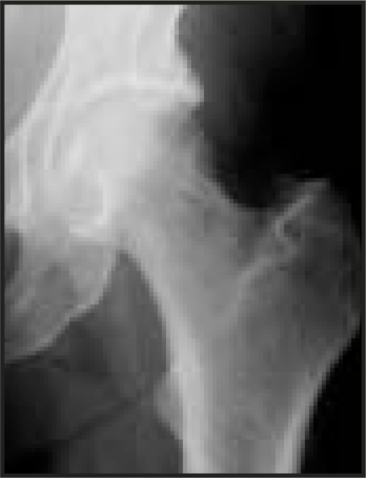
Additional images are shown on page 37. The diagnosis and discussion are presented on page 43.
IMAGING FINDINGS
The patient was taking steroids for asthma and a magnetic resonance image (MRI) was requested to exclude avascular necrosis of the femoral head (Figure 2). This demonstrated a small hip effusion, high signal on short tau inversion recovery in the femoral neck, which was consistent with edema and disruption of the femoral cortex.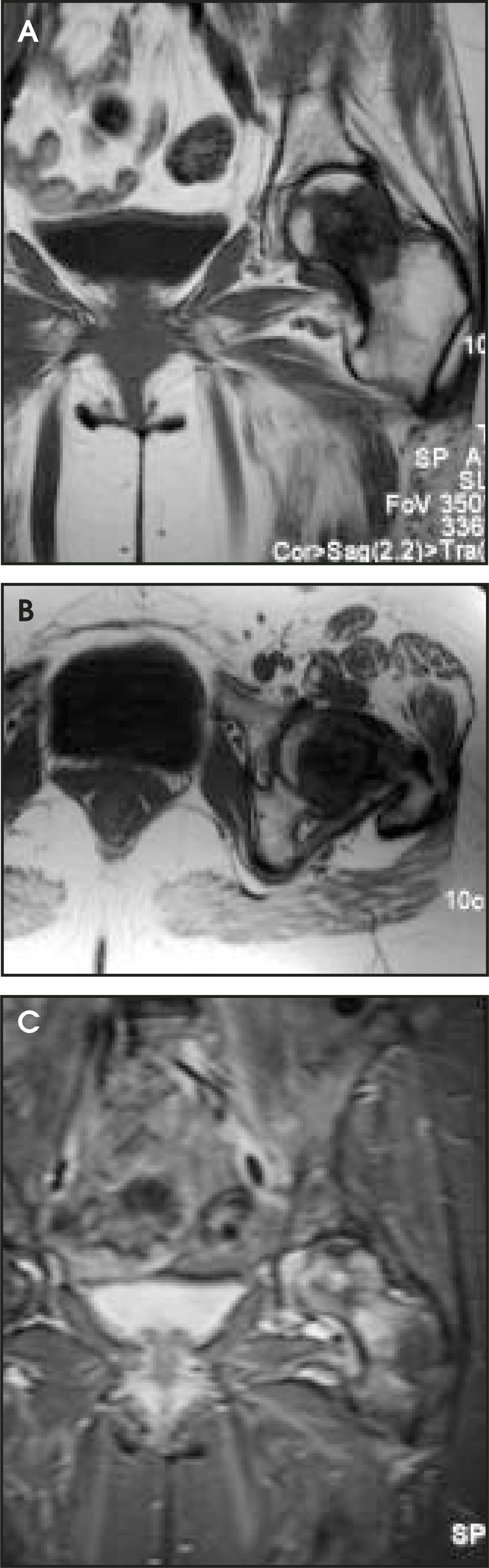
This raised the possibility of metastasis from the previous carcinoma of the cervix. A full-body bone scintogram was performed to look for further lesions (Figure 3), which showed a solitary area of increased tracer uptake in the left femoral neck/head.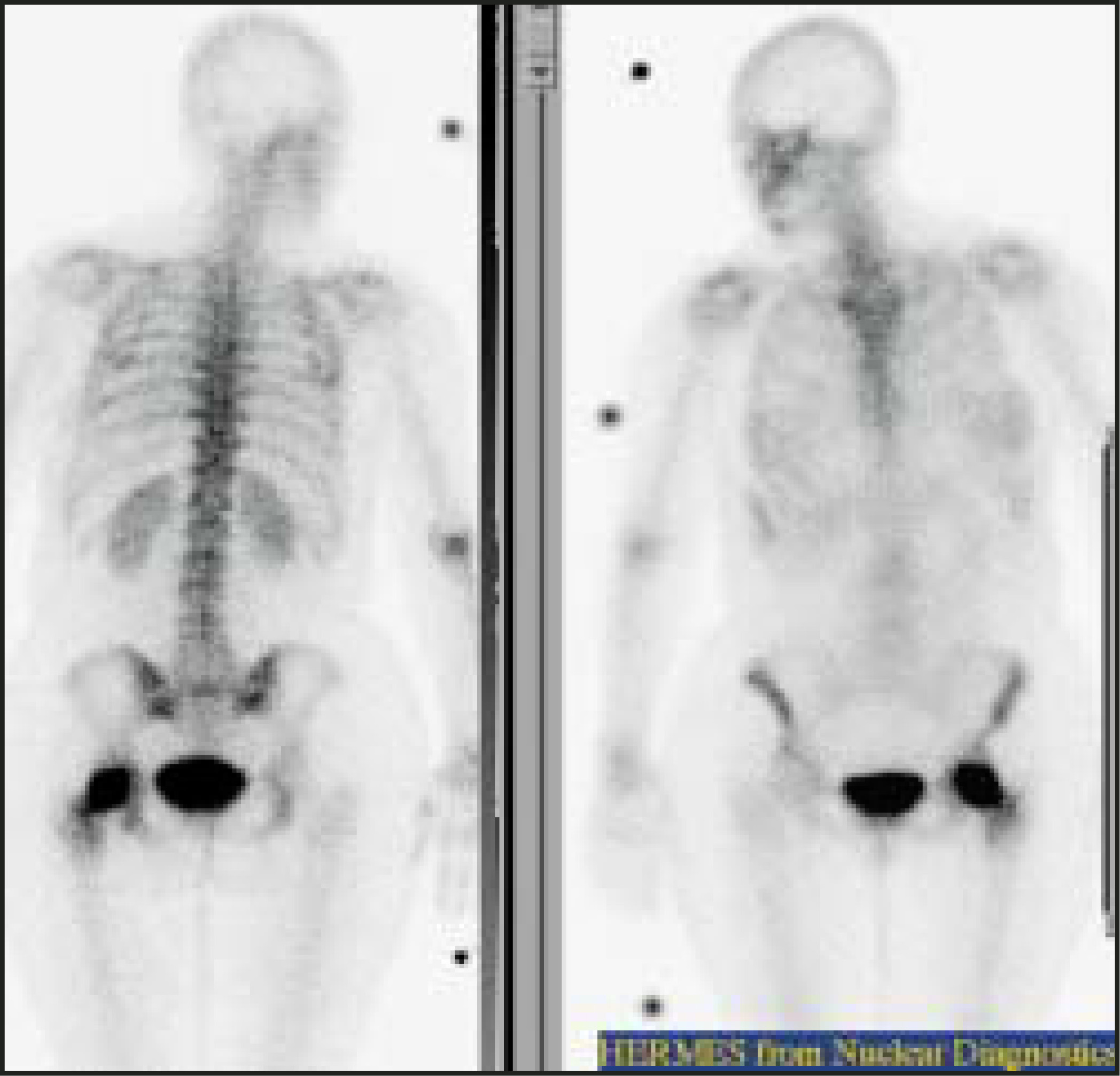
A CT-guided bone biopsy was performed to determine the nature of the lesion. The CT showed a diffuse lesion of mixed lytic and sclerotic appearance in the left femoral neck (Figure 4). The biopsy was reported as normal. This was discussed with the orthopedic surgeon, and as this did not correlate with the radiological and clinical findings, the patient was recalled for a second biopsy. This showed features of a diffuse large B cell lymphoma staged as 1E, an isolated lesion restricted to a single osseous site.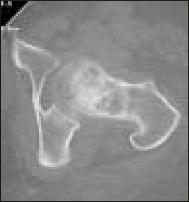
DIAGNOSIS
Primary bone lymphoma
DISCUSSION
Primary bone lymphoma is defined as a tumor involving a single focus with unequivocal evidence of lymphoma in the bone lesion.1 It is rare, accounting for <1% of all non-Hodgkin’s lymphomas and 5% of all primary bone tumors.2 Most cases are of the diffuse large B-cell category.3 The age distribution is bimodal with peaks in the second to third decade, and a second peak in the fifth to sixth decade with women more commonly affected in the older age group.4 There is a wide pattern of bone involvement with the spine forming the most frequent site of axial lesions, and the femur is the most common site overall.5 In many cases, the diagnosis is delayed because of nonspecific clinical signs.6 Chronic dull pain may be the only complaint.
Radiographic findings do not conform to a diagnostic pattern, but 58% show osteolysis, with soft tissue involvement in 70% of cases.6 Periosteal reaction is rare.1 The definitive diagnosis is made from biopsy and histology. Clinical stage follows the Ann Arbor classification, which determines the extent to which the tumor has metastasized. Grading is obtained from the tumor histology. The stage of the disease appears to be the most important prognostic indicator, with an overall 5-year survival rate of 54%.3
The patient’s initial presentation had been with left iliac fossa pain and her initial work-up was aimed at intra-abdominal/pelvic pathology in view of her medical history. This led to a significant delay before an orthopedic opinion was sought. This is very common and highlights the need to always consider bone pathology as a source of ill-defined pain, particularly when it disturbs the patient’s sleep.
CONCLUSION
The radiologic findings were nonspecific, but were suggestive of malignant disease. In particular, the lytic appearance on the plain radiograph and the loss of the femoral cortex on the MRI with the associated effusion were suspicious. Primary bone lymphoma typically has nonspecific radiologic findings. In cases such as this, biopsy is essential, after discussion with an orthopedic surgeon, to establish the diagnosis. If the clinicoradiologic findings and histology do not match, repeat biopsy is indicated.
References
- Edeiken-Monroe B, Ediken J, Kim E. Radiological concepts of lymphoma of bone. Rad Clin North Am. 1990;28:841-864.
- Chin H, McGuire M, Block M. Primary lymphoma of bone. Nebr Med J. 1990;75:303-306.
- Choi J, Hahn J, Suh C, Yang W. Primary lymphoma of bone—Survival and prognosis. Korean J Intern Med. 2002;17:191-197.
- Limb D, Dreghorn C, Murphy J, Mannion R. Primary lymphoma of bone. Int Orthop. 1994;18:180-183.
- Franczyk J, Samuels T, Rubenstein J. Skeletal lymphoma. Can Assoc Radiol J. 1989;40:75-79.
- Durr H, Muller P, Hiller E. Malignant lymphoma of bone. Arch Orthop Trauma Surg. 2002;122:10-16.
Citation
. CLINICAL QUIZ. Applied Radiology. 2004;33(3):36-44. doi:10.37549/AR1226.