CLINICAL QUIZ
Applied Radiology — Vol. 33 , Issue 11 , pp. 52 -75
DOI: 10.37549/AR1290
Published: November 1, 2004
Categories
CASE SUMMARY
A 28-year-old woman with a long-standing history of neck pain presented to her physicians with worsening symptoms and a new onset of parasthesia affecting the right arm.
Imaging findings, diagnosis, and discussion begin on page 74.
IMAGING FINDINGS
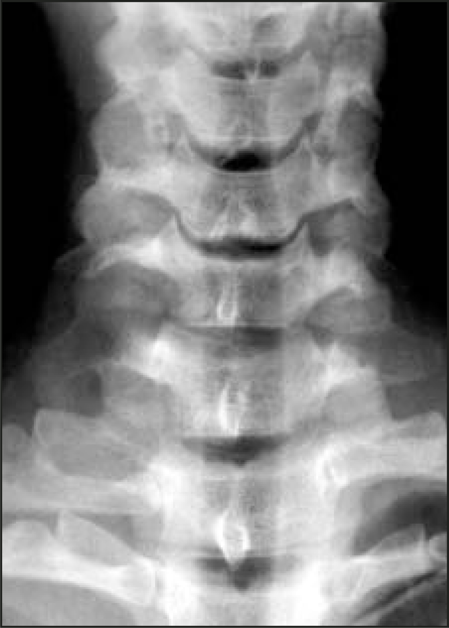
The lateral radiograph of the cervical spine revealed widening of the right C6/7 and C7/T1 intervertebral foramen (Figure 1). This widening was also seen on the anteroposterior film with an associated mass at the right apex, although there was no evidence of bony destruction (Figure 2).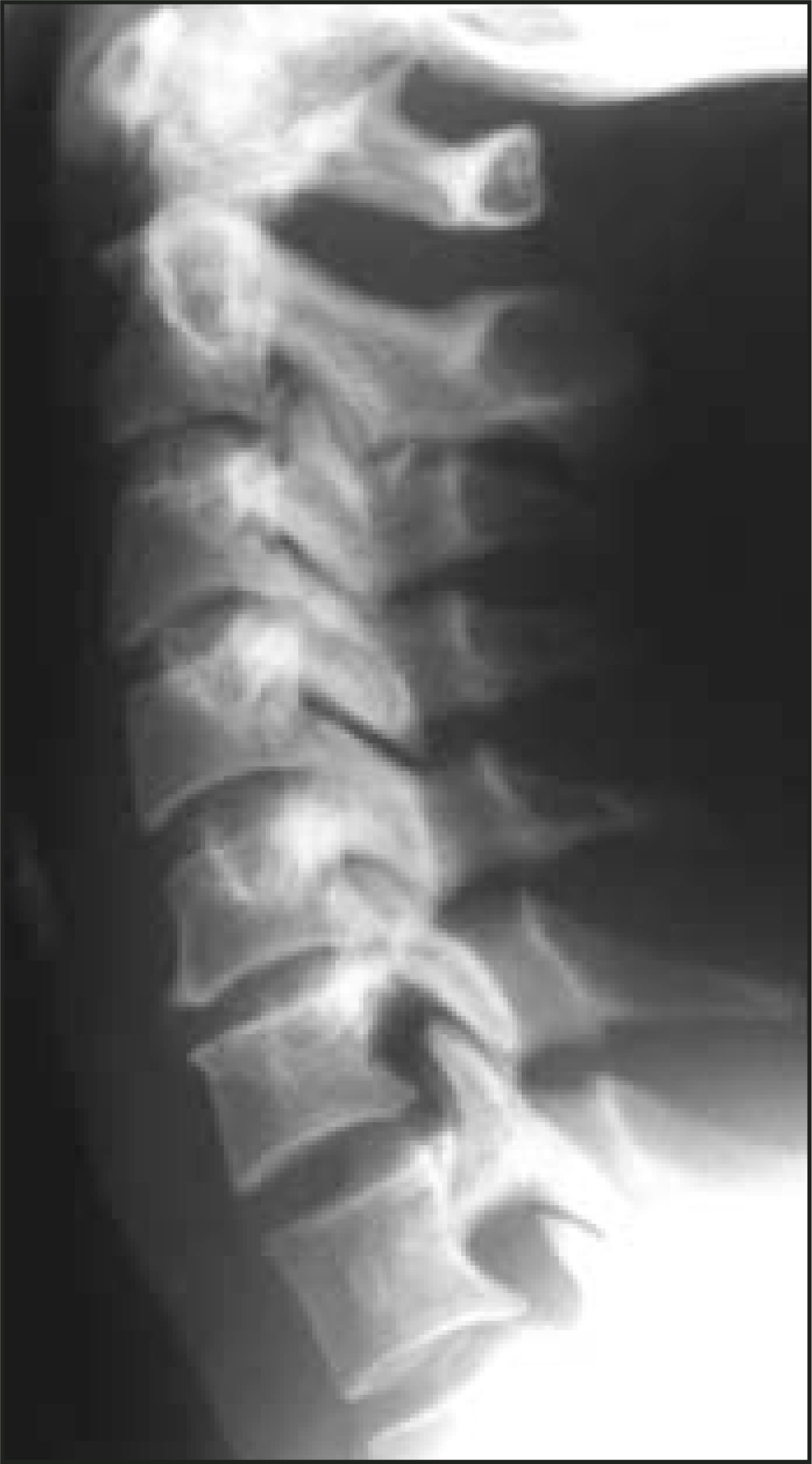
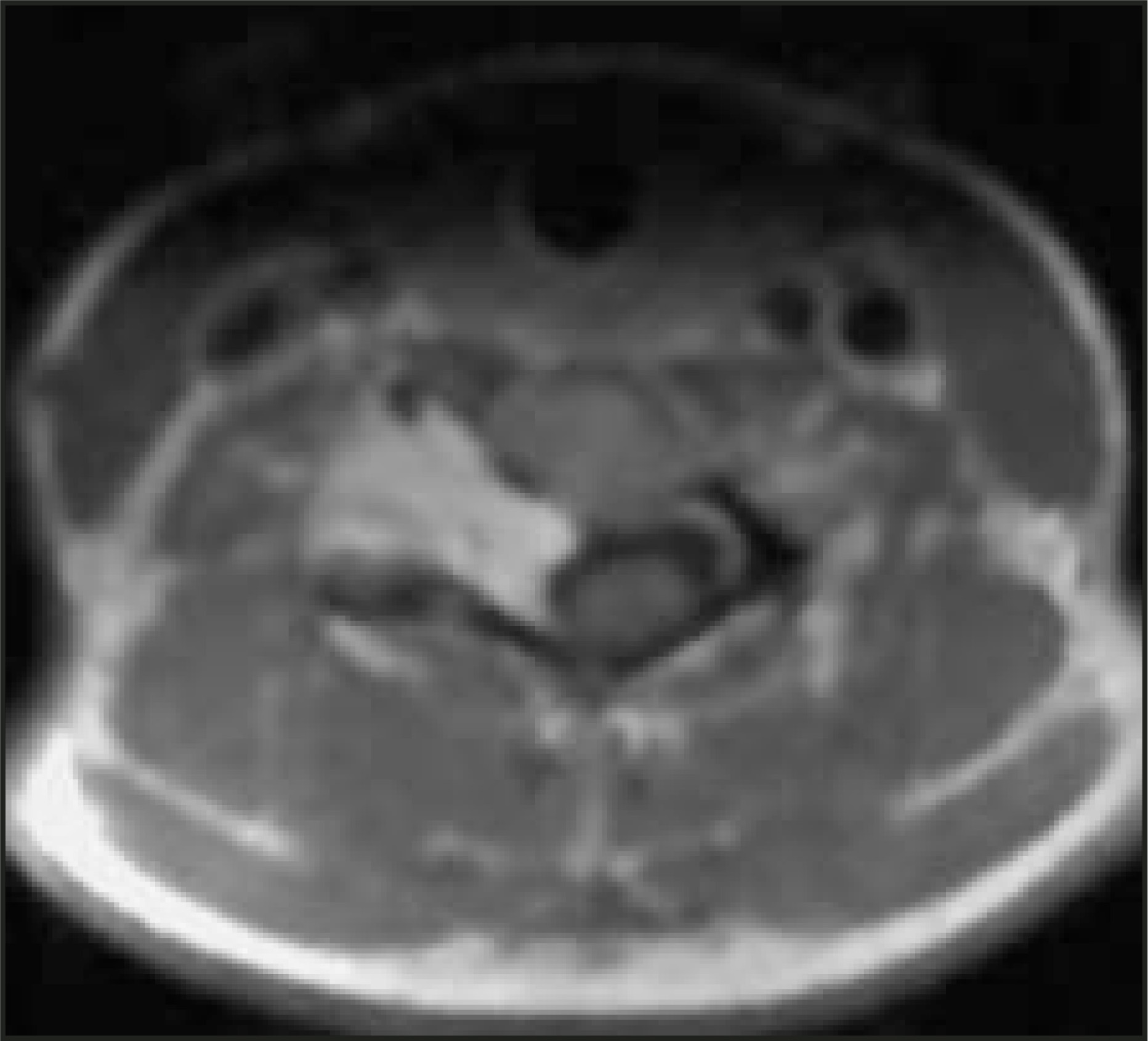
MRI revealed a soft-tissue mass in the right para-vertebral region extending into the lung apex (Figure 3). This originated from the right C6/7 intervertebral foramen with intracanal extension giving a dumb-bell appearance (Figure 4). On T1-weighted imaging (T1WI), the mass was well defined with an intermediate signal density, but demonstrated inhomogenous enhancement following injection of Gd-DTPA (Figure 5). The mass was of a heterogeneously high signal on T2-weighted (T2WI) sequences (Figure 6).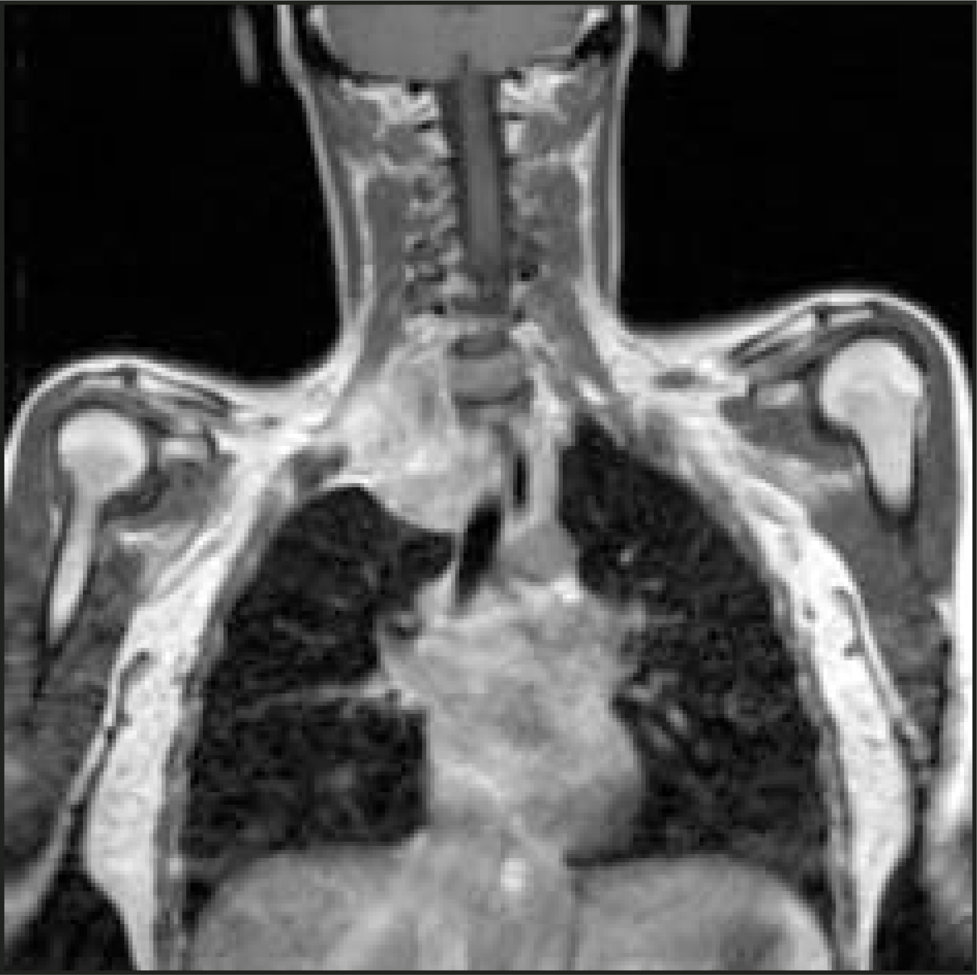

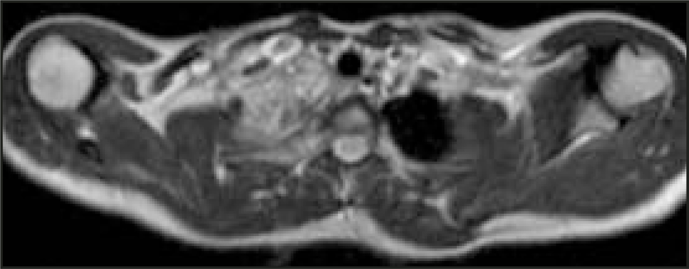
At operation, a 9 × 3 × 6-cm mass was visualized arising from the C6/C7 nerve root with extension into the superior mediastinum. The histology of the resected mass revealed packed ganglion and nerve sheath cells diagnostic of ganglioneuroma.
The patient made an uncomplicated recovery with resolution of her symptoms and no neurological deficit.
DIAGNOSIS
Ganglioneuroma
DISCUSSION
Ganglioneuroma is an uncommon benign neurogenic tumor that arises from the sympathetic ganglia. Ganglioneuromas can occur in any age group but are most common in patients <40 years of age and are usually located in the posterior mediastinum followed by the retroperitoneal and cervical regions.1 They can be considered to occupy the benign end of the spectrum of neurogenic tumors that include the more aggressive ganglioneuroblastoma and the malignant neuroblastoma. The other diagnoses to consider would be neurofibroma, schwannoma, and pheochromocytoma.1
Magnetic resonance imaging can be used to evaluate: 1) morphologic features, including size and capsule; 2) signal intensity and homogenicity; 3) contrast enhancement; and 4) dynamic enhancement pattern. These characteristics can be used in conjunction with the plain radiographs and clinical findings to attempt to differentiate between the above. Indeed, the image patterns usually correlate well with the histologic features.
Ganglioneuromas have a well-defined capsular margin. On T1WI, they have homogenous low/intermediate signal with heterogeneous high signal on T2WI and GD-DTPA–enhanced T1WI. The nodular low intensity areas within the high signal mass on T2WI correspond to interlacing areas of collagenous fibrous tissue and Schwann cells that can give a whorled appearance.
Neurofibromas also have a low/intermediate signal on T1WI, but the central aspect of the tumor may have a higher signal than the periphery, which correlates with the histologic findings of central packed nerve sheath cells and fibrous tissue with peripheral acellularity and myxoid degeneration. This is reflected on the T2WI as a bright peripheral signal with an intermediate signal of the central portion, reflecting the more solid component.
Schwannomas have similar characteristics on MRI to ganglioneuromas but sometimes have very high signal intensity within the tumor due to cystic degeneration.
Neuroblastomas and ganglioneuroblastomas are more aggressive than are ganglioneuromas and tend to occur in a younger age group with typical MRI appearances of irregular margins, vessel encasement, and invasion of adjacent organs. These more malignant tumors have a higher cellular component and less myxoid matrices, which results in a lower signal on T2WI.2-4
Pheochromocytomas, neuroblastomas, and ganglioneuroblastomas show marked early enhancement after administration of Gd-DTPA.5 However, ganglioneuromas are relatively avascular with abundant myxoid matrix that results in a lack of early enhancement on dynamic MR imaging.
CONCLUSION
Although the final arbiter for diagnosis is always histology, using the above imaging features, good radiological correlation can be expected, as was demonstrated in this case.
References
- Zhang Y, Nishimura H, Kato S. MRI of ganglioneuroma: Histologic correlation study. J Comput Assist Tomogr. 2001;25:617-623.
- Ichikwa T, Ohtomo K, Araki T. Ganglioneuroma: Computed tomography and magnetic resonance features. Br J Radiol. 1996;69:114-121.
- Sakai F, Sone S, Kiyonono K. Intrathoracic neurogenic tumours: MR-pathologic correlation. AJR Am J Roentgenol. 1992;159:279-283.
- Harkin J, Reed R. Atlas of tumour pathology 2nd series. Armed Forces Instit Pathol. 1969:29-96.
- Ichikawa T, Fujimoto H, Murakami K. Extra-adrenal phaeochromocytoma: Dynamic demonstration at 0.5 Tesla. Clin Imaging. 1992;16:187-189.
Citation
. CLINICAL QUIZ. Applied Radiology. 2004;33(11):52-75. doi:10.37549/AR1290.