Clinical update: Breath-hold 3D gadolinium-enhanced multiphasic abdominal MR
Applied Radiology — Vol. 32 , Issue 1 , pp. 9 -12
DOI: 10.37549/AR1152
Published: January 1, 2003
Categories
Improvements in gradient performance have made the rapid acquisition of three-dimensional (3D) volumetric gradient echo data possible. Thin contiguous images of the abdominal organs can be obtained with isotropic voxels in a single breath-hold with resultant decrease in respiratory motion artifact and improved anatomic sharpness.1 The volumetric image data can then be viewed in any desired plane.2 This technique results in higher signal-to-noise ratios (SNRs) (related to the longer repetition time) compared with spin-echo (SE) or fast-spin-echo (FSE) T2-weighted images or two-dimensional (2D) gradient-echo images.3 Contrast-to-noise levels for hepatic lesions are better than for spin-echo T2 and comparable to fast-spin-echo T2-weighted images.1
We have used this technique to obtain gadolinium-enhanced 3D spoiled gradient-echo images of the abdominal organs in multiple phases of enhancement (arterial, venous, delayed). This allows improved lesion detection and characterization by allowing assessment of lesion vascularity similar to that achieved with contrast-enhanced computed tomography (CT). This technique provides an alternative to conventional CT in cases in which iodinated contrast is contraindicated. This article will introduce the reader to this technique, including its technical facets, and to illustrate several cases of its use.
Technique
Images were obtained on a 1.5 T scanner (Signa, GE Medical Systems, Waukesha, WI) with a phased-array torso coil. Three plane-localizer images are first obtained. The patient is then timed to see how long he or she can maintain a breath-hold. Slice thickness and coverage area are adjusted based on the patient’s ability to hold a breath. Single breath-hold imaging of the organ of interest is achievable in the majority of patients imaged.
Our standard scan parameters include an echo time (TE) of 3 msec, representing a compromise between fat-water phase cancellation (occurring at 2.1 msec at 1.5 T) and signal loss due to T2* decay with longer TE times. Repetition time (TR) is set at minimum (usually approximately 7.6 to 7.7 msec) to reduce flow and susceptibility artifacts. Bandwidth is set to 31.25 kHz and is related indirectly to the other parameters. A wide bandwidth allows for a shorter TR and TE and therefore faster scanning but at the expense of lower SNR. A narrow bandwidth is preferred, resulting in longer scan times but higher SNR. We use a flip angle of 20°, which, in our experience, provides good tissue contrast. Others advocate higher flip angles of 30° to 60°.
Acquisition time is further reduced by using 0.5 number of excitations (NEX [fractional NEX]), which takes advantage of k-space symmetry, allowing sampling of just over half of k-space with mathematical reconstruction of the remaining data, resulting in a decrease in scan time by one-half.
Apparent spatial resolution is increased by using Zerofill Interpolation Processing (ZIP, GE Medical Systems). This post-processing technique improves scan resolution without increasing scan time. The trade-off is an increase in reconstruction time. When applied in the slice direction (Slice ZIP), there is no decrease in SNR. When applied in the x and y directions (512 ZIP), there is a small decrease in SNR. This technique improves the quality of reformations and maximum intensity projection (MIP) images and decreases volume-averaging artifacts. Gibbs ringing and truncation artifacts are increased with ZIP.
Noncontrast images are obtained first. Gadolinium contrast (20 mL) is administered through a ≥ 20-gauge needle placed in an antecubital vein at a rate of 1 to 1.5 mL/sec. The technologist instructs the patient on breathing while injecting the first 10 mL of contrast. The patient takes two deep breaths, then takes one deep breath and holds it. The first postcontrast scan is initiated and the remaining 10 mL of contrast is injected. This is followed by a 20-mL saline flush. Following the first scan, the patient takes a few breaths and then breath-holds for the venous phase and 5 minutes later for the delayed phase.
Clinical application
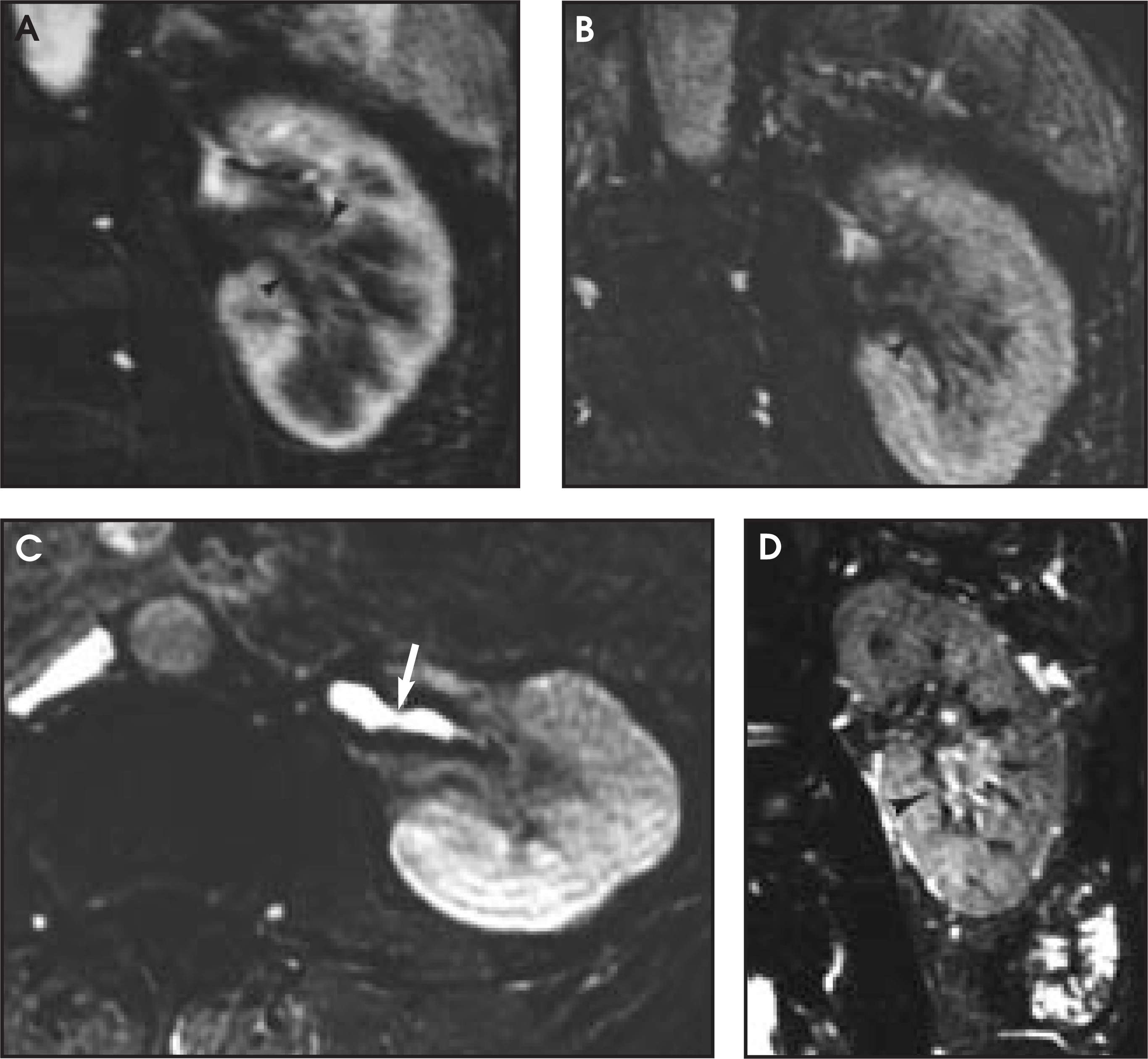
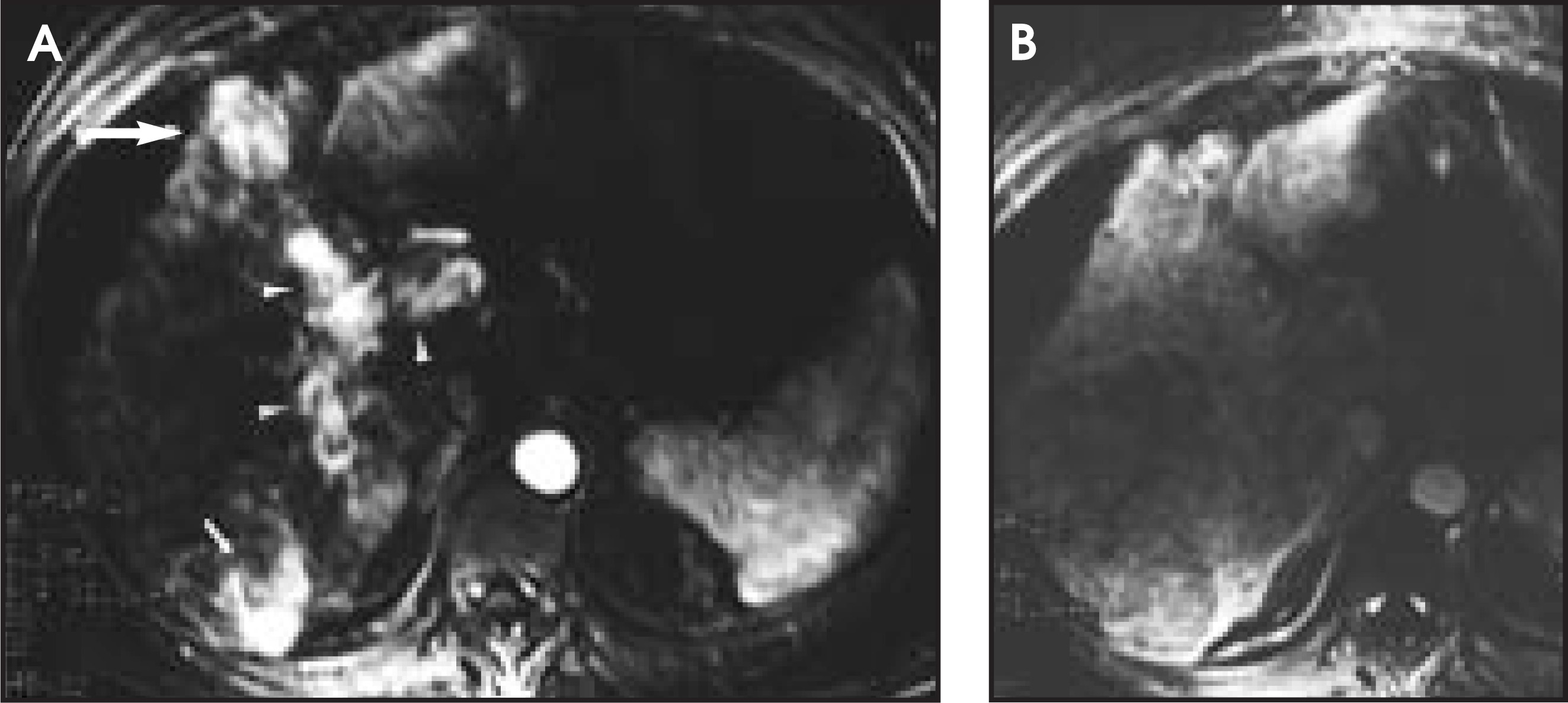
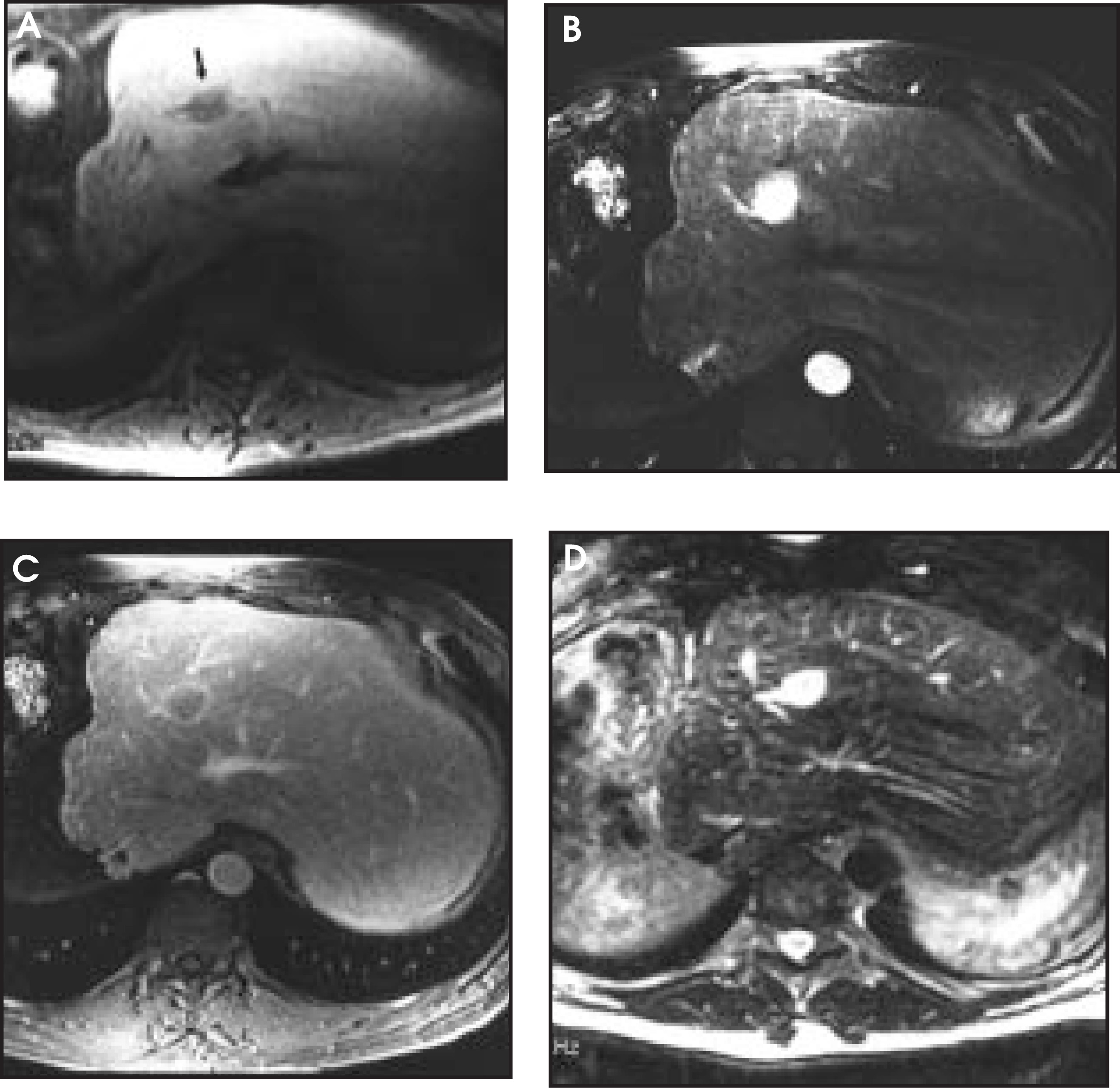
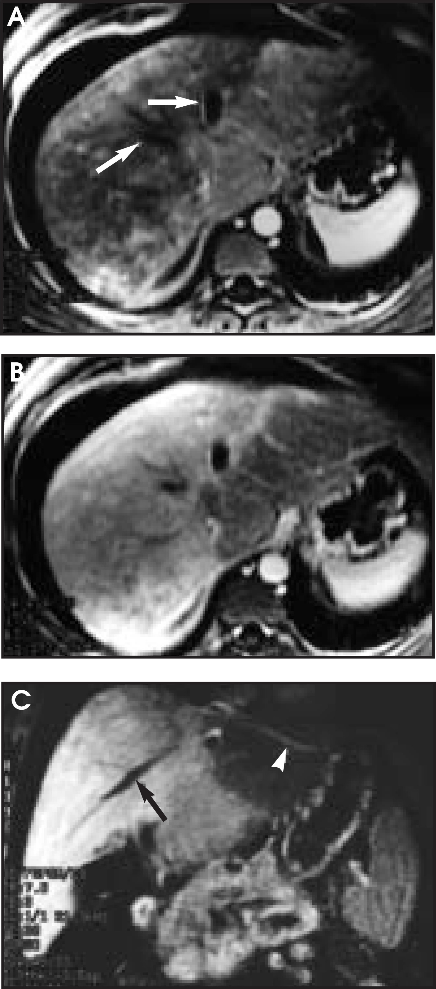
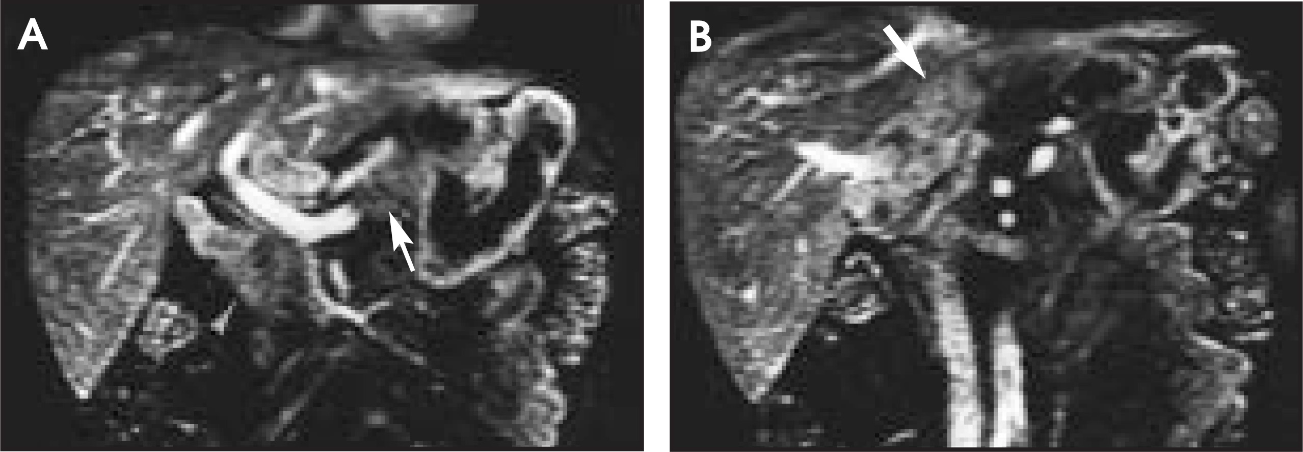
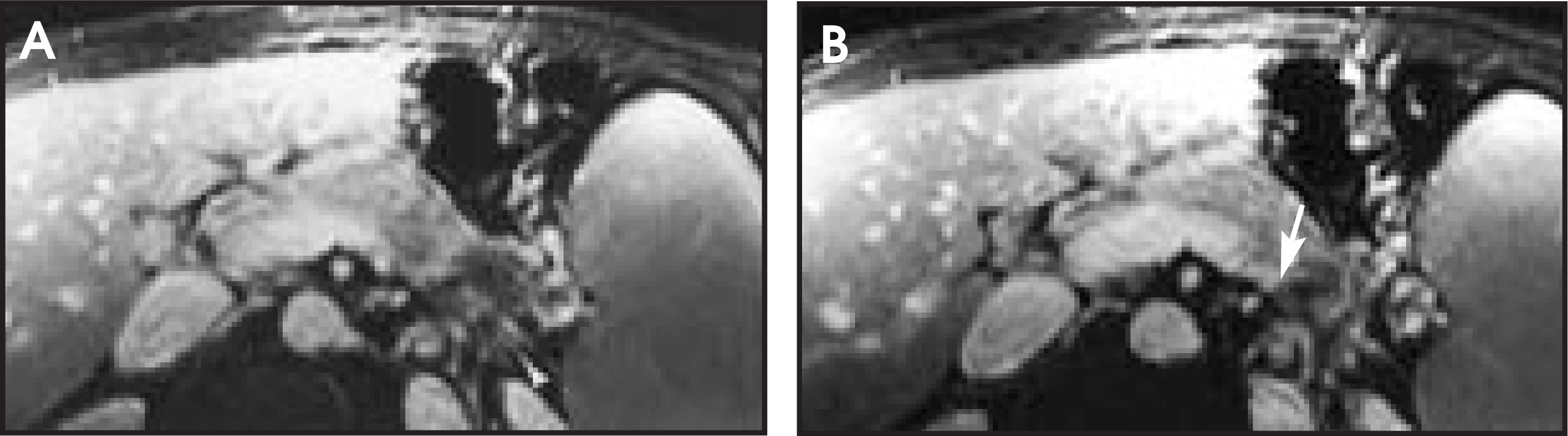
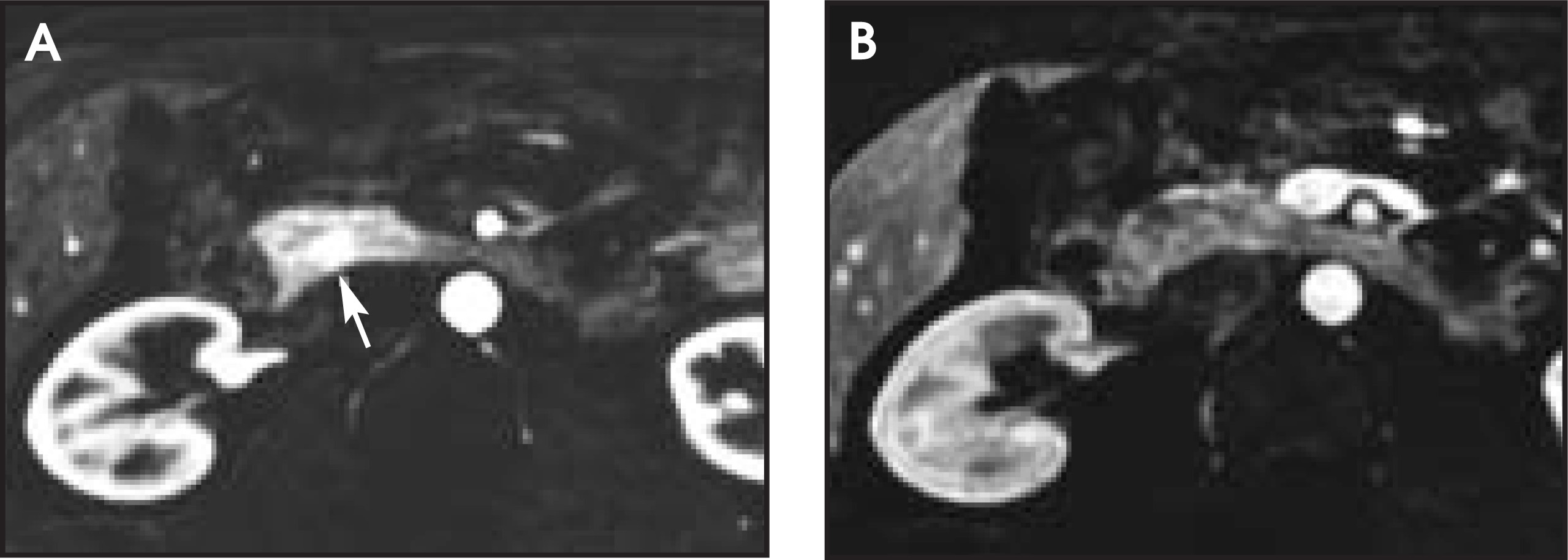
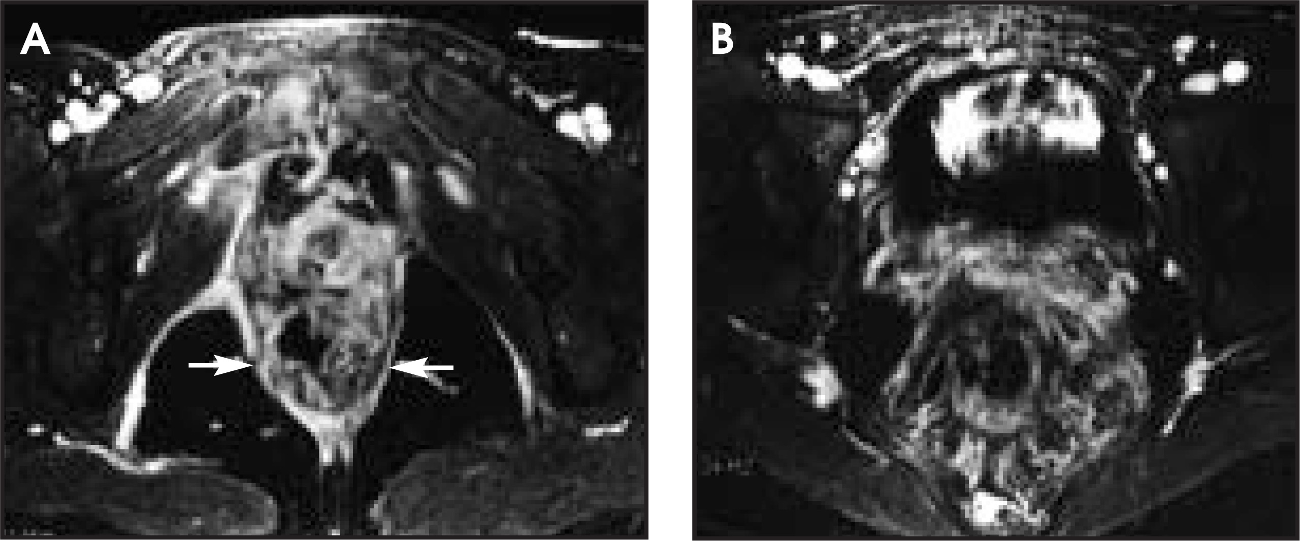
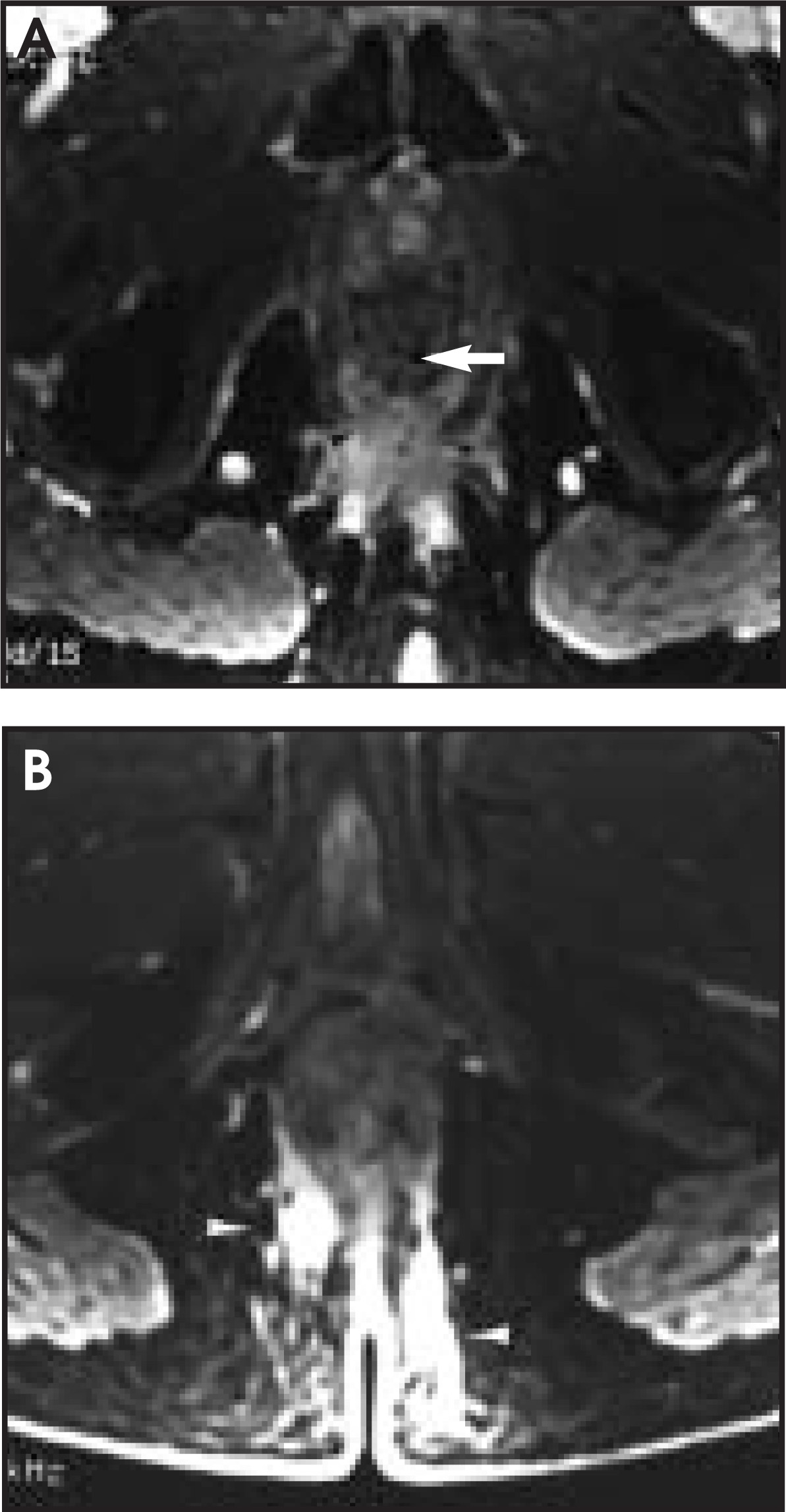
This technique can be used in place of CT to evaluate many abdominal and pelvic disease processes. The dynamic gadolinium-enhanced sequence is an adjunct to standard T1- and T2-weighted sequences and is helpful in characterizing renal masses (Figures 1 and 2), primary and secondary liver tumors (Figures 3 and 4), hepatic vascular disease (Figure 5), periportal masses (Figure 6), pancreatic masses (Figures 7 and 8), and rectal lesions (Figures 9 and 10).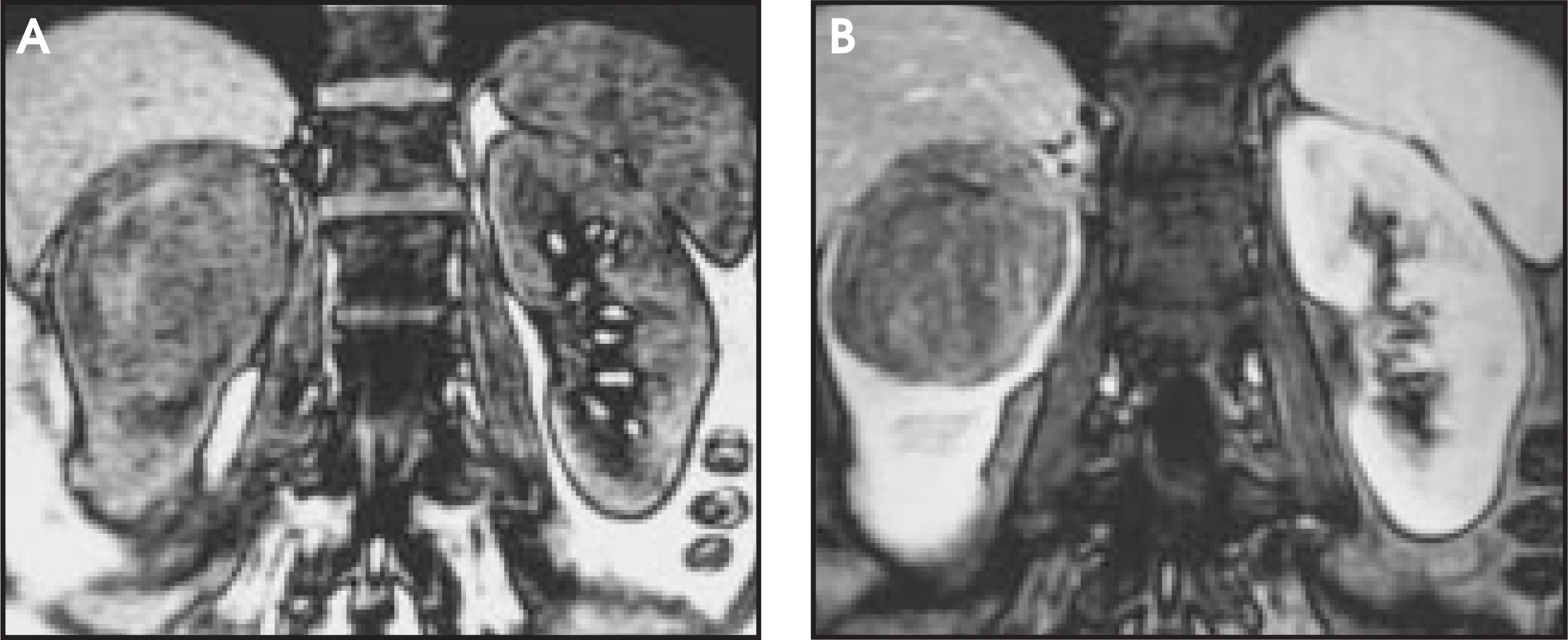
Conclusion
Breath-hold 3D gadolinium-enhanced multiphasic abdominal MR is a widely available technique that can be used, with excellent results, in the detection and characterization of a wide variety of abdominal and pelvic diseases. Breath-hold imaging eliminates respiratory motion and improves anatomic sharpness. The 3D data set can, when needed, be viewed in other planes. Lesion vascularity can be assessed similar to multiphasic CT. Its use is suggested when CT with iodinated contrast is either contraindicated or equivocal. AR
References
- Low R, Francis I, Sigeti J, Foo T. Abdominal MR imaging: Comparison of T2-weighted fast and conventional spin-echo, and contrast-enhanced fast multiplanar spoiled gradient-recalled imaging.. Radiology.. 1993;186:803-811.
- Price R. The AAPM/RSNA physics tutorial for residents: Contrast mechanisms in gradient-echo imaging and an introduction to fast imaging.. RadioGraphics.. 1995;15:165-178.
- Haacke E, Tkach J. Fast MR imaging: Techniques and clinical applications.. AJR Am J Roentgenol.. 1990;155:951-964.
Citation
. Clinical update: Breath-hold 3D gadolinium-enhanced multiphasic abdominal MR. Applied Radiology. 2003;32(1):9-12. doi:10.37549/AR1152.