1 University of Arizona College of Medicine–Phoenix, Phoenix, Arizona
2 Department of Radiology, Phoenix Children’s Hospital, Phoenix, Arizona
3 University of Cincinnati College of Medicine, Cincinnati, Ohio
4 Department of Radiology, Children’s Hospital Medical Center, Cincinnati, Ohio
* Corresponding author: Richard B. Towbin (rtowbin@gmail.com)
Abstract
Congenital Lipomatous Overgrowth, Vascular anomalies, Epidermal nevi, and Skeletal deformities (CLOVES) syndrome is caused by a gain-of-function mutation in PIK3CA, which leads to overactivity of the mammalian target of rapamycin complex pathway. Characteristic cutaneous malformations help suggest the diagnosis and differentiate it from other conditions. Clinicians should consider genetic testing for CLOVES syndrome when encountering key clinical features such as congenital lipomatous overgrowth, vascular malformations, epidermal nevi, and skeletal/spinal anomalies. Other characteristic features include segmental asymmetric limb growth, skin anomalies like capillary, venous, or lymphatic malformations, and renal anomalies. The presence of any of these signs should raise suspicion and warrant further investigation through genetic testing and imaging studies.
Keywords
syndrome, congenital, skeletal
Categories
Case Summary
A school-aged child presented with a history of Congenital Lipomatous Overgrowth, Vascular anomalies, Epidermal nevi, and Skeletal deformities (CLOVES) syndrome, with lipomatous overgrowth of her left leg and left great toe.
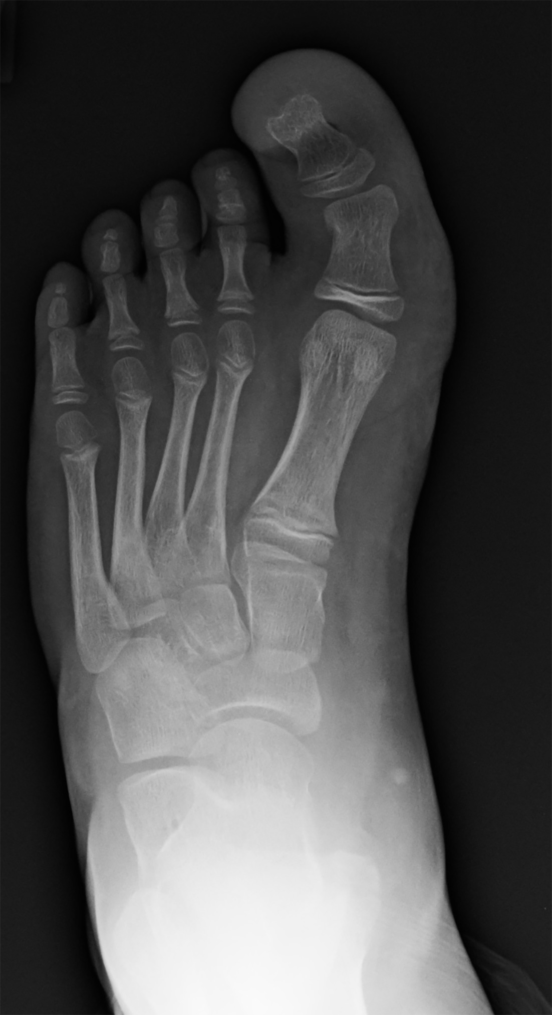
Imaging Findings
Anteroposterior radiograph of the left foot (Figure 1) showed overgrowth of the patient’s left great toe.
Diagnosis
CLOVES syndrome.
The spectrum of differential diagnosis for macrodactyly includes macrodystrophia lipomatosa, neurofibromatosis type 1, Klippel-Trenaunay syndrome (KTS), Proteus syndrome, and Parkes-Weber syndrome.
In KTS, characteristic imaging findings consist of unilateral lower extremity soft-tissue or osseous hypertrophy accompanied by low-flow malformations, typically devoid of arterial involvement. Proteus syndrome manifests similarly with low-flow malformations and tissue hypertrophy but distinguishes itself by the presence of cerebral manifestations, notably polymicrogyria and periventricular cysts. Conversely, PWS delineates itself through high-flow arteriovenous malformations (AVMs) discernible on A or MRA, facilitating differentiation from conditions such as KTS.
Discussion
CLOVES syndrome is a rare overgrowth syndrome. 1 While epidemiological data may be limited, CLOVES syndrome has been uniquely identified under the larger umbrella of phosphatidylinositol-4,5 bisphosphate 3 kinase catalytic subunit 3-kinase (PI3KCA)-related overgrowth syndromes (PROS). 2
PROS is characterized by mosaic somatic mutations in the PIK3CA gene, which commonly codes for the p110 alpha protein, a subunit of the enzyme PI3K. 3 The mutations eventually cause an upregulation of the mammalian target of rapamycin complex 1 (mTOR1) pathway. The pathological symptoms of CLOVES are thought to be caused by increased cellular proliferation, growth, and survival through the mTOR1 pathway. 1-3
While the phenotypical morphologies of patients affected by CLOVES syndrome will manifest in varying severities, activating mutations of PIK3CA predominately affect mesoderm-derived tissues (adipose tissues, vascular, lymphatic, muscle, and bone). The clinical diagnosis focuses on differentiating CLOVES syndrome from other types of overgrowth syndromes such as Proteus syndrome through genetic testing, physical examination, and imaging. For example, CLOVES is present at birth characterized by asymmetrical tissue overgrowth in the trunk, back, and limbs, while KTS has overgrowth primarily involving one or more limbs, usually a leg, and Proteus syndrome overgrowth is severe, progressive, and distorting. It usually appears postnatally, affecting bones, skin, and soft tissue. 4
Cutaneous manifestations often serve as the initial indicator of the underlying disease. They can also be used to help differentiate different conditions. Patients with CLOVES syndrome typically present with phlebectasia, lymphatic vesicles, and a linear epidermal nevus that can vary in color and texture. The nevus often appears as raised, thickened, or wart-like areas along a distinct linear pattern on the body. In addition to the nevus, patients with CLOVES syndrome commonly have capillary venous malformations in the trunk and extremities, and macrocytic or microcytic lymphatic malformations. 1,5 These features differ compared with patients with Proteus syndrome and KTS. For example, Proteus syndrome is characterized by soft-tissue overgrowth and cerebriform lesions, while KTS commonly exhibits capillary hemangiomas. 6,7
Other hallmarks of CLOVES include thoracic lipomatous growths and musculoskeletal anomalies that mainly affect the hands, feet, and spine. 1 These anomalies include macrodactyly, sandal gap toes, scoliosis, kyphosis, and spina bifida. 1,4,8 Additionally, spinal cord AVMs and arteriovenous shunts can cause patients to have radicular pain, sensitivity disorders, and motor dysfunction. 1,4
Skeletal radiographs can be used for initial assessment of affected body parts. 2,5 Common findings include bone and soft-tissue overgrowth. US and MRI are used to identify and characterize soft-tissue masses and vascular malformations. 5,6 These modalities can also be used to assess visceral anomalies such as renal hypoplasia or agenesis.
Currently, there is no cure for CLOVES syndrome. Management is largely focused on monitoring deformity and palliative care. 3,9,10 Debulking surgery is used to reduce the size of large masses and alleviate symptomatic deformities. Lower extremity epiphysiodesis can be used to prevent a leg length discrepancy. Finally, mTOR inhibitors such as rapamycin and alpelisib are used to block the activity of mTOR, the overactive PI3K/AKT/mTOR pathway, which is the driver of CLOVES syndrome. These drugs can reduce the size of the lipomatous and lymphatic masses, improve lymph flow, and possibly reduce the skeletal abnormalities. The life expectancy varies widely and depends on the severity and regions of the body that are involved. Generally, patients with severe respiratory issues, cardiac defects, and brain involvement have the worst outcomes.
Conclusion
CLOVES syndrome is caused by a gain-of-function mutation in PIK3CA, which leads to overactivity of the mTOR pathway. Characteristic cutaneous malformations help suggest the diagnosis and differentiate it from other conditions. Clinicians should consider genetic testing for CLOVES syndrome when encountering key clinical features such as congenital lipomatous overgrowth, vascular malformations, epidermal nevi, and skeletal/spinal anomalies. Other characteristic features include asymmetric limb growth and skin anomalies like capillary, venous, or lymphatic malformations. The presence of any of these signs should raise suspicion and warrant further investigation through genetic testing and imaging studies.
References
- Vahidnezhad H, Youssefian L, Baghdadi T. Phenotypic heterogeneity in PIK3CA-related overgrowth spectrum. Br J Dermatol. 2016;175(4):810-814. doi:10.1111/bjd.14618.
- Keppler-Noreuil K, Rios J, Parker V. PIK3CA-Related Overgrowth Spectrum (PROS): diagnostic and testing eligibility criteria, differential diagnosis, and evaluation. Am J Med Genet A. 2015;167(2):287-295. doi:10.1002/ajmg.a.36836.
- Lindhurst M, Sapp J, Teer J. A mosaic activating mutation in AKT1 associated with the Proteus syndrome. N Engl J Med. 2011;365(7):611-619. doi:10.1056/NEJMoa1104017.
- Martinez-Lopez A, Blasco-Morente G, Perez-Lopez I. CLOVES syndrome: review of a PIK3CA-Related Overgrowth Spectrum (PROS). Clin Genet. 2017;91(1):14-21. doi:10.1111/cge.12832.
- Bertino F, Braithwaite K, Hawkins C. Congenital limb overgrowth syndromes associated with vascular anomalies. Radiographics. 2019;39(2):491-515. doi:10.1148/rg.2019180136.
- Wang M, Kamel S, Elsayes K. Vascular anomaly syndromes in the ISSVA classification system: imaging findings and role of interventional radiology in management radiographics. Radiographics. 2022;42(6):1598-1620. doi:10.1148/rg.210234.
- Mahajan V, Gupta M, Chauhan P, Mehta K. Cloves syndrome: a rare disorder of overgrowth with unusual features—an uncommon phenotype?. Indian Dermatol Online J. 2019;10(4):447-452. doi:10.4103/idoj.IDOJ_418_18.
- Öztürk Durmaz E, Demircioğlu D, Yalınay Dikmen P. A review on cutaneous and musculoskeletal manifestations of CLOVES syndrome. Clin Cosmet Investig Dermatol. 2022;15(april):621-630. doi:10.2147/CCID.S351637.
- Alomari A. Characterization of a distinct syndrome that associates complex truncal overgrowth, vascular, and acral anomalies: a descriptive study of 18 cases of CLOVES syndrome. Clin Dysmorphol. 2009;18(1):1-7. doi:10.1097/MCD.0b013e328317a716.
- Venot Q, Blanc T, Rabia S. Targeted therapy in patients with PIK3CA-related overgrowth syndrome. Nature. 2018;558(7711):540-546. doi:10.1038/s41586-018-0217-9.
Disclosures
The authors have no conflicts of interest to disclose. None of the authors received outside funding for the production of this original manuscript and no part of this article has been previously published elsewhere.
Citation
. CLOVES Syndrome. Applied Radiology. 2025. doi:10.37549/JPCR-25-0046.