Clubfoot
Journal of Pediatric Case Reports — Vol. 1 , Issue 2
Published: May 1, 2026
1 UNTHSC Texas College of Osteopathic Medicine, Fort Worth, Texas
2 Department of Radiology, Phoenix Children’s Hospital, Phoenix, Arizona
3 Department of Radiology, Cincinnati Children’s Hospital, University of Cincinnati College of Medicine, Cincinnati, Ohio
* Corresponding author: Richard B. Towbin (rtowbin@gmail.com)
Abstract
Talipes equinovarus, or clubfoot, represents a spectrum of foot deformities characterized by hindfoot equinus and varus with forefoot adduction and supination. Most cases reflect congenital structural malalignment and are classified as idiopathic, syndromic, or neurogenic, while postural deformities are flexible and nonstructural. Prenatal diagnosis is typically established with US, and postnatal evaluation relies on clinical examination with selective use of radiographs. Weight-bearing or simulated weight-bearing anteroposterior and lateral radiographs allow measurement of the talocalcaneal and tibiocalcaneal angles, which aid in assessing severity and monitoring correction. The Ponseti method of serial casting remains the gold standard treatment, although relapse may occur and can be identified clinically or radiographically.
Keywords
mulsculoskeletal, foot, congenital
Categories
Case Summary
A young child with a known diagnosis of clubfoot presented to the orthopedic clinic for corrective management.
Imaging Findings
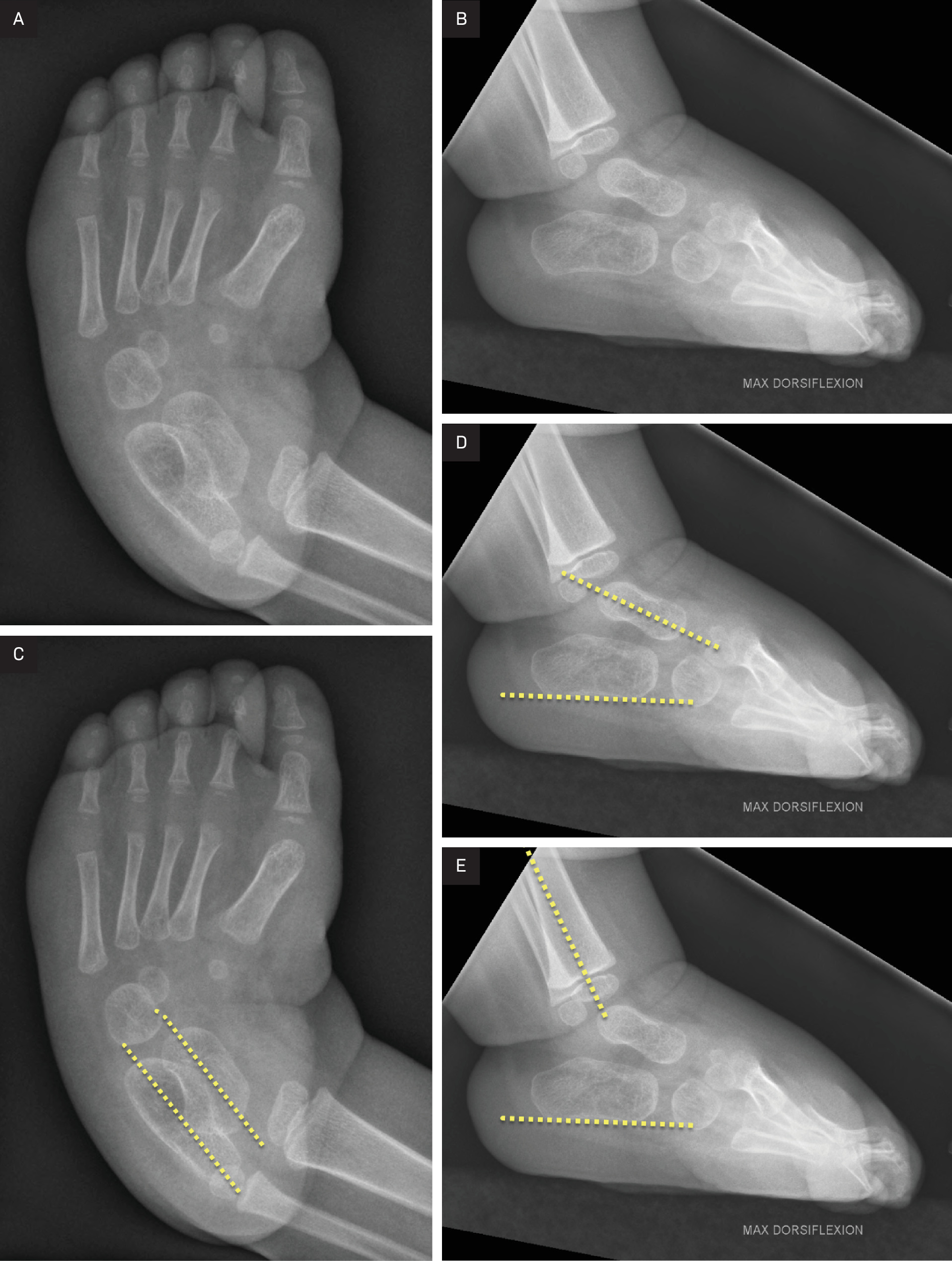
Anteroposterior and lateral simulated weight-bearing radiographs of the foot demonstrate findings consistent with clubfoot deformity. On the anteroposterior view, the talus and calcaneus are nearly parallel, reflecting hindfoot varus. On the lateral view, the talocalcaneal angle measures 16°, decreased relative to the normal range of 25-55°. The tibiocalcaneal angle measures 117° increased relative to the normal range of 25-60° in maximum dorsiflexion. These findings are consistent with hindfoot equinus and varus deformity (Figure 1).
Diagnosis
Clubfoot
Discussion
Talipes equinovarus, commonly referred to as clubfoot, describes a spectrum of foot deformities characterized by hindfoot equinus and varus with forefoot adduction and supination. The condition most commonly represents a congenital structural malalignment of the talus and adjacent hindfoot structures, with associated medial and posterior soft-tissue contractures, including shortening of the Achilles tendon and medial ligaments. Clubfoot is one of the most common congenital lower extremity deformities, occurring in approximately 0.5-3 per 1000 live births worldwide, with higher rates reported in certain populations and a male predominance of approximately 2:1.1-3
Clubfoot is broadly classified into 4 categories: postural, idiopathic, syndromic, and neurogenic.4 Postural clubfoot accounts for approximately 10% of cases and results from intrauterine positioning forces. These deformities are flexible and typically respond to stretching and casting alone.1,2 The remaining categories represent structural forms in which the deformity is rigid and not correctable with passive manipulation. Idiopathic clubfoot is the most common form and occurs in the absence of associated anomalies. Syndromic clubfoot is associated with chromosomal abnormalities, genetic syndromes, skeletal dysplasias, and connective tissue disorders. Common associations include arthrogryposis, Down syndrome, diastrophic dysplasia, and TARP (Talipes equinovarus, Atrial septal sefect, Robin sequence, Persistent left superior vena cava) syndrome.1,2,5 Neurogenic clubfoot results from disruption of the neuromuscular unit and may be seen in conditions such as myelomeningocele.1,2,5
Prenatal diagnosis of clubfoot is most commonly established during the first or second trimester using US. Reported diagnostic accuracy is approximately 86%.1,6 Fetal MRI may be considered when additional skeletal abnormalities are suspected on US. Following delivery, physical examination confirms or excludes the diagnosis. Postural deformities related to intrauterine constraint should be considered in the differential diagnosis, as these may mimic structural clubfoot on prenatal imaging but are typically reducible on postnatal examination.1,2
Radiographs may be used to assess deformity severity and to evaluate correction during and after intervention. Standard imaging includes anteroposterior and lateral weight-bearing or simulated weight-bearing views. On the anteroposterior view, the talocalcaneal angle is formed by the intersection of the longitudinal axes of the talus and calcaneus. On the lateral view, the talocalcaneal angle is measured using the same axes in the sagittal plane. The tibiocalcaneal angle, measured on the lateral view, is formed by the intersection of the longitudinal axis of the tibia and the longitudinal axis of the calcaneus (Figure 1).
Reported normal ranges are 15-55° for the anteroposterior talocalcaneal angle, 25-55° for the lateral talocalcaneal angle, and 25-60° for the tibiocalcaneal angle in maximum dorsiflexion.7,8 In clubfoot, the lateral talocalcaneal angle is typically decreased and the tibiocalcaneal angle increased, reflecting hindfoot varus and equinus.
The gold standard treatment for clubfoot is the Ponseti method of serial casting, with Achilles tenotomy required in up to 90% of cases.2,3,5 Early intervention prior to ambulation and continued bracing through early childhood are associated with improved outcomes. Treatment begins with correction of the cavus deformity by supinating the forefoot and applying pressure beneath the first metatarsal head. Hindfoot varus, forefoot adduction, and hindfoot equinus are then sequentially corrected over several casting sessions. Following tenotomy and application of a final long-leg cast, patients are transitioned to a foot abduction brace to reduce the risk of relapse.
Relapse is defined as the recurrence of any component of the deformity after treatment. It may be identified clinically or radiographically, with lateral radiographs helping assess persistent equinus or varus deformity.5-7 Although initial correction rates approach 92% in patients treated with the Ponseti method, relapse occurs in approximately one-third of patients, with reported long-term recurrence rates ranging from 3% to 62.5%.2,5,8 Greater initial deformity severity and poor brace compliance are associated with increased relapse risk. Recurrent or residual deformity may result in overcorrection, undercorrection, bunion formation, ankle impingement, and degenerative changes, sometimes requiring additional intervention to preserve function and alleviate pain.9
Conclusion
Talipes equinovarus, or clubfoot, represents a spectrum of foot deformities characterized by hindfoot equinus and varus with forefoot adduction and supination. Most cases reflect congenital structural malalignment and are classified as idiopathic, syndromic, or neurogenic, while postural deformities are flexible and nonstructural. Prenatal diagnosis is typically established with US, and postnatal evaluation relies on clinical examination with selective use of radiographs. Weight-bearing or simulated weight-bearing anteroposterior and lateral radiographs allow measurement of the talocalcaneal and tibiocalcaneal angles, which aid in assessing severity and monitoring correction. The Ponseti method of serial casting remains the gold standard treatment, although relapse may occur and can be identified clinically or radiographically.
References
- Martin S. In: Obstetric imaging: fetal diagnosis and care. 2018.
- Rieger M, Dobbs M. Clubfoot. Clin Podiatr Med Surg. 2022;39(1):1-14. doi:10.1016/j.cpm.2021.08.006.
- Williams M, Dobbs M. Clubfoot. Clin Podiatr Med Surg. 2024;41(1):17-25. doi:10.1016/j.cpm.2023.06.009.
- Green A. The pediatric foot and ankle. Pediatr Clin North Am. 2020;67(1):169-183. doi:10.1016/j.pcl.2019.09.007.
- Dobbs M, Gurnett C. Update on clubfoot: etiology and treatment. Clin Orthop Relat Res. 2009;467(5):1146-1153. doi:10.1007/s11999-009-0734-9.
- Fantasia I, Dibello D, Di Carlo V. Prenatal diagnosis of isolated clubfoot: diagnostic accuracy and long-term postnatal outcomes. Eur J Obstet Gynecol Reprod Biol. 2021;264:60-64. doi:10.1016/j.ejogrb.2021.07.009.
- Moerman S, Zijlstra-Koenrades N, Reijman M. The predictive value of radiographs and the pirani score for later additional surgery in ponseti-treated idiopathic clubfeet, an observational cohort study. Children. 2022;9(6). doi:10.3390/children9060865.
- Zhang G, Zhang Y, Li M. A modified PONSETI method for the treatment of rigid idiopathic congenital clubfoot. J Foot Ankle Surg. 2019;58(6):1192-1196. doi:10.1053/j.jfas.2019.04.003.
- Johnson J, Fortney T, Luk P. Late effects of clubfoot deformity in adolescent and young adult patients whose initial treatment was an extensive soft-tissue release: topic review and clinical case series. J Am Acad Orthop Surg Glob Res Rev. 2020;4(5). doi:10.5435/JAAOSGlobal-D-19-00126.
Disclosures
The authors have no conflicts of interest to disclose. None of the authors received outside funding for the production of this original manuscript and no part of this article has been previously published elsewhere.
Citation
. Clubfoot. Journal of Pediatric Case Reports. 2026;1(2). doi:10.37549/JPCR-26-0083.