Comprehensive Review of Clinical Presentation, Multimodality Imaging, and Therapeutic Strategies for Inflammatory Breast Cancer
Applied Radiology — Vol. 54 , Issue 4 , pp. 1 -9
Published: June 1, 2025
1 Department of Breast Imaging, University of Texas MD Anderson Cancer Center, Houston, Texas
2 Department of Breast Medical Oncology, University of Texas MD Anderson Cancer Center, Houston, Texas
3 Department of Breast Surgical Oncology, University of Texas MD Anderson Cancer Center, Houston, Texas
4 Department of Breast Radiation Oncology, University of Texas MD Anderson Cancer Center, Houston, Texas
* Corresponding author: Huong T. Le-Petross (hlepetross@mdanderson.org)
Categories
Introduction
Inflammatory breast cancer (IBC) is a rare, locally advanced invasive cancer that commonly presents with skin redness (erythema) and swelling (edema). Diagnosing IBC is difficult, and prompt identification is crucial when a patient presents with a reddened or inflamed breast. According to the National Cancer Institute’s Surveillance, Epidemiology, and End Results data, the incidence of IBC was 2.76 cases per 100,000 from 1973 to 2015. 1 IBC has a higher mortality rate compared with noninflammatory advanced breast cancers, 1,2 with an overall relative 5-year survival rate of approximately 40.5%, which is even lower among Black patients. 1
Specific clinical criteria for diagnosing IBC include rapid onset of erythema, edema, and/or peau d’orange with a duration of less than 6 months; inflammation occupying more than one-third of the breast; and pathological confirmation of invasive carcinoma. 3 The rapid onset distinguishes IBC from noninflammatory, locally advanced cancer. 4 In addition to clinical diagnosis, multimodality imaging tests improve IBC detection and confirmation, with the aim of improving overall survival. Breast MRI has been found to be most beneficial for identifying primary lesions and evaluating treatment response, while 18FFDG PET/CT plays a significant role in detecting distant metastases at initial diagnosis for appropriate treatment selection. Here, we offer an overview of current approaches to diagnosis and treatment of IBC.
Clinical Diagnosis
The clinical presentation of IBC arises from the diffuse and rapid obstruction of lymphatics in the breast by tumor emboli, leading to edema and hyperemia of the blood vessels in the skin. 5 The most commonly used definition of IBC is based on the American Joint Committee on Cancer (AJCC) Staging System, 8th edition. This system defines IBC, stage T4d, as a clinicopathological entity characterized by diffuse erythema and edema involving approximately a third or more of the skin of the breast. 6 The system requires a pathological diagnosis of invasive cancer in less than 6 months from initial symptom presentation 6 ; however, while pathological identification of dermal lymphatic emboli is pathognomonic, it is not required for diagnosis. In addition, although skin erythema is a mandatory criterion for diagnosis under the staging system, it may not be present or may fluctuate or diminish over time.
The difficulty of diagnosing IBC was highlighted in an extensive external review of medical photographs and records of 270 patients with IBC across 6 sites in Egypt, Tunisia, and Morocco. 7 The clinical diagnosis was based on an expert panel consensus statement. Among the cases, 76% met the consensus criteria, but only 36% adhered to the AJCC 8th edition. 6,8 Nevertheless, 86% of the cases were confirmed as IBC through photographic review adherence to the consensus statement by independent, external experts. 7 An expert panel convened by the Susan G. Komen Foundation recently validated a more formal and quantitative definition of IBC that incorporates clinical, pathological, and imaging features that may improve diagnosis. 9
The incidence of IBC among women with breast cancer is low, typically estimated at 2-3%. 1,10 However, according to the available references, IBC’s incidence among women presenting with breast inflammation ranges from 5-50%. 11-13 Dabi et al proposed a diagnostic algorithm emphasizing the importance of identifying and treating IBC as an oncologic emergency. According to this algorithm, all nonlactating patients with inflammatory symptoms should undergo imaging. If malignancy is suspected but no focal mass abnormality is amenable to biopsy, a skin punch biopsy of the most involved skin should be obtained. A negative biopsy indicates the need for MRI with biopsy. 11 In lactating patients with strongly suspected acute mastitis, beginning with a “test and treat” strategy is a reasonable approach. If no improvement is observed within 2 weeks of antibiotic therapy, further imaging studies should be obtained. 11 At one tertiary surgical referral center, IBC accounted for 50% of cases presenting with inflammatory symptoms. 12 Thus, a high index of suspicion is warranted. Patients with presumed benign mastitis that does not resolve quickly with medical therapy should undergo imaging and image-guided biopsy.
Radiological Diagnosis of IBC
Mammography
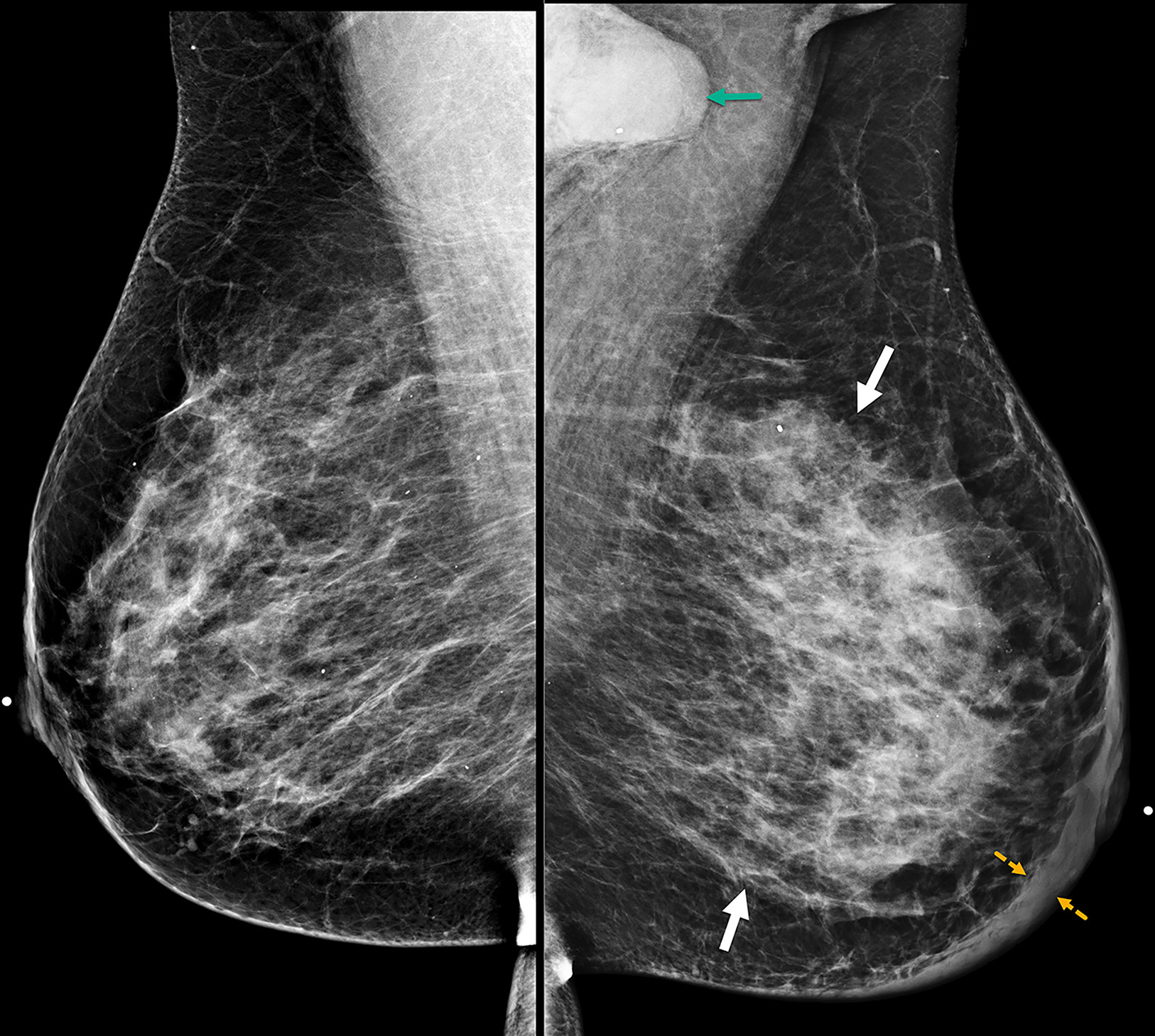
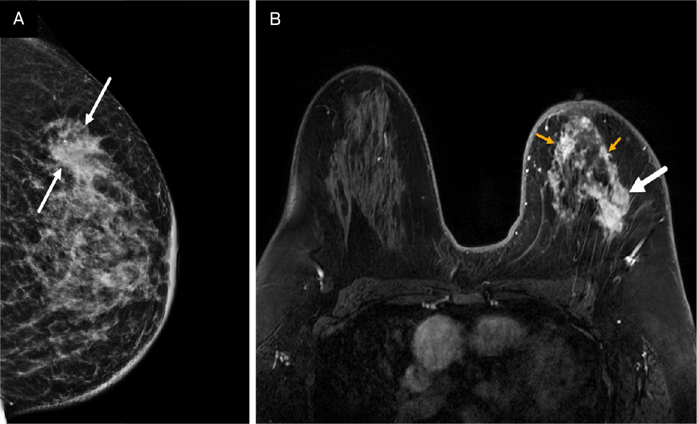
Patients with suspected IBC are often initially referred for mammography despite the modality’s limitation in detecting lesions in dense breast parenchyma. In many cases, no identifiable mass may be observed and/or the mammogram may be interpreted as normal. 13 The features most associated with IBC include diffuse breast enlargement, trabecular thickening, global skin thickening, and ipsilateral axillary lymphadenopathy (Figure 1). 14-16 Skin and trabecular thickening, although nonspecific, are subtle early findings observed in 80% of IBC cases. 15-17 Less commonly seen mammographic findings include a visible irregular mass, architectural distortion, or calcifications (Figures 2, 3). 14,17
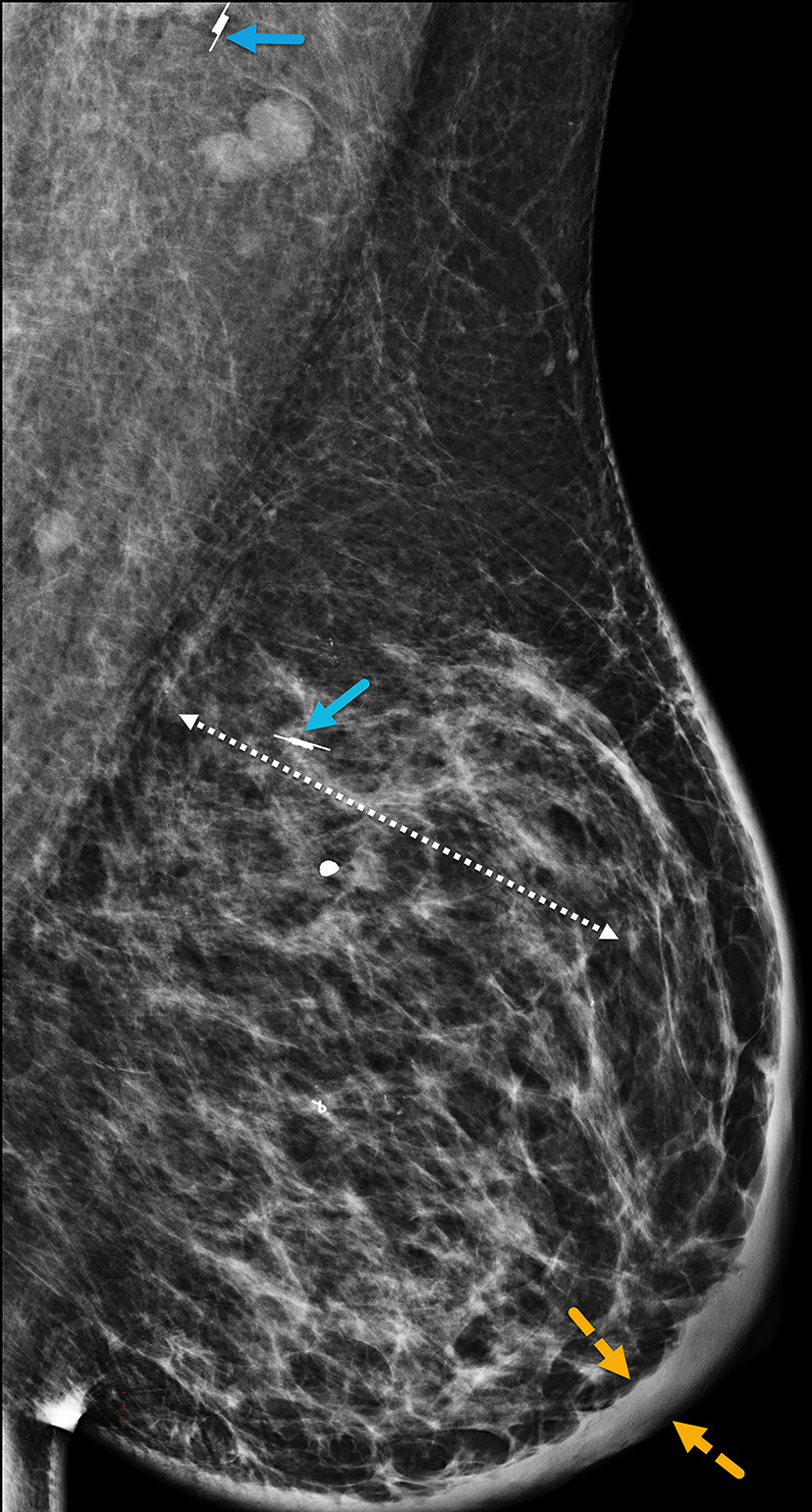
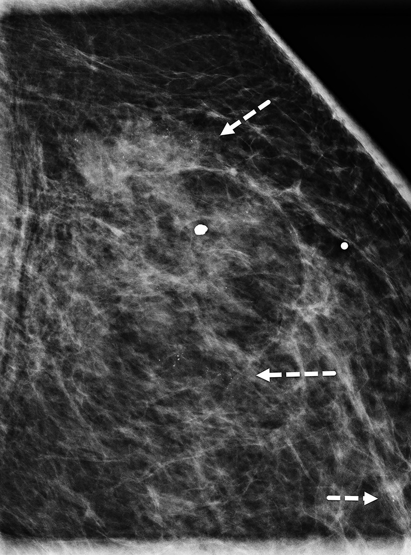
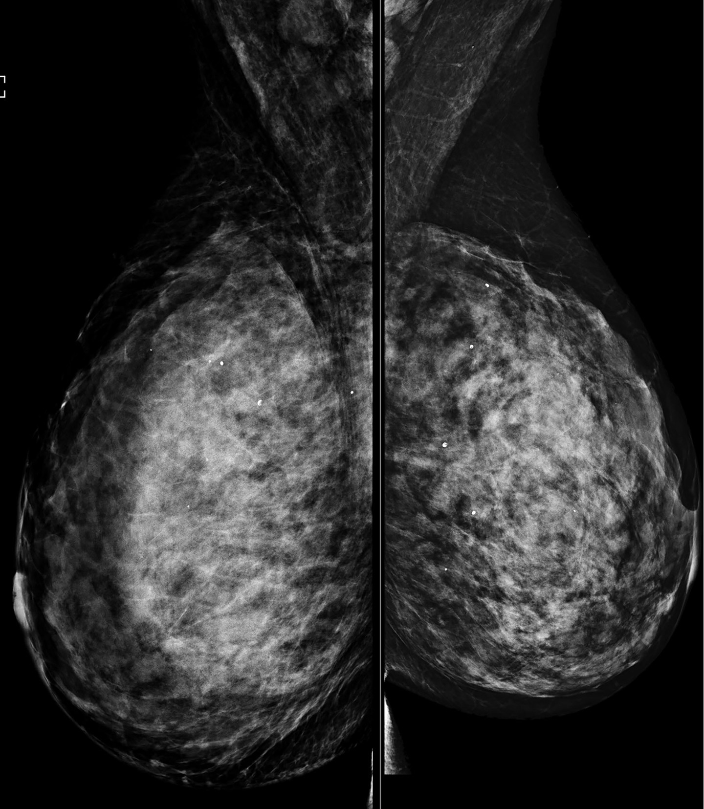
The ability of mammography to detect a primary breast lesion in patients with IBC is limited; one retrospective study found that only 20% of cases demonstrated a detectable primary lesion on mammography. 15 97% of subjects in the same study had nonfatty breasts, leading the authors to suggest that the dense breast parenchyma background likely contributed to the poor visibility of lesions (Figures 4, 5). 15 Another study observed that findings of skin thickening, axillary adenopathy, trabecular thickening, and nipple-areolar swelling were significantly more frequent in IBC than in non-IBC cases, while the presence of a mass was more commonly associated with non-IBC cases. 18 Compared with other imaging modalities, mammography is the least sensitive to multifocal and multicentric disease in patients with IBC. 15
Ultrasound
Common sonographic features of IBC include one or more masses, skin thickening, tissue edema with lymphatic dilation, and regional lymphadenopathy. 15-20 However, these are nonspecific features that overlap with findings seen in benign conditions such as mastitis and in other malignancies such as locally advanced breast cancer (Figure 6). As a result, diagnosis with ultrasound alone can be challenging. If an index mass is identified, its common sonographic morphology includes hypoechoic mass with lobulated or irregular margins and posterior acoustic shadowing. 21-23 Ultrasound does not reliably detect microcalcifications, for which mammography remains the most sensitive modality for identification. Architectural distortion and diffuse posterior acoustic shadowing (Figure 7), often multifocal and multicentric in distribution, are observed in over 80% of cases. 15,16
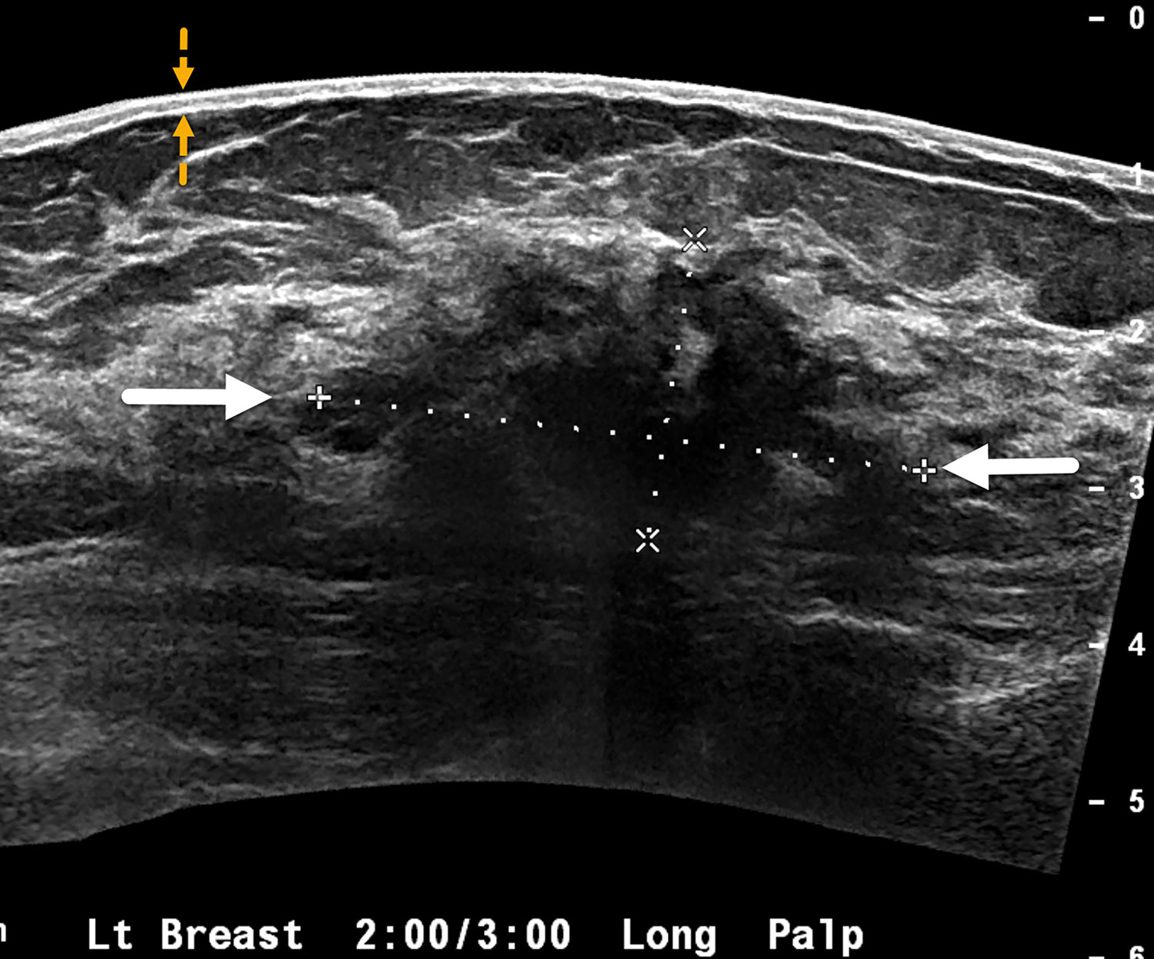
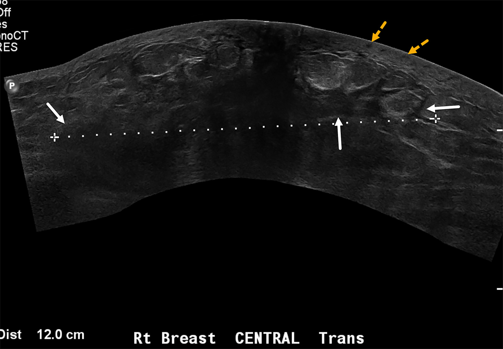
The inflammation seen in IBC is associated with increased vascularity of the breast lesions and surrounding parenchyma, which correlates with the erythema observed on physical examination. Diffuse skin thickening, a hallmark of IBC, can be quantified on ultrasound. Skin and breast edema are also commonly noted (Figure 7). The dermal emboli and dermal lymphatic involvement characteristic of IBC may account for sonographic findings of diffuse hypoechoic and thickened skin with an indistinct dermal-subcutaneous fat interface. 20,24 Breast edema can extend into the chest wall and pleural spaces and may require multimodality imaging for complete evaluation.
MRI
MRI is superior to mammography and ultrasound in identifying index tumor masses (Figure 8), which are also referred to as primary breast parenchymal lesions (BPLs). 15,19 In a study at our center, MRI successfully identified all BPLs, while ultrasound identified 95%, and mammography 80%, of lesions in patients with a clinical diagnosis of IBC. 19 Owing to its high sensitivity, MRI is recommended early in the evaluation of patients with clinical suspicion for IBC, particularly when mammography and ultrasound fail to detect lesions. Additionally, MRI findings can guide biopsy procedures.
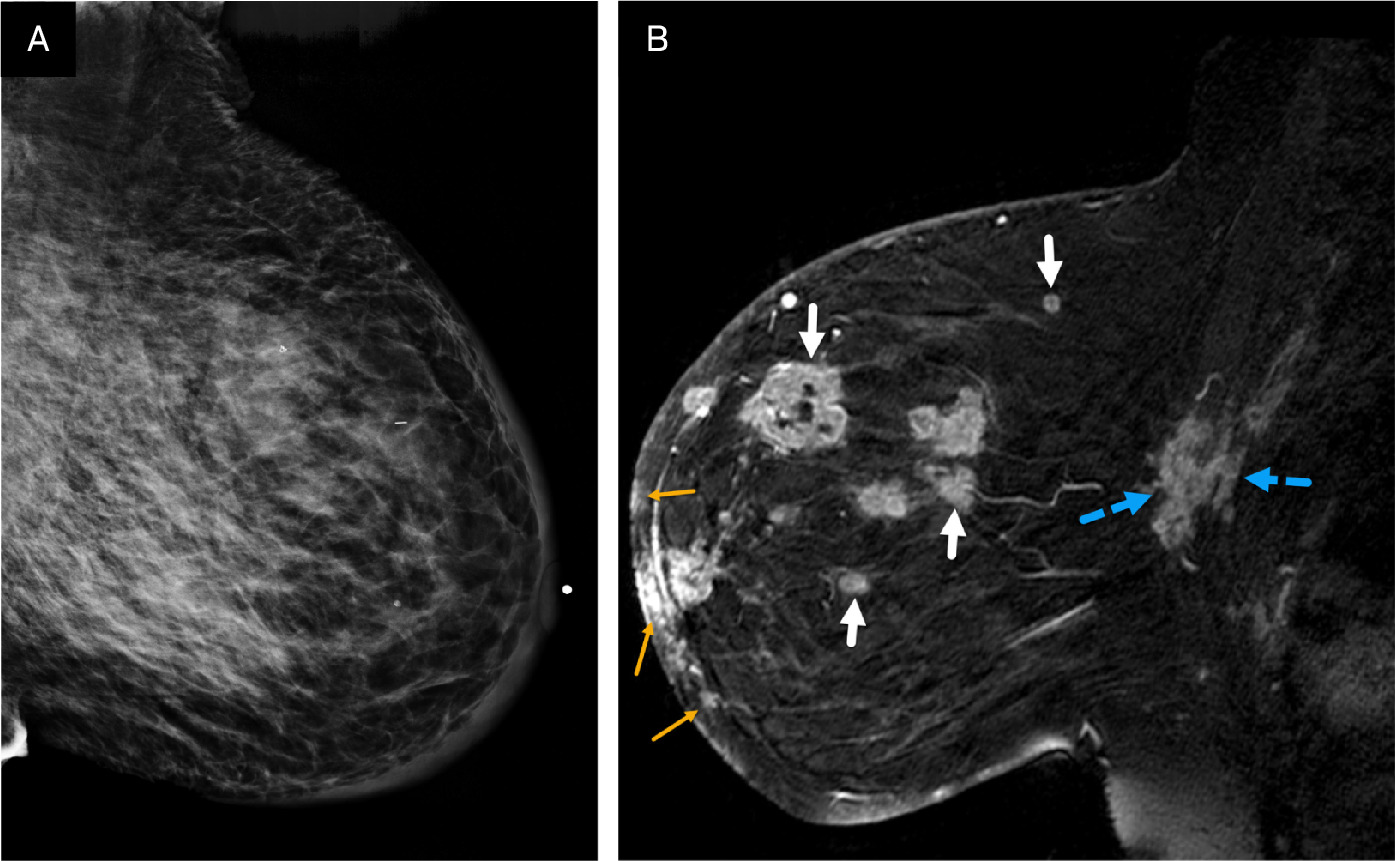
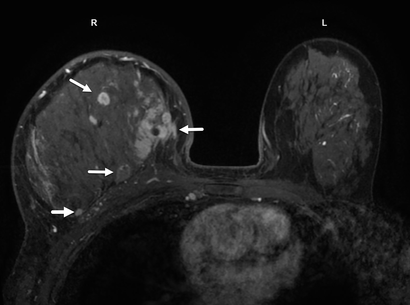
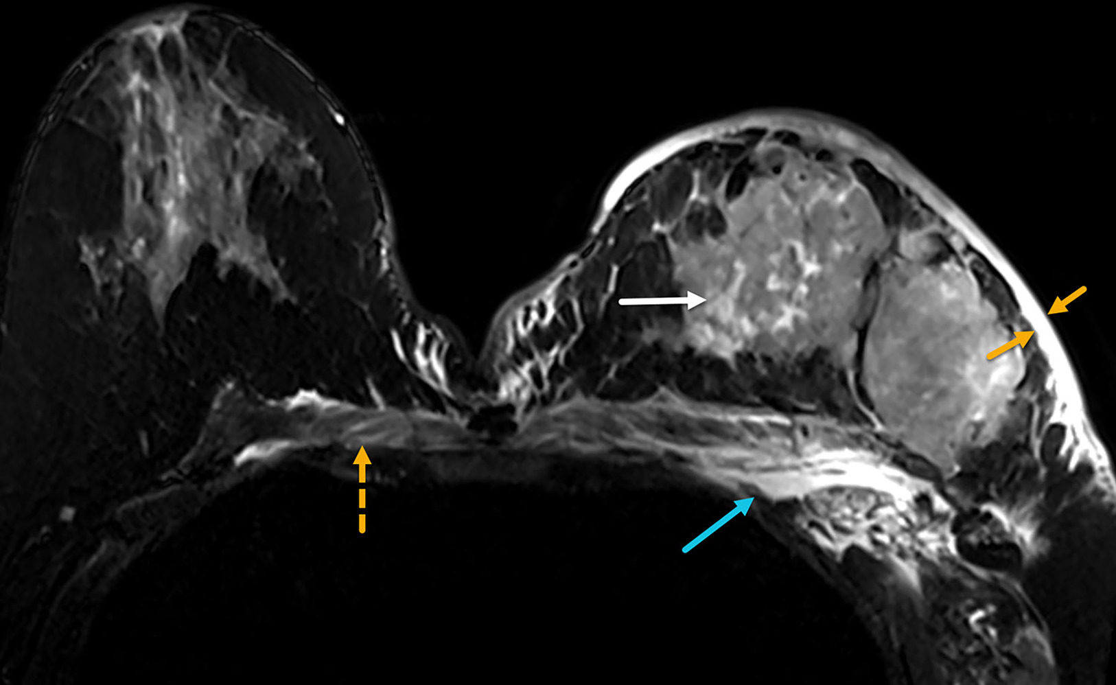
The multicentric distribution commonly associated with IBC, especially in dense enlarged and inflamed breasts (Figure 9), may best be appreciated on MRI. Edema of the subdermal breast, pre-pectoral region, and chest wall is more commonly seen in IBC than in other breast cancers and is most evident on T2 images (Figure 10). 19 The presence of pre-pectoral edema has been suggested as a prognostic factor in breast cancer. 25,26
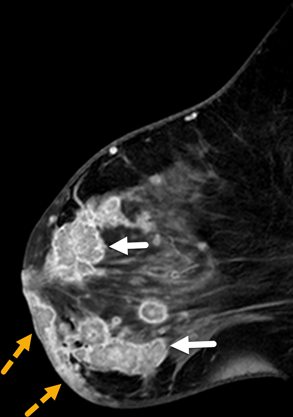
Diffuse skin thickening, observed in 90-100% of patients with IBC, may be present with or without skin enhancement or focal-enhancing skin lesions (Figure 11). 19 Skin thickening typically involves the entire breast and may extend across the midline or into the contralateral breast. Focal skin thickening adjacent to the BPL is more often associated with locally advanced or neglected carcinoma than with IBC. 27 Enhancing skin foci detected on MRI may represent tumor emboli or dermal lymphatic invasion, pathological hallmarks of IBC.
Nodal Staging
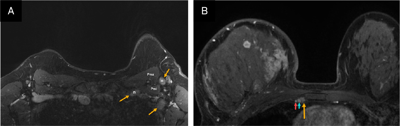
Bilateral nodal basins are visible on MRI despite artifact resulting from cardiac motion and respiration. The axillary level I and II nodal regions, as well as the internal mammary nodal basins, are often well visualized (Figure 12), while the supraclavicular and medial infraclavicular or axillary level III regions are better evaluated with nodal ultrasound.
18FFDG PET/CT
PET/CT is recommended for patients with IBC at initial presentation, particularly when standard staging studies are inconclusive, and for identifying extra-axillary lymph node metastases (Figure 13) and occult distant disease. 28,29 Since at least one-third of these patients present with distant metastases, the modality is also valuable for guiding treatment selection and determining prognosis. 30 PET/CT has demonstrated a sensitivity of 96-100% for PBL in IBC. 31,32 However, false-positive findings are possible in such cases as mastitis, which may show FDG avidity similar to that of IBC. 32
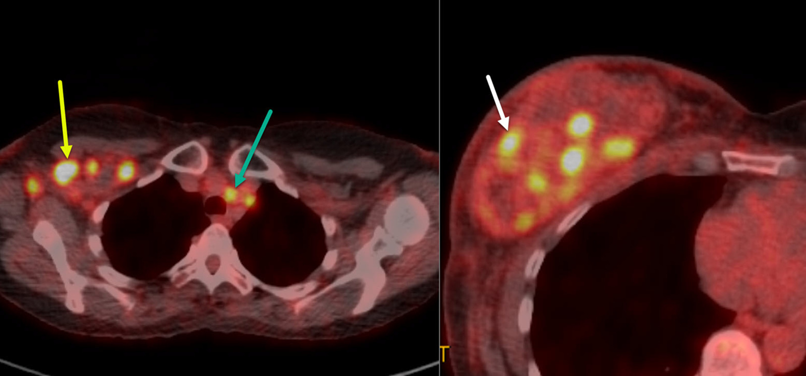
PET/CT is particularly valuable for evaluating regional lymph node metastases in patients with IBC. Studies by Alberini, Groheux, and Caracki et al have highlighted its utility in identifying nodal disease in the axilla, as well as in the subpectoral, internal mammary, and supraclavicular lymph nodes. 31-34 In addition, PET/CT may identify metastatic disease not evident on clinical examination and/or other imaging modalities.
In addition to regional lymph node assessment, PET/CT is useful in identifying distant nodal disease (eg, mediastinal or contralateral axillary adenopathy) and distant metastases. Studies by Groheux et al and Carkaci et al reported that distant metastases were detected by the modality in 46% and 49% of patients with IBC, respectively. 22,23 It was also superior to CT for distant lymph node, bone, and liver metastases, and it outperformed bone scans in identifying metastases in these tissues. 22 However, chest CT was more sensitive for lung and pleural metastases. 22 In certain subsets of patients with IBC, such as those with triple-negative breast cancer, visceral metastases, or young age at diagnosis, up to 30% of cases can present with brain metastasis. Therefore, brain MRI is recommended for these patients. 34
Treatment
Systemic Chemotherapy
IBC is highly aggressive, with poor survival rates. Before the introduction of systemic chemotherapy, fewer than 5% of patients treated with surgery and/or radiation therapy alone survived beyond 5 years, with a median survival under 15 months. 35,36 Local recurrence rates were high, at approximately 50%, and many patients were candidates for surgery. 37 Over the past 2 decades, the consensus treatment for IBC has evolved to include systemic chemotherapy (with trastuzumab and endocrine therapy when indicated), followed by surgery and radiation therapy. Although IBC has been excluded from most prospective chemotherapy trials, retrospective trials have demonstrated the efficacy of systemic chemotherapy in IBC.
Anthracycline-based chemotherapy, introduced in the 1970s, significantly improved outcomes for IBC, achieving clinical response rates up to 72% and increasing 5- and 10-year survival rates compared with earlier treatments. 38-44 Combining anthracycline-based chemotherapy with taxanes has further enhanced these responses. Studies at the MD Anderson Cancer Center that added paclitaxel to standard regimens increased pathological complete response (pCR) rates. 39-43 Achieving pCR, particularly in the axillary lymph nodes, remains the most significant prognostic factor for long-term survival. 44
Approximately 17-30% of IBC cases are triple-negative; that is, the tumor lacks estrogen and progesterone receptors and HER2 overexpression. 45,46 For these patients, adding pembrolizumab to neoadjuvant anthracycline- and taxane-based systemic chemotherapy has demonstrated improved pCR rates. 47 This combination is now widely used for triple-negative IBC.
HER2-positive IBC accounts for 36-60% of cases and benefits significantly from trastuzumab-based regimens. 48,49 The NOAH trial demonstrated that adding trastuzumab to standard chemotherapy significantly increased the pCR rate in patients with HER2-positive IBC. 50 Other studies corroborate these findings, suggesting that trastuzumab is essential to treatment in these cases. 51,52
No standard IBC-specific treatments currently exist for patients with advanced or metastatic disease. This highlights the importance of clinical trials, including those focusing on exploratory or novel targeted therapies for patients with advanced or metastatic IBC.
Surgery
Total mastectomy with axillary lymph node dissection (modified radical mastectomy) is recommended for patients with IBC. The optimal timing for surgery is 3-6 weeks after the completion of neoadjuvant systemic therapy. The primary goal is to excise all macroscopic diseases and obtain pathologically clear margins. Excising grossly abnormal skin is also recommended; in patients where primary closure is not possible, advanced wound coverage techniques, such as skin grafting or myocutaneous flap closure with assistance from plastic surgery, may be indicated. Because breast skin excision is necessary for patients with IBC, immediate reconstruction is contraindicated. An MD Anderson Cancer Center study evaluating long-term outcomes in patients who completed trimodal therapy (neoadjuvant systemic therapy, modified radical mastectomy, and radiation) showed durable survival and a local recurrence rate of 6.9%, which is comparable to that of non-IBC patients. 52 Metastatic spread to the regional nodes is noted at presentation in most patients; axillary dissection is recommended in these cases. De-escalation measures such as breast conservation and limited axillary surgeries, which have not been well studied in patients with IBC, may be associated with increased rates of local recurrence. 53 As such, total mastectomy with axillary dissection remains the recommended approach to these cases.
Radiation Therapy
All patients should be offered radiation therapy regardless of treatment response. Radiation targets the chest wall and undissected draining lymphatics, with an extra dose (boost) to the chest wall and any undissected clinical stage N3 disease (infraclavicular, supraclavicular, or internal mammary lymph nodes). Dose fractionation details can be found online. 54 While historical locoregional control rates are low, at approximately 80% over 5 years, recent data show significant improvement with a risk-stratified approach. 54 It is important to note that patients with IBC were excluded in recently presented clinical trials of de-escalation treatment, including hypofractionation and observation after pCR. Therefore, these approaches should be avoided in these patients.
Conclusion
IBC is a rare and aggressive form of advanced breast cancer characterized by rapid progression and distinct skin findings. Accurate, timely diagnosis is essential as skin findings overlap with benign pathologies such as infection and mastitis. Standard-of-care confirmatory imaging tests consist of mammography, breast and nodal ultrasound, and MRI. PET/CT is instrumental in detecting distant metastases and disease staging. Standard-of-care treatment involves a multimodal approach with chemotherapy, mastectomy with axillary lymph node dissection, and radiation therapy. Despite advances in imaging technology and emerging chemotherapies, IBC survival rates remain poor, underscoring the importance of continued research and encouraging participation in clinical trials.
References
- Abraham H, Xia Y, Mukherjee B, Merajver S. Incidence and survival of inflammatory breast cancer between 1973 and 2015 in the SEER database. Breast Cancer Res Treat. 2021;185(1):229-238. doi:10.1007/s10549-020-05938-2.
- Hance K, Anderson W, Devesa S, Young H, Levine P. Trends in inflammatory breast carcinoma incidence and survival: the surveillance, epidemiology, and end results program at the National Cancer Institute. J Natl Cancer Inst. 2005;97(13):966-975. doi:10.1093/jnci/dji172.
- Menta A, Fouad T, Lucci A. Inflammatory breast cancer: what to know about this unique, aggressive breast cancer. Surg Clin North Am. 2018;98(4):787-800. doi:10.1016/j.suc.2018.03.009.
- Dawood S, Merajver S, Viens P. International expert panel on inflammatory breast cancer: consensus statement for standardized diagnosis and treatment. Ann Oncol. 2011;22(3):515-523. doi:10.1093/annonc/mdq345.
- Lê M, Arriagada R, Contesso G. Dermal lymphatic emboli in inflammatory and noninflammatory breast cancer: a French-Tunisian joint study in 337 patients. Clin Breast Cancer. 2005;6(5):439-445. doi:10.3816/CBC.2005.n.049.
- Amin M, Edge S, Greene F. AJCC Cancer Staging Manual. 2017.
- Schairer C, Hablas A, Eldein I. Clinico-pathologic and mammographic characteristics of inflammatory and non-inflammatory breast cancer at six centers in North Africa. Breast Cancer Res Treat. 2019;176(2):407-417. doi:10.1007/s10549-019-05237-5.
- Edge S, Byrd D, C C. AJCC Cancer Staging Manual. 2010:345-376.
- Lynce F, Niman S, Kai M. Development of a multi-institutional dataset to validate a novel inflammatory breast cancer diagnostic score. J Natl Cancer Inst. 2025. doi:10.1093/jnci/djaf088.
- Inflammatory Breast Cancer. 2022.
- Dabi Y, Darrigues L, Pons K. Incidence of inflammatory breast cancer in patients with clinical inflammatory breast symptoms. PLoS One. 2017;12(12). doi:10.1371/journal.pone.0189385.
- Froman J, Landercasper J, Ellis R, De Maiffe B, Theede L. Red breast as a presenting complaint at a breast center: an institutional review. Surgery. 2011;149(6):813-819. doi:10.1016/j.surg.2010.12.013.
- Kamal R, Hamed S, Salem D. Classification of inflammatory breast disorders and step by step diagnosis. Breast J. 2009;15(4):367-380. doi:10.1111/j.1524-4741.2009.00740.x.
- Yeh E, Jacene H, Bellon J. What radiologists need to know about diagnosis and treatment of inflammatory breast cancer: a multidisciplinary approach. Radiographics. 2013;33(7):2003-2017. doi:10.1148/rg.337135503.
- Yang W, Le-Petross H, Macapinlac H. Inflammatory breast cancer: PET/CT, MRI, mammography, and sonography findings. Breast Cancer Res Treat. 2008;109(3):417-426. doi:10.1007/s10549-007-9671-z.
- Günhan-Bilgen I, Ustün E, Memiş A. Inflammatory breast carcinoma: mammographic, ultrasonographic, clinical, and pathologic findings in 142 cases. Radiology. 2002;223(3):829-838. doi:10.1148/radiol.2233010198.
- Yamauchi H, Woodward W, Valero V. Inflammatory breast cancer: what we know and what we need to learn. Oncologist. 2012;17(7):891-899. doi:10.1634/theoncologist.2012-0039.
- Le-Petross H, Cristofanilli M, Carkaci S. MRI features of inflammatory breast cancer. AJR Am J Roentgenol. 2011;197(4):W769-W776. doi:10.2214/AJR.10.6157.
- Chow C. Imaging in inflammatory breast carcinoma. Breast Dis. 2005;22:45-54. doi:10.3233/bd-2006-22106.
- Wright F, Zubovits J, Gardner S. Optimal assessment of residual disease after neo-adjuvant therapy for locally advanced and inflammatory breast cancer—clinical examination, mammography, or magnetic resonance imaging?. J Surg Oncol. 2010;101(7):604-610. doi:10.1002/jso.21559.
- Groheux D, Giacchetti S, Delord M. 18F-FDG PET/CT in staging patients with locally advanced or inflammatory breast cancer: comparison to conventional staging. J Nucl Med. 2013;54(1):5-11. doi:10.2967/jnumed.112.106864.
- Carkaci S, Macapinlac H, Cristofanilli M. Retrospective study of 18F-FDG PET/CT in the diagnosis of inflammatory breast cancer: preliminary data. J Nucl Med. 2009;50(2):231-238. doi:10.2967/jnumed.108.056010.
- Bonnier P, Charpin C, Lejeune C. Inflammatory carcinomas of the breast: a clinical, pathological, or a clinical and pathological definition?. Int J Cancer. 1995;62(4):382-385. doi:10.1002/ijc.2910620404.
- Harada T, Uematsu T, Nakashima K. Evaluation of breast edema findings at T2-weighted breast MRI is useful for diagnosing occult inflammatory breast cancer and can predict prognosis after neoadjuvant chemotherapy. Radiology. 2021;299(1):53-62. doi:10.1148/radiol.2021202604.
- Uematsu T, Kasami M, Watanabe J. Is evaluation of the presence of prepectoral edema on T2-weighted with fat-suppression 3T breast MRI a simple and readily available noninvasive technique for estimation of prognosis in patients with breast cancer?. Breast Cancer. 2014;21(6):684-692. doi:10.1007/s12282-013-0440-z.
- Hirko K, Soliman A, Banerjee M. A comparison of criteria to identify inflammatory breast cancer cases from medical records and the surveillance, epidemiology and end results data base, 2007–2009. Breast J. 2014;20(2):185-191. doi:10.1111/tbj.12234.
- Bertagna F, Treglia G, Orlando E. Prevalence and clinical significance of incidental F18-FDG breast uptake: a systematic review and meta-analysis. Jpn J Radiol. 2014;32(2):59-68. doi:10.1007/s11604-013-0270-0.
- Gradishar W, Moran M, Abraham J. Breast cancer, version 3.2022, NCCN clinical practice guidelines in oncology. J Natl Compr Canc Netw. 2022;20(6):691-722. doi:10.6004/jnccn.2022.0030.
- Fouad T, Kogawa T, Liu D. Erratum to: overall survival differences between patients with inflammatory and noninflammatory breast cancer presenting with distant metastasis at diagnosis. Breast Cancer Res Treat. 2015;152(2):417. doi:10.1007/s10549-015-3477-1.
- Patel M, Le-Petross H. Baseline FDG PET-CT imaging is necessary for newly diagnosed inflammatory breast cancer patients: a narrative review. Chin Clin Oncol. 2021;10(6):56. doi:10.21037/cco-21-82.
- Alberini J, Lerebours F, Wartski M. 18F-fluorodeoxyglucose positron emission tomography/computed tomography (FDG-PET/CT) imaging in the staging and prognosis of inflammatory breast cancer. Cancer. 2009;115(21):5038-5047. doi:10.1002/cncr.24534.
- Warren L, Niman S, Remolano M. Incidence, characteristics, and management of central nervous system metastases in patients with inflammatory breast cancer. Cancer. 2022;128(23):4085-4094. doi:10.1002/cncr.34441.
- Han S, Choi J. Impact of 18F-FDG PET, PET/CT, and PET/MRI on staging and management as an initial staging modality in breast cancer: a systematic review and meta-analysis. Clin Nucl Med. 2021;46(4):271-282. doi:10.1097/RLU.0000000000003502.
- Bozzetti F, Saccozzi R, De Lena M, Salvadori B. Inflammatory cancer of the breast: analysis of 114 cases. J Surg Oncol. 1981;18(4):355-361. doi:10.1002/jso.2930180405.
- Zucali R, Uslenghi C, Kenda R, Bonadonna G. Natural history and survival of inoperable breast cancer treated with radiotherapy and radiotherapy followed by radical mastectomy. Cancer. 1976;37(3):1422-1431. doi:10.1002/1097-0142(197603)37:3<1422::aid-cncr2820370325>3.0.co;2-y.
- Macdonald S, Harris E, Arthur D. ACR appropriateness criteria® locally advanced breast cancer. Breast J. 2011;17(6):579-585. doi:10.1111/j.1524-4741.2011.01150.x.
- Thoms W, McNeese M, Fletcher G. Multimodal treatment for inflammatory breast cancer. Int J Radiat Oncol Biol Phys. 1989;17(4):739-745. doi:10.1016/0360-3016(89)90060-6.
- Ueno N, Buzdar A, Singletary S. Combined-modality treatment of inflammatory breast carcinoma: twenty years of experience at M. D. Anderson Cancer Center. Cancer Chemother Pharmacol. 1997;40(4):321-329. doi:10.1007/s002800050664.
- Blumenschein G, Montague E, Eckles N. Sequential combined modality therapy for inflammatory breast cancer. Breast. 1976;2:16-20.
- Koh E, Buzdar A, Ames F. Inflammatory carcinoma of the breast: results of a combined-modality approach--M. D. Anderson Cancer Center experience. Cancer Chemother Pharmacol. 1990;27(2):94-100. doi:10.1007/BF00689090.
- Singletary S, Ames F, Buzdar A. Management of inflammatory breast cancer. World J Surg. 1994;18(1):87-92. doi:10.1007/BF00348197.
- Buzdar A, Singletary S, Booser D. Combined modality treatment of stage III and inflammatory breast cancer. Surg Oncol Clin N Am. 1995;4(4):715-734. doi:10.1016/S1055-3207(18)30426-5.
- Baldini E, Gardin G, Evagelista G. Long-term results of combined-modality therapy for inflammatory breast carcinoma. Clin Breast Cancer. 2004;5(5):358-363. doi:10.3816/cbc.2004.n.042.
- Li J, Gonzalez-Angulo A, Allen P. Triple-negative subtype predicts poor overall survival and high locoregional relapse in inflammatory breast cancer. Oncologist. 2011;16(12):1675-1683. doi:10.1634/theoncologist.2011-0196.
- Bertucci F, Finetti P, Rougemont J. Gene expression profiling identifies molecular subtypes of inflammatory breast cancer. Cancer Res. 2005;65(6):2170-2178. doi:10.1158/0008-5472.CAN-04-4115.
- Schmid P, Cortes J, Pusztai L. Pembrolizumab for early triple-negative breast cancer. N Engl J Med. 2020;382(9):810-821. doi:10.1056/NEJMoa1910549.
- Guérin M, Sheng Z, Andrieu N, Riou G. Strong association between c-myb and oestrogen-receptor expression in human breast cancer. Oncogene. 1990;5(1):131-135.
- Parton M, Dowsett M, Ashley S. High incidence of HER-2 positivity in inflammatory breast cancer. Breast. 2004;13(2):97-103. doi:10.1016/j.breast.2003.08.004.
- Gianni L, Eiermann W, Semiglazov V. Neoadjuvant chemotherapy with trastuzumab followed by adjuvant trastuzumab versus neoadjuvant chemotherapy alone, in patients with HER2-positive locally advanced breast cancer (the NOAH trial): a randomised controlled superiority trial with a parallel HER2-negative cohort. Lancet. 2010;375(9712):377-384. doi:10.1016/S0140-6736(09)61964-4.
- Hurley J, Doliny P, Reis I. Docetaxel, cisplatin, and trastuzumab as primary systemic therapy for human epidermal growth factor receptor 2-positive locally advanced breast cancer. J Clin Oncol. 2006;24(12):1831-1838. doi:10.1200/JCO.2005.02.8886.
- Van Pelt A, Mohsin S, Elledge R. Neoadjuvant trastuzumab and docetaxel in breast cancer: preliminary results. Clin Breast Cancer. 2003;4(5):348-353. doi:10.3816/cbc.2003.n.040.
- Adesoye T, Everidge S, Chen J. Low rates of local-regional recurrence among inflammatory breast cancer patients after contemporary trimodal therapy. Ann Surg Oncol. 2023;30(10):6232-6240. doi:10.1245/s10434-023-13906-5.
- Layman R, Lucci A, Woodward W. Breast Cancer—Inflammatory (IBC). 2023.
- Rosso K, Tadros A, Weiss A. Improved locoregional control in a contemporary cohort of nonmetastatic inflammatory breast cancer patients undergoing surgery. Ann Surg Oncol. 2017;24(10):2981-2988. doi:10.1245/s10434-017-5952-x.
Disclosures
The authors have no conflicts of interest to disclose. None of the authors received outside funding for the production of this original manuscript and no part of this article has been previously published elsewhere.
Citation
. Comprehensive Review of Clinical Presentation, Multimodality Imaging, and Therapeutic Strategies for Inflammatory Breast Cancer. Applied Radiology. 2025;54(4):1-9. doi:10.37549/AR-D-25-0082.