Congenital Toxoplasmosis
Applied Radiology — Vol. 55 , Issue 5
Published: June 1, 2026
1 Vanderbilt University School of Medicine, Nashville, Tennessee
2 Department of Pediatrics, Division of Infectious Diseases, Monroe Carell Jr. Children’s Hospital, Vanderbilt University Medical Center, Nashville, Tennessee
3 Department of Radiology, Division of Pediatric Radiology, Monroe Carell Jr. Children’s Hospital, Vanderbilt University Medical Center, Nashville, Tennessee
* Corresponding author: Asha Sarma (asha.sarma@vumc.org)
Abstract
Congenital toxoplasmosis is a parasitic infection caused by vertical transmission of Toxoplasma gondii. Our case describes an early-term neonate presenting with poor feeding, abnormal movements, and hypothermia. Neuroimaging revealed hydrocephalus, randomly distributed intracranial calcifications, leptomeningeal and ependymal enhancement, and intraocular plaques. Diagnosis was confirmed by maternal and infant serologies. The patient was treated with pyrimethamine, sulfadiazine, and folinic acid, in addition to adjunctive neurosurgical and ophthalmologic interventions. Of note, there was no maternal exposure to cats or ingestion of undercooked meats, which are the typical risk factors associated with toxoplasmosis, but there was reported exposure to soil excavated in the yard during pregnancy. This case illustrates the variable presentation of congenital toxoplasmosis, which may be asymptomatic at birth and later present with the classic triad of hydrocephalus, chorioretinitis, and intracranial calcifications. Early recognition of imaging findings is critical for timely initiation of therapy, which improves neurodevelopmental and visual outcomes.
Keywords
congenital toxoplasmosis, TORCH infections, intracranial calcifications, pediatric neuroradiology
Categories
Case Summary
A neonate born at early term presented to their pediatrician with decreased oral intake and a 3-day history of abnormal eye and neck movements. Pregnancy was uncomplicated, with unremarkable prenatal US, and the neonate had done well in the first 2 postnatal weeks. Owing to a core temperature of 93°F, the neonate was referred to the emergency department. Physical exam demonstrated dry mucous membranes, decreased interactivity, and 30-second episodes of rightward gaze deviation. Lumbar puncture was performed, followed by empiric treatment with ampicillin, gentamicin, and acyclovir. Analysis of cerebrospinal fluid demonstrated elevated protein, low glucose, and neutrophilic pleocytosis. The patient’s complete blood count with differential and liver function panel were unremarkable. Noncontrast CT of the head and contrast MRI of the brain were performed.
Imaging Findings
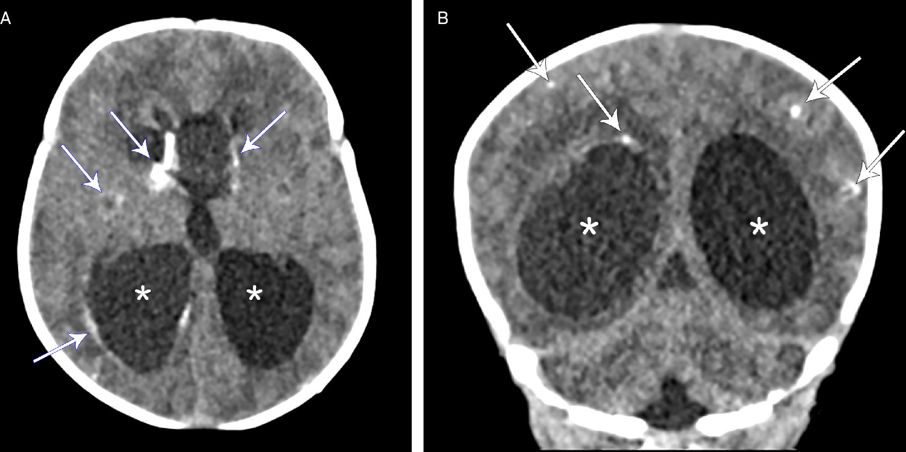
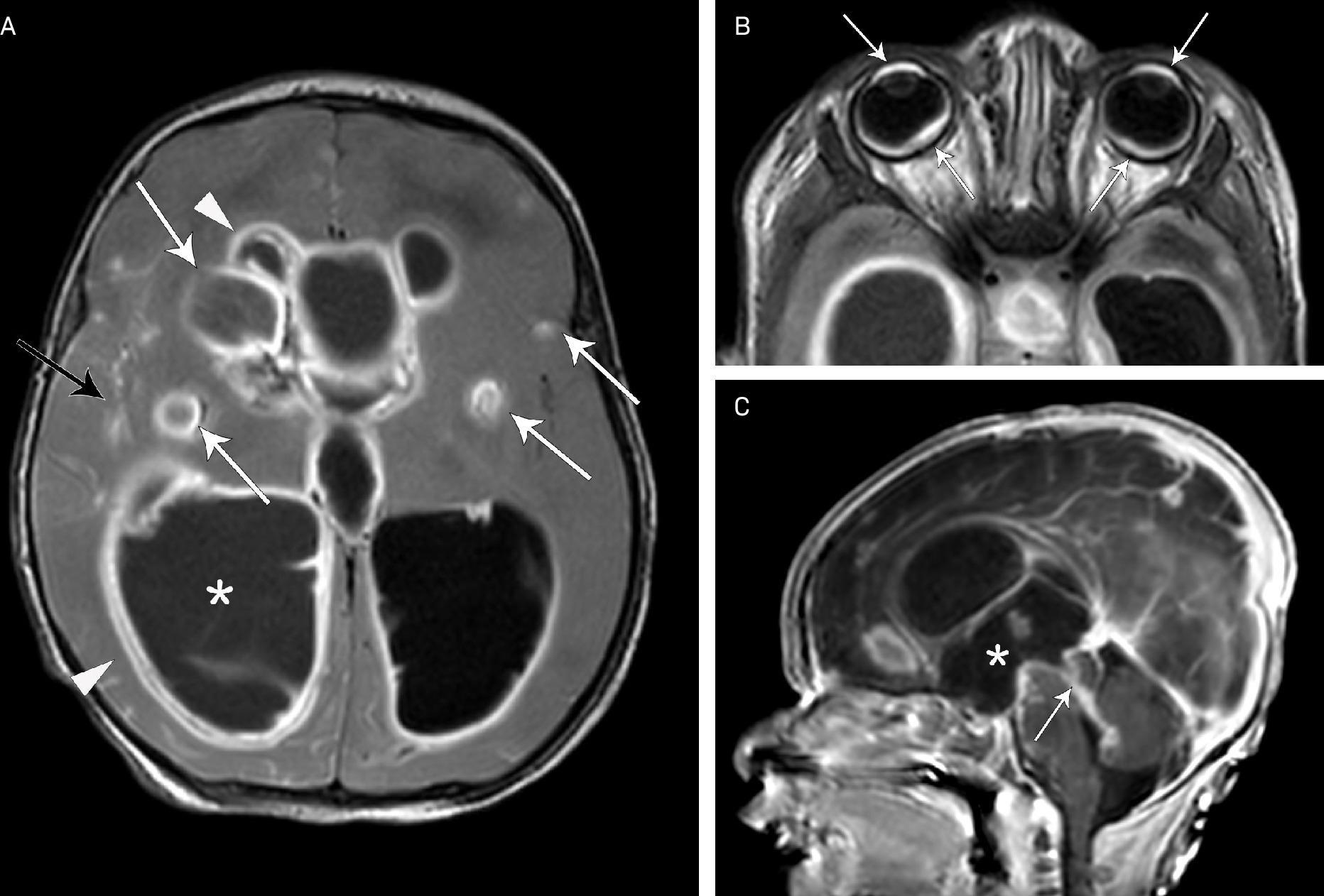
Noncontrast head CT (Figure 1) demonstrated marked enlargement of the lateral and 3rd ventricles and extensive parenchymal calcifications; these included basal ganglia, subcortical, and periventricular calcifications, raising concerns for a TORCH infection. The subsequent T2 FLAIR sequence on brain MRI (Figure 2) similarly demonstrated hydrocephalus and multifocal enhancing parenchymal lesions in the basal ganglia and subcortical white matter. This sequence further revealed leptomeningeal enhancement, ependymal enhancement, and enhancing plaque-like intraocular thickenings in the anterior and posterior segments. The T1 sequence demonstrated dilatation of the 3rd ventricle with dilated 3rd ventricular recesses and aqueductal stenosis with ependymal enhancement.
Diagnosis
Congenital toxoplasmosis.
The differential diagnosis included congenital toxoplasmosis, congenital cytomegalovirus (CMV), congenital lymphocytic choriomeningitis virus, Escherichia coli meningitis, and Aicardi-Goutières syndrome.
Discussion
Congenital toxoplasmosis is a parasitic infection caused by the vertical transmission of Toxoplasma gondii following acute maternal infection. In the United States, approximately 85% of women of childbearing age are susceptible to primary infection, with an incidence of 400-4000 cases annually.1
T. gondii is primarily transmitted through ingestion of raw or undercooked meat containing tissue cysts or inadvertent ingestion of oocysts found in cat feces, either from a litter box or soil.2 In this case, there was no maternal exposure to cats or ingestion of undercooked meats, but maternal exposure to soil excavated from the patient’s yard (presumably containing cat feces) throughout the pregnancy was reported.
Maternal infection with T. gondii is usually asymptomatic, but nonspecific symptoms such as cervical lymphadenopathy, fever, malaise, and myalgias may occur following an incubation period of 5-18 days. In immunocompetent adults, the clinical course is benign and self-limited. The overall risk of congenital infection from acute maternal infection during pregnancy is 20-50%. While this risk increases with more advanced gestational age, congenital toxoplasmosis is more severe when infection is acquired in the first trimester.3 Since this patient had normal US and was asymptomatic at birth, transmission possibly occurred in the third trimester.
Congenital toxoplasmosis presents with a wide spectrum of outcomes ranging from a healthy neonate to significant neurological and ocular findings, including chorioretinitis, intracranial calcifications, hydrocephalus, seizures, hepatosplenomegaly, and jaundice.4 Clinical findings may not be present at birth, as in the case of our patient. Typically, preterm infants with toxoplasmosis demonstrate neurological and ocular manifestations in the first 3 months of life, while term infants demonstrate milder disease with lymphadenopathy and hepatosplenomegaly within the first 2 months of life.4
Pre- or postnatal imaging may suggest a diagnosis of congenital toxoplasmosis. Prenatal US can reveal intracranial hyperechogenic nodular foci, ventriculomegaly, hepatosplenomegaly, and ascites.5 Postnatally, bulging fontanelles or seizures can prompt head CT and MRI. While the findings can overlap with other congenital infections, several features help distinguish toxoplasmosis from CMV and Zika virus. Congenital toxoplasmosis typically demonstrates randomly distributed intracranial calcifications (often involving the cortex and basal ganglia), hydrocephalus (with various patterns such as aqueductal stenosis and ependymitis), chorioretinitis, and normo- or macrocephaly. In contrast, congenital CMV presents with periventricular calcifications, ventriculomegaly, and migrational abnormalities such as polymicrogyria. Congenital Zika virus infection is characterized by striking microcephaly, periventricular and subcortical calcifications, and brain surface smoothness.6 The clinical diagnosis is made with serological testing but, as in our case, empiric treatment was initiated based on imaging. The diagnosis was confirmed a few weeks later, with maternal T. gondii IgG values of 1:8000, and corresponding values of 1:2048 (normal ≤1:16) in the infant.
Treatment consists of pyrimethamine, sulfadiazine, and folinic acid for 12 months, along with steroids for severe chorioretinitis or CNS inflammation. Early initiation of therapy is recommended, as it is associated with a lower incidence of retinochoroidal lesions and improved neurodevelopmental outcomes.4 Signs and symptoms of infection tend to resolve within the first month of treatment.7 Management of hydrocephalus may require surgical CSF diversion.8 In addition to antiparasitic therapy, our patient was treated with levetiracetam for seizure management, intravitreal clindamycin injections for chorioretinal scarring, and 2 ventriculoperitoneal shunts. The prognosis is generally favorable in children who receive early diagnosis and treatment, with most remaining asymptomatic or experiencing only mild sequelae, although a lifelong risk of ocular complications and neurological impairment remains, especially in cases of delayed diagnosis or inadequate therapy.7
Conclusion
Congenital toxoplasmosis is a parasitic infection that can present in neonates with the classic triad of chorioretinitis, hydrocephalus, and diffuse intracranial calcifications. The condition may be identified on prenatal US with intracranial echogenic nodular foci and ventriculomegaly. It can also be diagnosed postnatally with CT and MR images that demonstrate randomly distributed calcifications, hydrocephalus with aqueductal stenosis, ependymitis, and chorioretinitis, followed by confirmatory serological testing. Management involves antiparasitic therapy with pyrimethamine, sulfadiazine, and folinic acid and, depending on severity, steroids, intravitreal injections, and neurosurgical intervention.
References
- Jones J, Lopez A, Wilson M. Congenital toxoplasmosis. Am Fam Physician. 2003;67(10):2131-2138.
- Elmore S, Jones J, Conrad P. Toxoplasma gondii: epidemiology, feline clinical aspects, and prevention. Trends Parasitol. 2010;26(4):190-196. doi:10.1016/j.pt.2010.01.009.
- Practice bulletin no.151: cytomegalovirus, parvovirus B19, varicella zoster, and toxoplasmosis in pregnancy. Obstet Gynecol. 2015;125(6):1510-1525. doi:10.1097/01.AOG.0000466430.19823.53.
- Khan K, Khan W. Congenital toxoplasmosis: an overview of the neurological and ocular manifestations. Parasitol Int. 2018;67(6):715-721. doi:10.1016/j.parint.2018.07.004.
- Codaccioni C, Picone O, Lambert V. Ultrasound features of fetal toxoplasmosis: a contemporary multicenter survey in 88 fetuses. Prenat Diagn. 2020;40(13):1741-1752. doi:10.1002/pd.5756.
- Werner H, Daltro P, Fazecas T, Zare Mehrjardi M, Araujo Júnior E. Neuroimaging findings of congenital toxoplasmosis, cytomegalovirus, and Zika virus infections: a comparison of three cases. J Obstet Gynaecol Can. 2017;39(12):1150-1155. doi:10.1016/j.jogc.2017.05.013.
- McLeod R, Boyer K, Karrison T. Outcome of treatment for congenital toxoplasmosis, 1981-2004: the national collaborative Chicago-based, congenital toxoplasmosis study. Clin Infect Dis. 2006;42(10):1383-1394. doi:10.1086/501360.
- Caceres A, Caceres-Alan A, Caceres-Alan T. Toxoplasma gondii infections in pediatric neurosurgery. Childs Nerv Syst. 2024;40(2):295-301. doi:10.1007/s00381-023-05915-2.
Disclosures
The authors have no conflicts of interest to disclose. None of the authors received outside funding for the production of this original manuscript and no part of this article has been previously published elsewhere.
Citation
. Congenital Toxoplasmosis. Applied Radiology. 2026;55(5). doi:10.37549/AR-D-25-0121.