Congenital urethroperineal fistula
Applied Radiology — Vol. 37 , Issue 7 , pp. 30 -31
DOI: 10.37549/AR1628
Published: July 1, 2008
Categories
CASE SUMMARY
An 82-year-old man presented for evaluation and management of chronic dysuria and recurrent urinary tract infections. He indicated that he had urinary tract difficulties all of his life. He also stated that he sometimes wet himself during urination. His surgical history included transurethral resection of the prostate (TURP) for benign prostatic hypertrophy, approximately 10 years prior to his current presentation. Physical examination revealed normal male genitalia and perineum. Voiding cysto urethrography (VCUG) was requested.
IMAGING FINDINGS
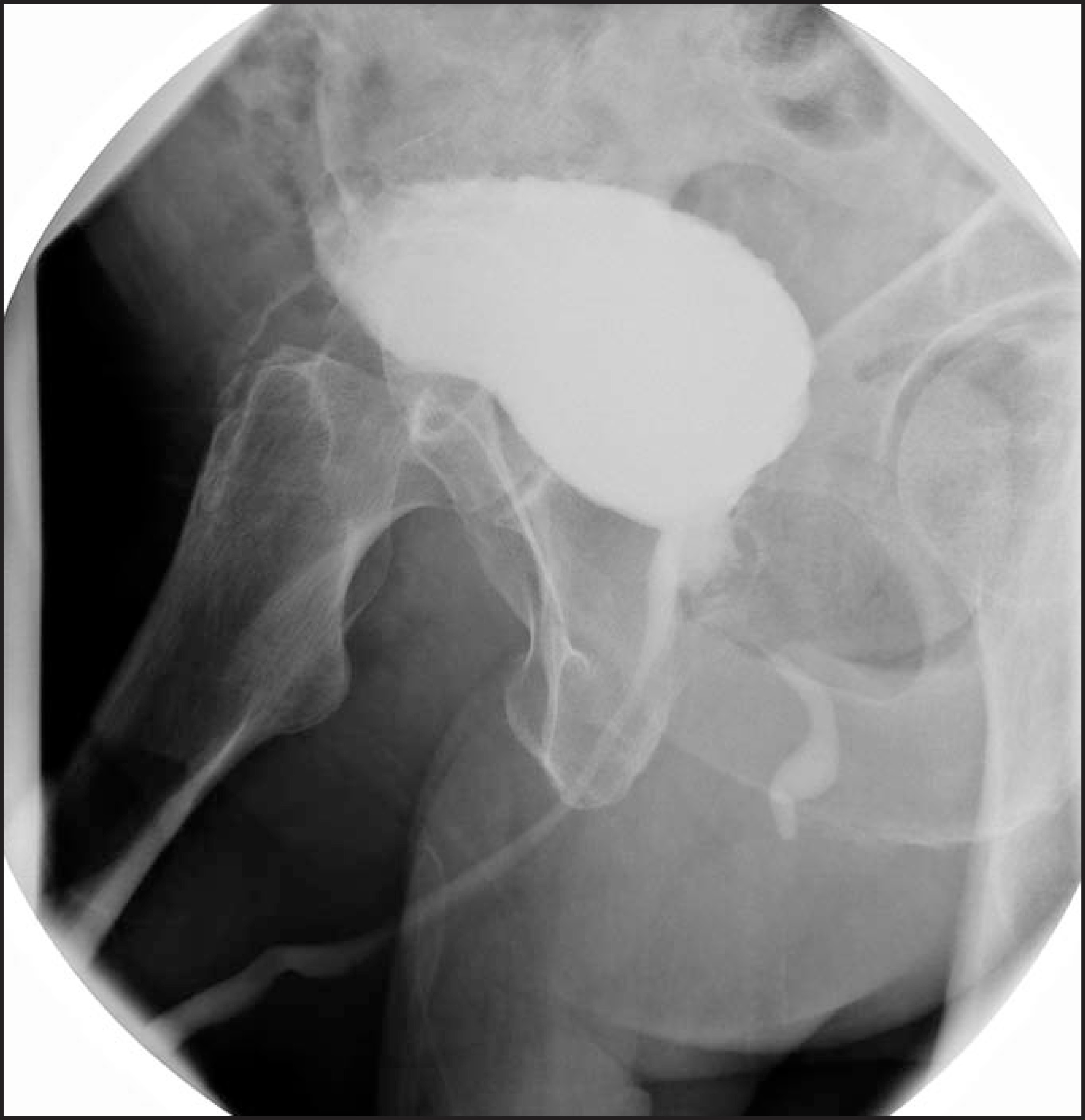
With the patient in an oblique position, VCUG images obtained during voiding revealed a TURP defect. The main route of contrastevacuation was via a normal urethra. A second channel was identified, however. This channel originated near the upper level of the TURP defect, extended posteroinferiorly toward the anus, and opened onto the perineum (Figure 1).
DIAGNOSIS
Congenital urethroperineal fistula
DISCUSSION
Differential diagnostic considerations in this case included the hypospadiac form of urethral duplication, urethroperineal fistula, and iatrogenic fistula development related to the TURP. Although a fistula is a potential complication of TURP, this patient’s symptoms predated the surgery by many years, and this was believed to be an unlikely source.
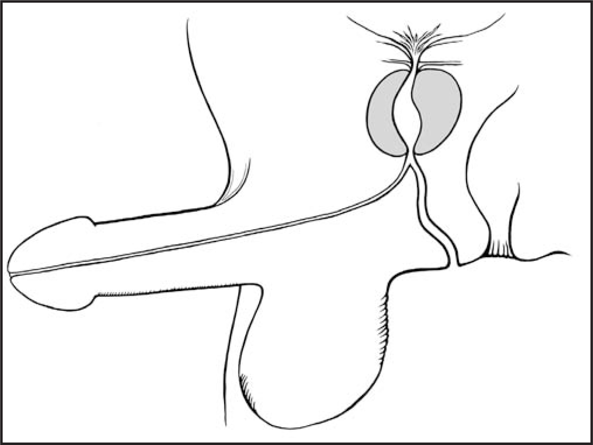
Urethral duplication is a rare anomaly that is commonly classified as epispadiac, normally positioned, or hypospadiac.1Regardless of theposition of the duplicated urethra, the ventral urethra is always the more normal in structure and function.1,2 In the hypospadiac form of urethral duplication, the normal ventral channel opens onto the perineum, while the dorsal, duplicated urethra exits the penis (Figure 2). A patient with this anomaly typically presents early in life with most urine exiting onto the perineum. Radiographic findings in this condition include a dominant channel that exits onto the perineum.2,3 Management is difficult, as the dorsal urethra is hypoplastic and resection of the ventral urethracan have devastating effects.2,4 Surgical correction is most commonly performed by a 2-part, staged hypospadia correction procedure.2,5
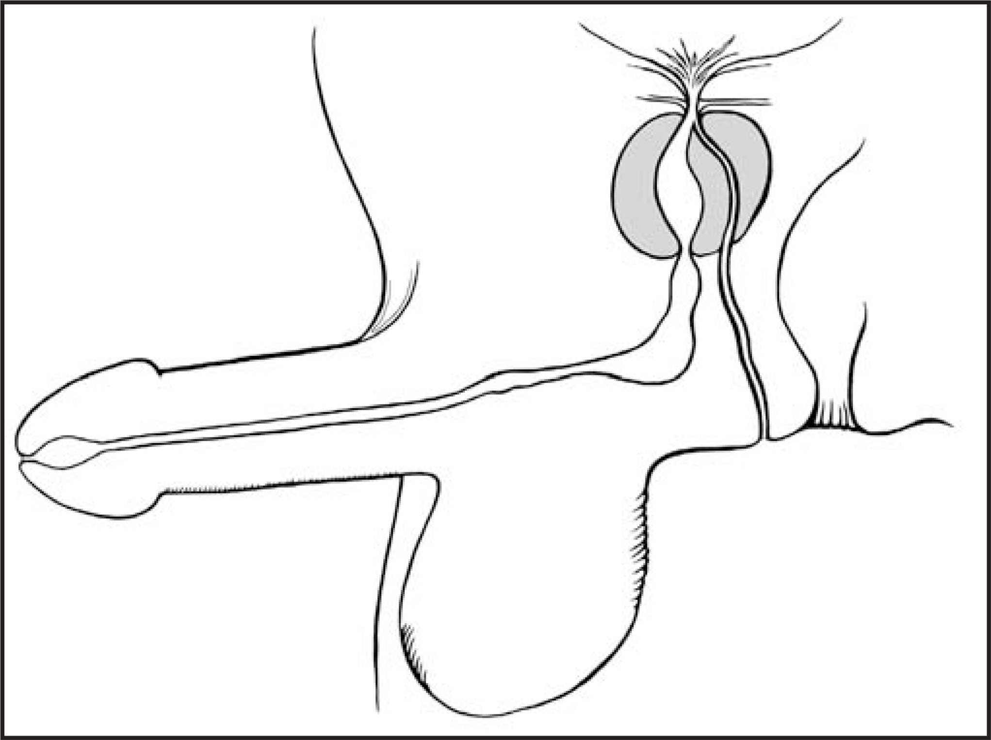
Congenital urethroperineal fistula is manifested by a normal penile urethra with a second ventral channel arising from the prostatic urethra and opening onto the perineum (Figure 3). These patients usually present later in life. Symptoms include moisture on the perineum after voiding or recurrent urinary tract infections, typically with gastrointestinal flora. Radiographic findings may mimic that of urethral duplication.2,5 However, with congenital urethroperineal fistula, the dominant and normal urethra is dorsal in position and exits the penis, while a small, irregular channel extends posteriorly from the prostatic urethra onto the perineum. Such was the case in the patient reported here. In patients with a congenital urethroperineal fistula, surgical resection of the ventral channel is the treatment of choice and usually results in a good outcome in the small number of patients reported.2,5
CONCLUSION
While both are rare entities, differentiation of the hypospadiac form of urethral duplication and congenital urethroperineal fistula is critical for treatment planning. Both channels should be visualized by VCUG and/or retrograde urethrography. Identification of the dominant channel is integral to the diagnosis. Simple resection of the ventral channel of the urethroperineal fistula is curative in most patients, whereas a similar procedure in hypospadiac urethral duplication can have a devastating outcome.
References
- Effmann E, Lebowitz R, Colodny A. Duplication of the urethra. Radiology. 1976;119:179-185.
- Senocak M, Hicsonmez A, Buyukpamukcu N. Congenital urethroperineal fistula: A case report and literature review. Z Kinderchir. 1990;45:192-196.
- deVries P, Friedland G. Congenital “H-type” ano-urethral fistula. Radiology. 1974;113:397-407.
- Tanaka Y, Sasaki Y, Matsumura M. Cystic lesion of the buttock communicating with the lateral side of the posterior urethra. J Urol. 1994;151:1363-1364.
- Bates G, Lebowitz R. Congenital urethroperineal fistula. Radiology. 1995;194:501-504.
Citation
. Congenital urethroperineal fistula. Applied Radiology. 2008;37(7):30-31. doi:10.37549/AR1628.