Constrictive pericarditis
Applied Radiology — Vol. 37 , Issue 5 , pp. 35 -37
DOI: 10.37549/AR1611
Published: May 1, 2008
Categories
CASE SUMMARY
The patient was a 38-year-old man with a 1-year history of bilateral leg edema and persistent blisters over the tibia bilaterally. He presented to the emergency department with acute onset shortness of breath, dizziness, and pleuritic chest pain that started 1 week previously and resolved slowly over 3 to 4 days. Previous medical evaluations had investigated the possibility of inferior vena cava thrombosis. The review of systems was positive for significant weight gain over the last month. His medical history was unremarkable other than a documented episode of viral pericarditis 4 years prior that resolved with nonsteroidal anti-inflammatory medication. Laboratory data revealed moderate hypoalbuminemia, a slight elevation in total bilirubin, and a low platelet count. A nephrology evaluation showed no kidney disease.
IMAGING FINDINGS
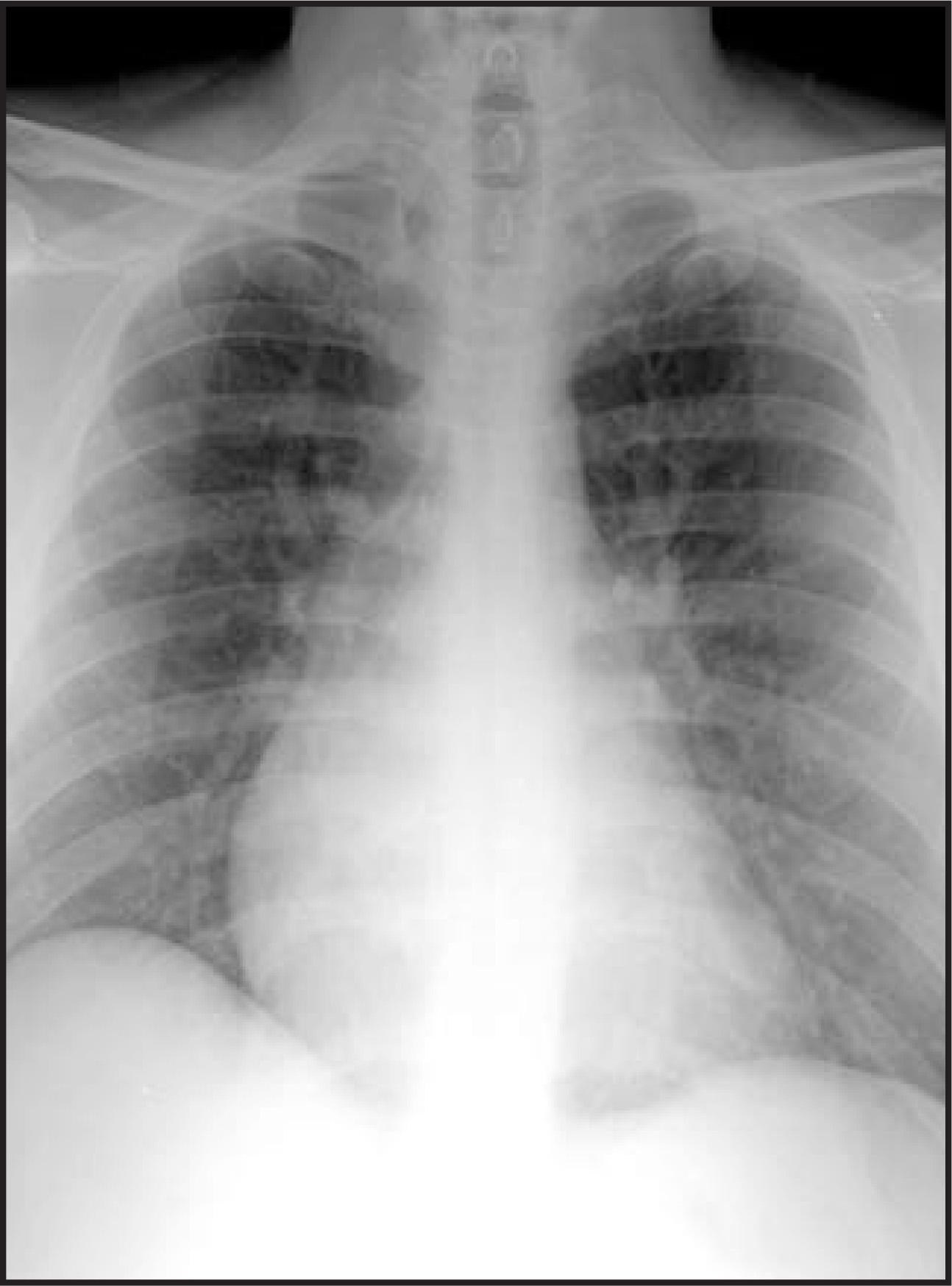
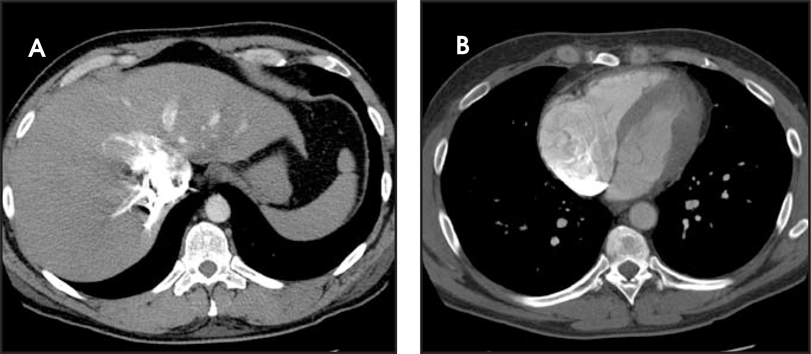
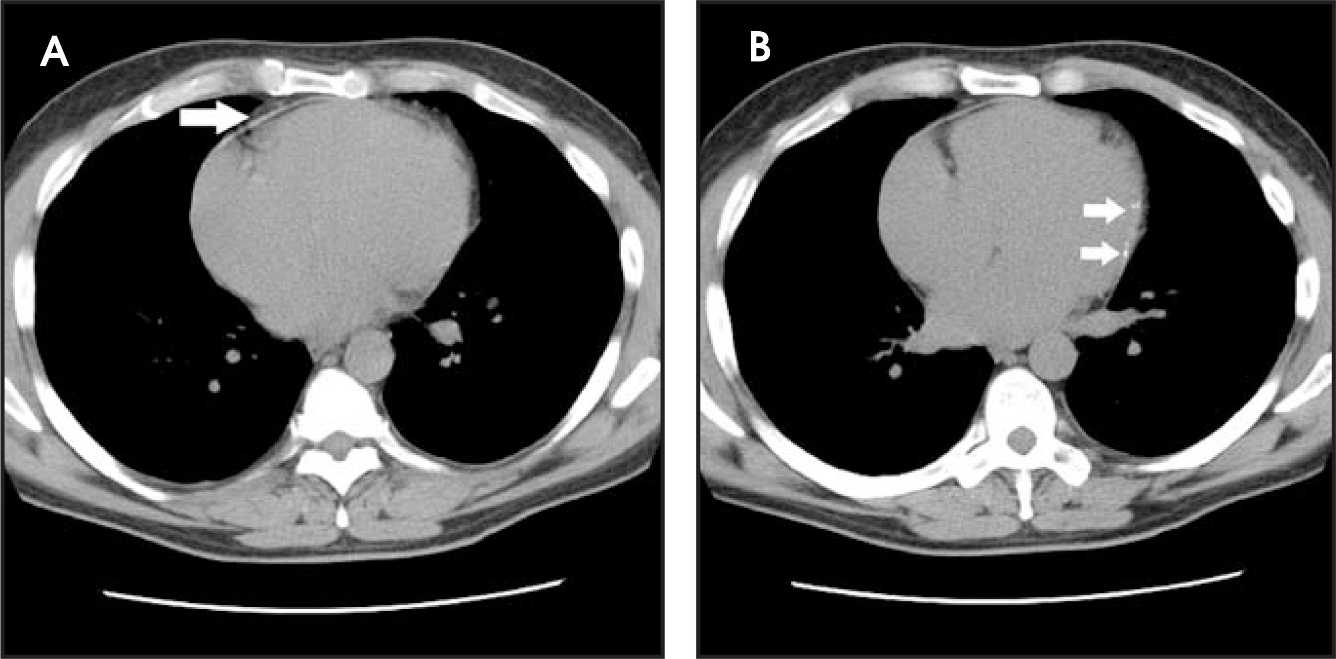
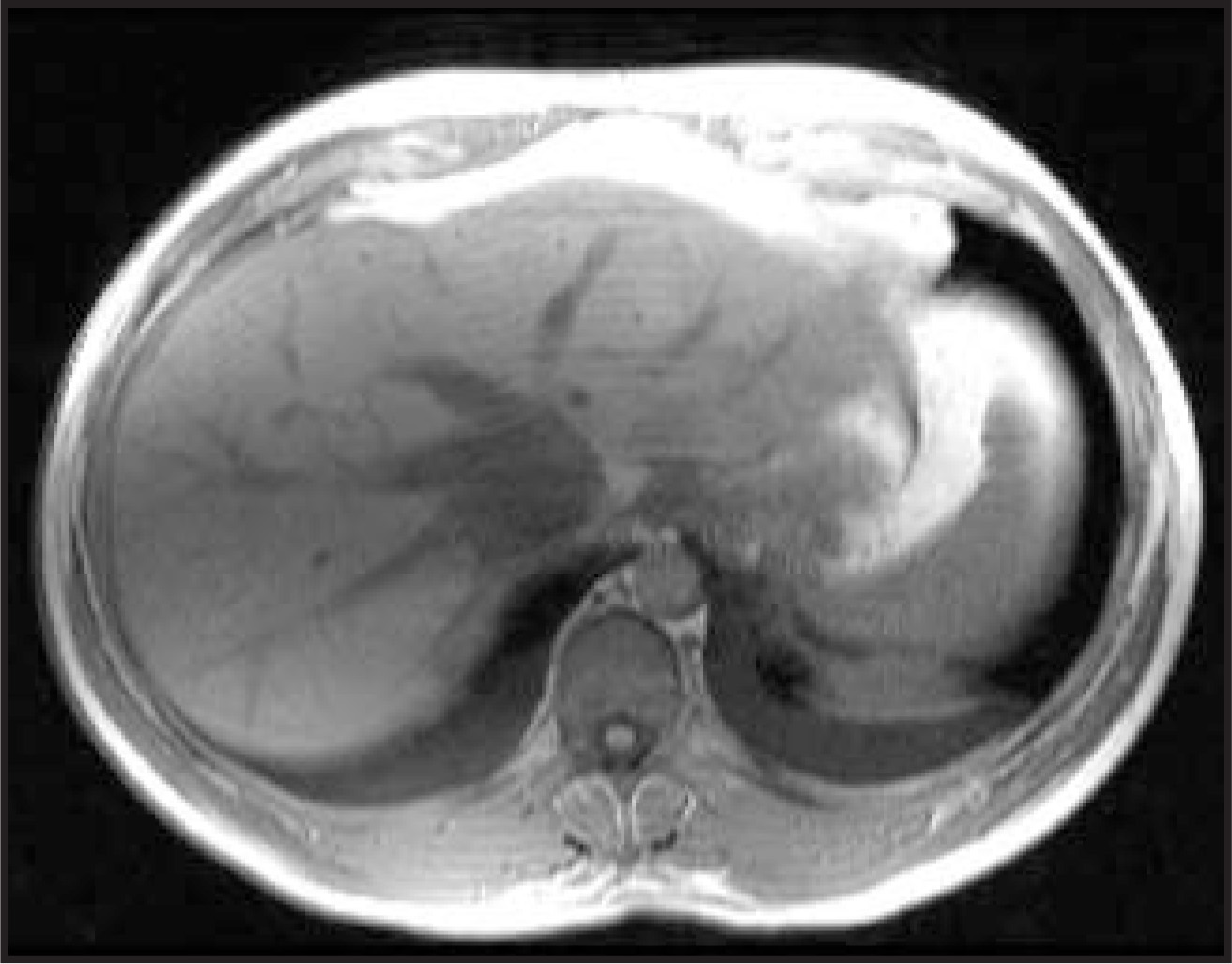
An anteroposterior portable chest radiograph showed a prominent heart (Figure 1). Using a LightSpeed Ultra multidetector computed tomography (CT) scanner (GE Healthcare, Waukesha, WI), a CT pulmonary angiogram was performed with intravenous infusion of contrast (Visipaque 320 mgI/mL, GE Healthcare, Princeton, NJ). The study included 2.5-mm–thick axial slices at 1.5-mm intervals obtained through the chest. No pulmonary artery or segmental artery embolism was identified. The right atrium was enlarged, and the inferior vena cava was prominent, which was suggestive of poor heart function (Figure 2). Intravenous reflux into the azygos vein and the inferior vena cava to the level of the hepatic venous system (Figure 2) was seen. The anterior pericardium was noted to be within the upper limits of normal for thickness, in association with punctate areas of calcification involving the anterior and left lateral pericardium (Figure 3). A radio graphic diagnosis of possible con strictive pericarditis was conveyed to his primary medical team. A magnetic resonance angiogram (MRA) of the abdomen was performed (Siemens 1.5T Vision MRI scanner, Siemens Medical Solutions, Malvern, PA), which showed a heterogeneous and enlarged proximal inferior vena cava, dilated hepatic veins, and bilateral pleural effusions (Figure 4). A cardiology consult, repeat echo cardiogram, and cardiac catheterization confirmed the diagnosis of moderately severe constrictive pericarditis. The patient subsequently underwent a pericardiectomy without complications. A postoperative visit found that he had made a full recovery and that his symptoms had improved.
DIAGNOSIS
Constrictive pericarditis
DISCUSSION
Constrictive pericarditis is an uncommon disorder. However, its incidence may begin to increase with the growing population of immunocompromised patients susceptible to tuberculosis infection and reactivation.1 Because chronic constrictive pericarditis can present with signs and symptoms similar to other disease processes such as chronic pulmonary emboli, Budd-Chiari syndrome, cirrhosis of the liver, or restrictive cardiomyopathy, the identification of diagnostic radiographic signs has become increasingly valuable.1-3 We report the clinical and imaging findings of a patient with unexplained leg edema and shortness of breath who eventually was diagnosed with constrictive pericarditis.
The normal pericardium is a double-layered sac that encases the heart and plays a role in ventricular filling.2 In the case of constrictive pericarditis, the pericardium becomes thickened and noncompliant.4,5 These pathologic changes to the pericardium cause the pressure in both ventricles to rise early in diastole, with elevation of the end-diastolic pressure and reduction in ventricular filling.2,5,6 The changes in normal heart physiology are the basis for increased central venous pressure.2,5 Although the cause of pericardial disease is often undetermined, it is important to remember that an asymptomatic or forgotten episode of viral pericarditis could have been the inciting event.5 Presenting signs may include intractable ascites, hepato megaly, and derangement of liver enzymes.1,5,7 This may lead a physician to suspect abdominal pathology and may lead to a mistaken diagnosis of cirrhosis of the liver, Budd-Chiari syndrome, or abdominal carcinomatosis.2,3 Given a clinical picture resembling the above disease processes, but with the added feature of distended neck veins and an elevated serumascites albumin gradient (≥1.1 g/dL), a careful search for pathophysiologic changes that involve the heart and lungs should be carried out.1,5
Given the limitations of cardiac catheterization and echocardiography, CT has become an important complementary and even first-line imaging tech nique for evaluating disease processes involving the pericardium.3,4,8 When compared with echocardiography, CT scanning is more accurate in establishing or excluding the presence of a thickened pericardium.3,5 An observed ≥3-mm thickness of the pericardium with the appropriate clinical signs on patient history and physical examination is virtually diagnostic of constrictive pericarditis.3 Since pericardial thickening can occasionally occur without seriously impairing ventricular filling, and since clinically significant thickening can go undiagnosed or be misinterpreted on CT imaging as motion artifact of the right ventricle in the presence of disease, secondary signs of constrictive pericarditis have become important in confirming its presence.3,5 This was especially true in the currently reported case in which impaired cardiac filling with dilatation of the inferior and superior vena cava and early reflux of contrast medium into dilated inferior vena cava and hepatic veins were seen but the pericardial thickness was within the upper limits of normal. Other secondary signs of constrictive pericarditis on CT include pleural effusions, intracardiac thrombi, and interventricular septal deformities.3,7 The presence of inferior and superior vena cava dilatation illustrates the elevated right atrial end-diastolic and end-systolic pressures present in constrictive pericarditis and right-sided heart failure.3 These findings should raise the clinical suspicion of constrictive pericarditis even when the pericardial thickening is not impressive.7 Lastly, the observation of passive hepatic congestion in a centrilobular distribution illustrates the impaired hepatic venous drainage.7 This is directly related to the elevated central venous pressure secondary to increased right atrial filling pressure. The poor compliance of the right side of the heart with diminished diastolic filling manifests as contrast reflux into the inferior vena cava and hepatic veins on CT.7 Other causes of contrast reflux include chronic pulmonary emboli, pulmonary hypertension, tricuspid regurgitation, and pulmonary valve stenosis.7 In the case of our patient, the presence of contrast medium reflux indicated a possible cardiac etiology in light of his abdominal signs and symptoms. This secondary sign of constrictive pericarditis, in conjunction with suspect characteristics of the pericardium and a medical history of an episode of viral pericarditis, led to the diagnosis of constrictive pericarditis.
CONCLUSION
The use of CT to evaluate disease of the heart and the pericardium is becoming more commonplace. With a clinical presentation of ascites, hepatomegaly, and minor derangement of liver enzymes, a cardiac cause can often be overlooked.6 By recognizing a simple sign, like contrast reflux into the inferior vena cava and hepatic veins, a clinician can confidently focus on a cardiac etiology. With pericardial thickness within the upper limits of normal, a clinician should consider constrictive pericarditis as part of the differential diagnosis when appropriate secondary signs are present.
References
- Van der Merwe S, Dens J, Daenen W. Pericardial disease is often not recognised as a cause of chronic severe ascites. J Hepatol. 2000;32:164-169.
- Brockington G, Zebede J, Pandian N. Constrictive pericarditis. Cardiol Clin. 1990;8:645-661.
- Suchet I, Horwitz T. CT in tuberculous constrictive pericarditis. J Comput Assist Tomogr. 1992;16:391-400.
- Hoit B. Imaging the pericardium. Cardiol Clin. 1990;8:587-600.
- Braunwald E, Fauci A, Kasper D. Harrison’s Principles of Internal Medicine. 2001:1370-1372.
- Wise D, Conti C. Constrictive pericarditis. Cardiovasc Clin. 1976;7:197-209.
- Holley H, Koslin D, Berland L, Stanley R. Inhomogeneous enhancement of liver parenchyma secondary to passive congestion: Contrast-enhanced CT. Radiology. 1989;170:795-800.
- Gouliamos A, Andreou J, Steriotis J. Detection of pericardial heart disease by computed tomography. Clin Radiol. 1984;35:397-400.
Citation
. Constrictive pericarditis. Applied Radiology. 2008;37(5):35-37. doi:10.37549/AR1611.