Contemporary treatment of varicose veins
Applied Radiology — Vol. 33 , Issue 12 , pp. 19 -22
DOI: 10.37549/AR1299
Published: December 1, 2004
Categories
New minimally invasive techniques for ablation of the greater saphenous vein (GSV) and short saphenous vein (SSV) are an exciting part of interventional radiology practice today. An increasing number of devices are becoming available to accomplish this task. Both radiofrequency (RF) ablation and laser energy are being used effectively to ablate incompetent segments of the GSV and SSV with less pain and morbidity than surgical ligation and stripping. This overview should help to familiarize interventionalists with the major devices now commercially available for percutaneous treatment of varicose veins.
Saphenous vein (SV) valvular incompetence is one of the most common causes of venous hypertension and symptomatic varicosities (Figure 1). Along with the visible varicosities, patients will frequently present with associated complaints of lower extremity aching, pain, fatigue, swelling, throbbing, and superficial thrombophlebitis. More chronic sequelae of venous hypertension that are also indications for treatment include lipodermatosclerosis, hyperpigmentation, and skin ulceration.1 The object of treatment in these situations is elimination of the refluxing, incompetent portions of the SV.2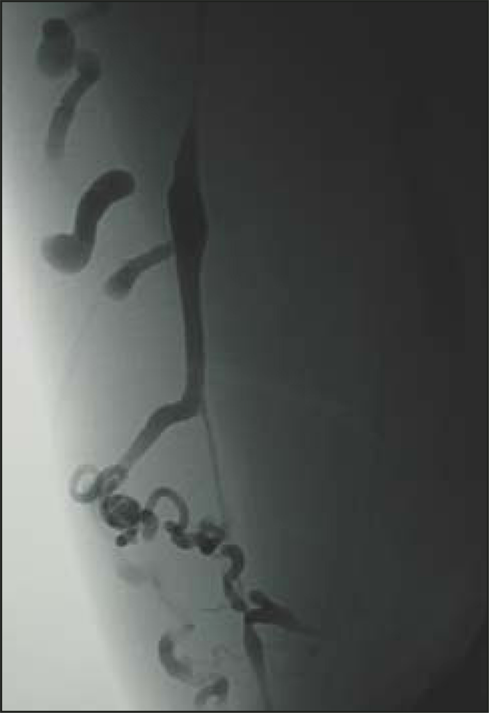
Before treatment can be performed, confirmation of valvular dysfunction and overall evaluation of superficial and deep venous systems must be completed. These objectives are best achieved with duplex ultrasound. While the absolute necessity of duplex ultrasound in the workup of chronic venous insufficiency has been debated, it offers great accuracy and efficiency.3 It is likely to enjoy much greater usage with the expansion of image-guided procedures in venous disease.
Duplex ultrasound evaluation achieves some crucial goals.3,4 First, it confirms the diagnosis of valvular incompetence and reflux. Second, the incompetent venous segments and branches are mapped for identification and future intervention (Figure 2). Finally, deep veins are evaluated to exclude thrombosis or occlusion, verifying that the refluxing superficial pathways are not the primary route of blood return for the extremity.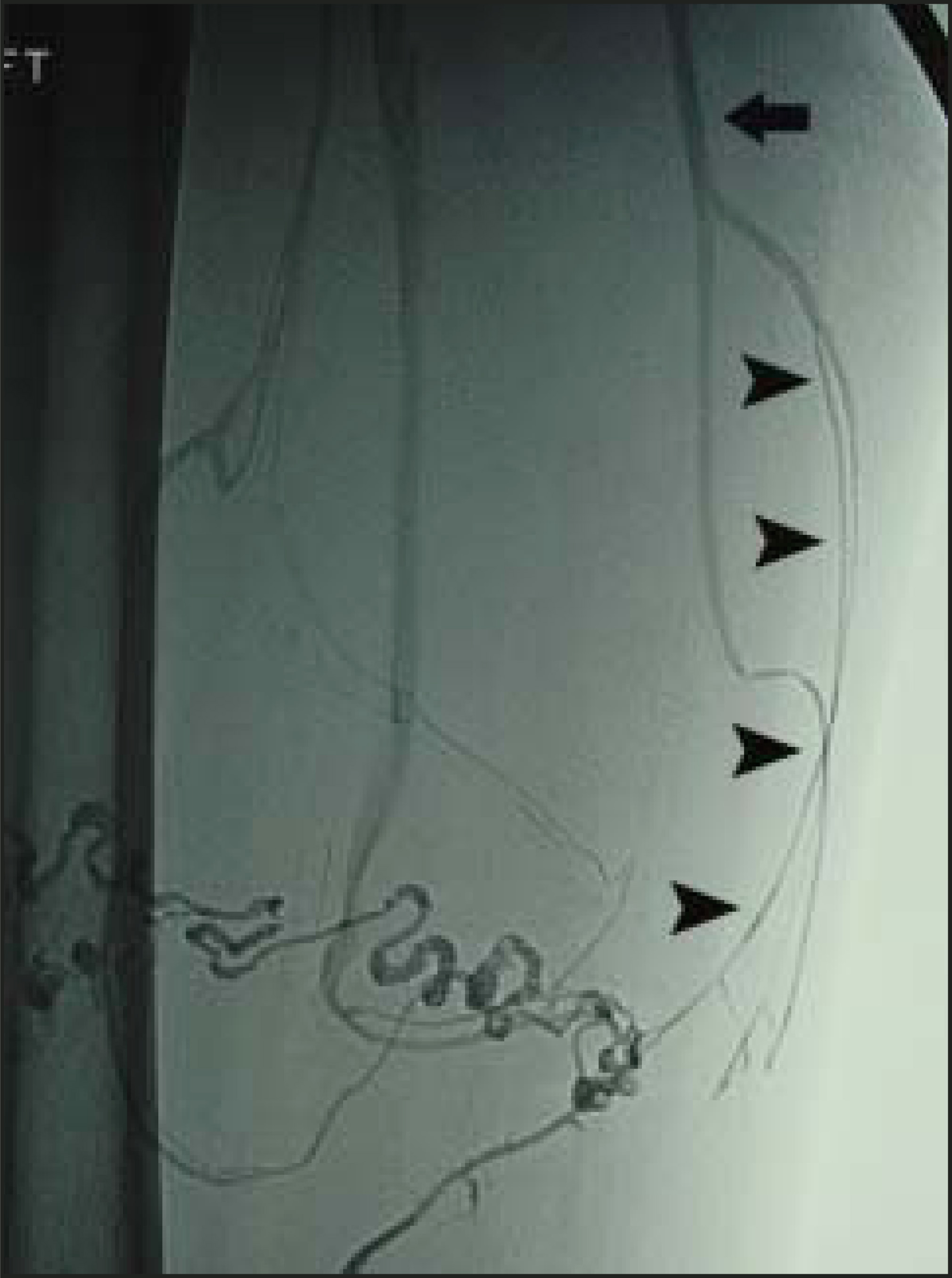
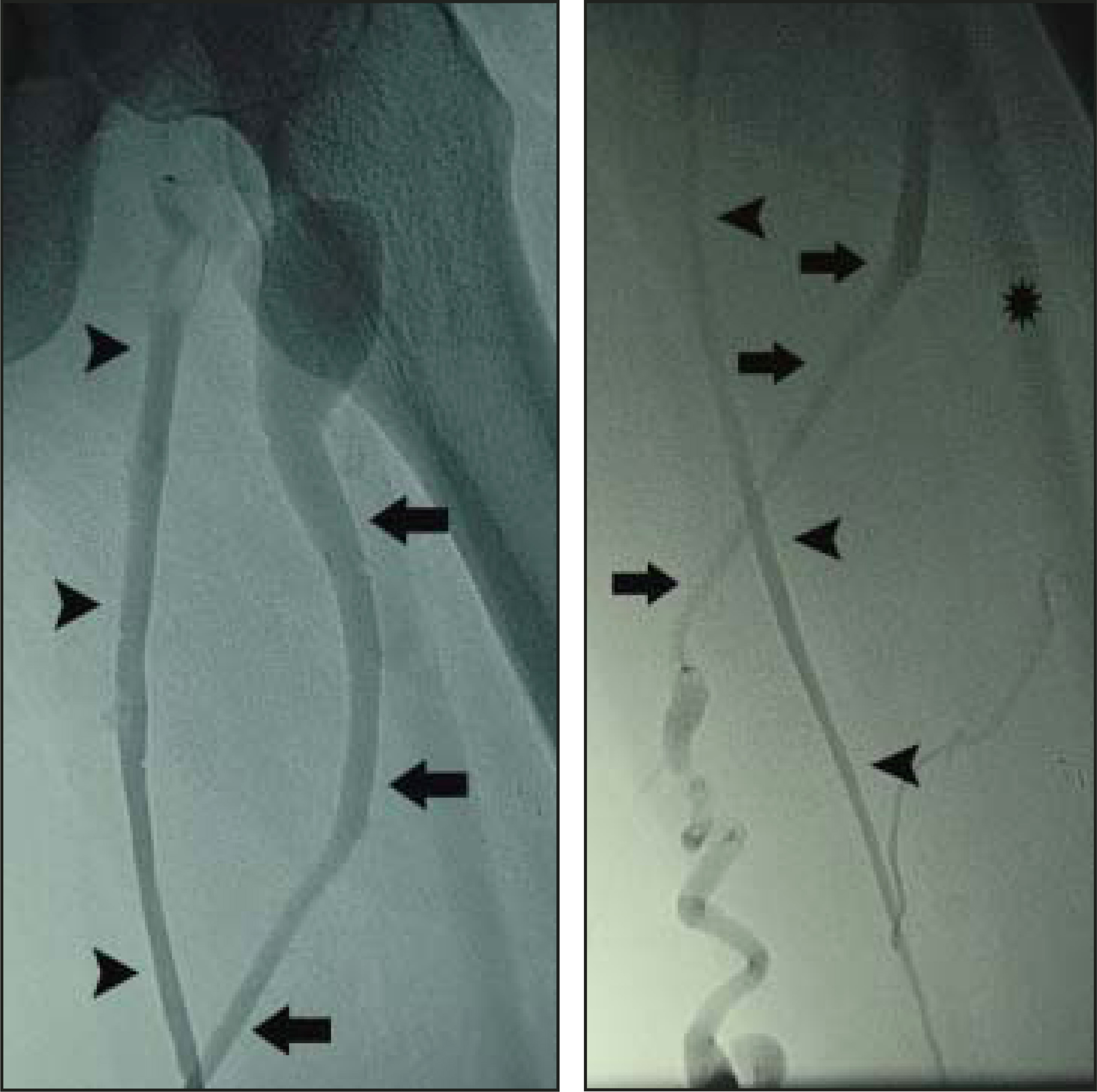
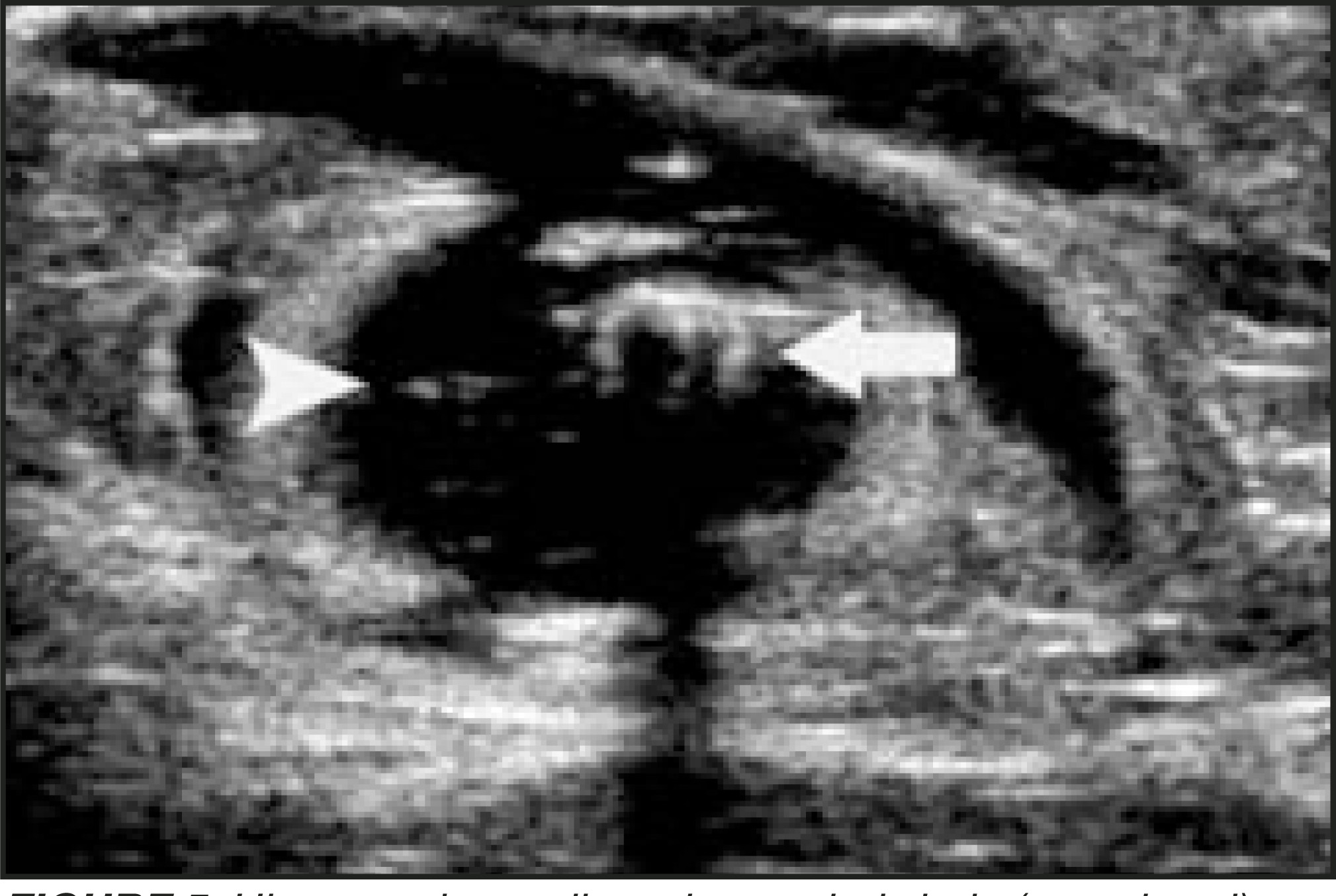
Laser and RF techniques for ablation of the SV share some important common features. In particular, delivery of perivenous tumescent anesthesia after venous cannulation and device placement is a cornerstone of safe and effective treatment.5 The incompetent SV is marked on the skin from saphenofemoral junction (SFJ) or saphenopopliteal junction (SPJ) to the intended entry site, which is usually just above or below the knee or mid-calf. Ultrasound (US)-guided (7 to 10 MHz) percutaneous access into the SV is performed through a small skin nick after initial local anesthesia is provided. Sheath placement (5F to 8F, depending on device) is accomplished over a guidewire, and the device tip is positioned just below (5 to 10 mm) the SFJ or SPJ (Figure 3). Occasionally, fluoroscopy and contrast venography is useful in difficult situations (Figure 4). A total of 100 to 200 mL of 0.1 to 0.2% lidocaine is infiltrated with a 25-g needle under US guidance into the perivenous space. Correct delivery results in a hypoechoic halo that expands the perivenous space around the compressed SV (Figure 5). This achieves 3 objectives: 1) The device activation is essentially painless; 2) the vein wall is effectively compressed around the device for direct delivery of heat energy so that one is not just heating blood alone; and 3) the perivenous tissues are insulated from the heat source to prevent damage to the surrounding structures, especially skin and nerves.5 A class II (30 to 40 mm Hg) compression stocking is applied for at least 1 week immediately after the procedure to exclude blood from the superficial venous system and to maintain coaptation of the vein walls in the treated venous segment during the initial healing process.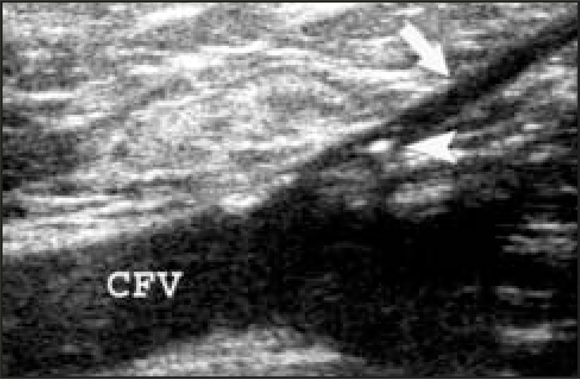
Laser ablation
Since endovascular delivery of laser energy to ablate large varicosities was first described by Dr. Carlos Boné6 in 1999, a number of laser devices have become available for use. They share the common goal of causing thermal damage to the vein wall, resulting in endothelial denudation, vein wall thickening, and, finally, fibrosis. A key mechanism of such heat injury has been shown to be intravascular blood serving as the chromophore for absorption of specific wavelengths of energy. Proebstle et al7 have reported that laser wavelengths of 810, 940, and 980 nm similarly generate steam bubbles in intravascular blood that effectively transfer heat to the vein wall.
Correspondingly, 3 laser devices are currently available for use. The most extensive clinical results are currently reported for the 810-nm diode laser (Diomed, Andover, MA). This 600-μm laser fiber is inserted through a 5F sheath and pulled back through the SV at 2 to 3 mm/sec (12 to 18 cm/min) with a power of 14 W in continuous mode. Of 500 limbs reported by Min et al,5,8 499 were available for follow-up. The treated segment of GSV was closed in 98% at 1 month, 97.5% at 1 year, and 93.4% at 2 years. A total of 49 GSVs were followed for 3 years with no new recurrences. No skin burns, paresthesias, or complications of deep vein thrombosis (DVT) were reported. Earlier reports by the same author in 84 patients for a 9-month period showed only a transient paresthesia at the mid-calf in 1 patient, which resolved after 6 weeks.9
Another device available for GSV closure is a 940-nm diode laser (Dornier MedTech, Kennesaw, GA). A bare-tipped 600-μm fiber is used through a 5F catheter or sheath in the manner already described. Energy can be delivered in a pulsed or continuous mode at a 15-W power setting. The safety and 1-month efficacy were shown in 31 limbs with a 97% 1-month occlusion rate.10 There were no major complications. Minor complications included 2 patients with superficial thrombophlebitis at 5 days status after endovenous laser therapy and 1 patient with hyperpigmentation that was still apparent at 4 weeks. A more recent study used a pulsed/intermittent pullback technique in 203 limbs (154 GSVs, 37 lesser saphenous veins, and 12 accessory saphenous veins) to achieve a 97% clinical success rate.11 No DVT, nerve injury, or skin burn resulted. Additional in vitro and in vivo investigation of the mechanism of laser injury to the vein wall has been elegantly performed with the 940-nm device. This work has elucidated the key role of intravascular blood and steam bubble formation in distributing thermal damage to all parts of the inner vein wall.7
The third major laser device currently available is the 980-nm diode laser (Angiodynamics, Queensbury, NY). Energy is also delivered through a bare 600-μm fiber placed through a 5F sheath. Parameters are a power of 14 W delivered with continuous mode and pullback at 2 to 3 mm/sec. Based on the mechanism of action already described, one would not expect significant differences from above data regarding device safety and efficacy. An initial study of 13 GSVs treated showed 100% closure rate at 3 months postintervention with no significant complications.12 This device received 510(k) exemption in 2002 and has been available for use since that time.
Endovenous RF ablation
Endovenous RF ablation is the second major technique for minimally invasive obliteration of the GSV. There is a single device available—the Closure System (VNUS Medical Technologies, San Jose, CA). The Closure catheter employs a collapsible electrode, which contacts and slightly embeds itself into the vein wall.1 Dilute heparin solution is infused through the catheter lumen to inhibit coagulum formation on the electrodes. A 5F catheter is used for veins 2 to 8 mm in diameter. An 8F system is reserved for larger GSVs up to 12 mm diameter. Tumescent anesthesia is placed and is a key aspect of safety and effectiveness, similar to endovenous laser therapy. The RF unit has a feedback mechanism that allows it to deliver the minimum heat necessary (85˚C) localized around the active electrode, where it maintains contact with the vein wall. The catheter tip is positioned just below the SFJ and an impedance and temperature check performed. Impedance of the vein wall should be 200 to 300 ohms with a baseline temperature of 33°C to 37° C. A target temperature of 85° C is reached in approximately 15 seconds. The Closure catheter is then withdrawn 4 cm during a period of 3 minutes, followed by a rate of 2.5 cm/min (impedance of 150 to 200 ohms and transmitted temperature of 85°C ± 3°C). The mechanism of action is collagen contraction in the vein wall, which immediately and significantly reduces the lumen of the vein. Endothelial denudation also occurs. The controlled injury ultimately produces a fibrotic rather than thrombotic seal of the vein lumen. Early reports of Closure treatment have had a 90% success rate at 4.7 months (6% recurrent reflux and 4% recurrent varices).13 Initial complications included skin burns (3%), DVT (3%), 1 pulmonary embolus, and transient paresthesia (thigh 9%, leg 51%).
The addition of generous tumescent anesthesia in a more recent report of longterm data has resulted in significant improvements. Weiss14 reported a 90% successful outcome at 2-year follow-up (19 of 21 patients) with no incidents of skin burn or DVT. Transient paresthesia rate was reduced to 8.5%.
Comparison of techniques
While RF and laser-mediated ablation of the GSV have both shown safety and efficacy, some experts disagree over which technique is preferable. Proponents of RF ablation favor the feedback mechanism of the Closure device to precisely control the energy delivered to the vein wall. This feature may be worth the slightly longer pullback time required, as compared with the lasers. On the other hand, some critics point out that the risk of heat injury to perivenous tissues (ie, skin and nerve) is higher with RF energy due to direct conduction from the vein walls in contact with the electrode.5 A potential disadvantage of the laser is nonuniform distribution of heat energy to the vein wall that has been identified in some histopathologic data.15 This could possibly lead to recanalization of the vein and less reliable results than could be achieved with RF. Issues of microperforation (with a pulsed technique) causing increased postprocedure bruising and pain have also been raised. Additional recent evidence has found more uniform circumferential thermal damage to the vein wall due to generation of steam bubbles.7 Refinement in technique with continuous, rather than intermittent, withdrawal of the laser fiber has led to decreased ecchymosis and postprocedural pain. Additional laser research with a diffusing fiber that distributes its heat in a more uniform cylindrical fashion may completely resolve this potential issue in the future.16
Conclusion
Discussion of minor potential drawbacks of either device is intriguing; however, these points should not be overshadowed by the evidence that both RF and laser obliteration of the GSV have shown effectiveness at 2 years, with results that are equivalent to surgery with a much less invasive approach and with decreased morbidity. There will likely be continued refinements in the technology and application of both of these modalities, including the use of tumescent anesthesia. We can all feel encouraged by the present data and look forward to advancements in treating this extremely prevalent medical condition.
References
- Weiss R, Feied C, Weiss M. Vein Diagnosis & Treatment—A Comprehensive Approach. 2001:28-30.
- Bergan J, Kumins N, Owens E, Sparks S. Surgical and endovascular treatment of lower extremity venous insufficiency. J Vasc Interv Radiol. 2002;13:563-568.
- Khilnani N, Min R. Duplex ultrasound for superficial venous insufficiency. Tech Vasc Interv Radiol. 2003;6:111-115.
- Zwiebel W. Introduction to Vascular Ultrasonography. 2000:287-296.
- Min R, Khilnani N. Endovenous laser treatment of saphenous vein reflux. Tech Vasc Interv Radiol. 2003;6:125-131.
- Navarro L, Min R, Bone C. Endovenous laser: A new minimally invasive method of treatment for varicose veins—Preliminary observations using an 810 nm diode laser. Dermatol Surg. 2001;27:117-122.
- Proebstle T, Sandhofer M, Kargl A. Thermal damage of the inner vein wall during endovenous laser treatment: Key role of energy absorption by intravascular blood. Dermatol Surg. 2002;28:596-600.
- Min R, Khilnani N, Zimmet S. Endovenous laser treatment of saphenous vein reflux: Long-term results. J Vasc Interv Radiol. 2003;14:991-996.
- Min R, Zimmet S, Isaacs M, Forrestal M. Endovenous laser treatment of the incompetent greater saphenous vein. J Vasc Interv Radiol. 2001;12:1167-1171.
- Proebstle T, Lehr H, Kargl A. Endovenous treatment of the greater saphenous vein with a 940-nm diode laser: Thrombotic occlusion after endoluminal thermal damage by laser-generated steam bubbles. J Vasc Surg. 2002;35:729-736.
- Perkowski P, Ravi R, Gowda R. Endovenous laser ablation of the saphenous vein for treatment of venous insufficiency and varicose veins: Early results from a large single-center experience. J Endovasc Ther. 2004;11:132-138.
- Oh C, Jung D, Jang H, Kwon K. Endovenous laser surgery of the incompetent greater saphenous vein with a 980-nm diode laser. Dermatol Surg. 2003;29:1135-1140.
- Manfrini S, Gasbarro V, Danielsson G. Endovenous management of saphenous vein reflux. J Vasc Surg. 2000;32:330-342.
- Weiss R, Weiss M. Controlled radiofrequency endovenous occlusion using a unique radiofrequency catheter under duplex guidance to eliminate saphenous varicose vein reflux: A 2-year follow-up. Dermatol Surg. 2002;28:38-42.
- Weiss R. Comparison of endovenous radiofrequency versus 810 nm diode laser occlusion of large veins in an animal model. Dermatol Surg. 2002;28:56-61.
- Parente E, Rosenblatt M. Endovenous laser treatment to promote venous occlusion. Lasers Surg Med. 2003;33:115-118.
Citation
. Contemporary treatment of varicose veins. Applied Radiology. 2004;33(12):19-22. doi:10.37549/AR1299.