Current status of breast ultrasound
Applied Radiology — Vol. 33 , Issue 9 , pp. 16 -21
DOI: 10.37549/AR1278
Published: September 1, 2004
Categories
The purpose of this review is to highlight important historical points in the development of breast ultrasound. The topics addressed will include determining the sonographic criteria for simple cysts, the differentiation of benign and malignant solid breast lesions, and the value of screening ultrasound to detect breast cancer.
The simple cyst
The use of ultrasound in the management of breast masses began in the 1970s. One of several studies at that time was performed by Texidor1 with B-mode ultrasound. This study determined that ultra- sound was safe and highly accurate in differentiating simple cysts > 1 cm from solid masses of the breast.
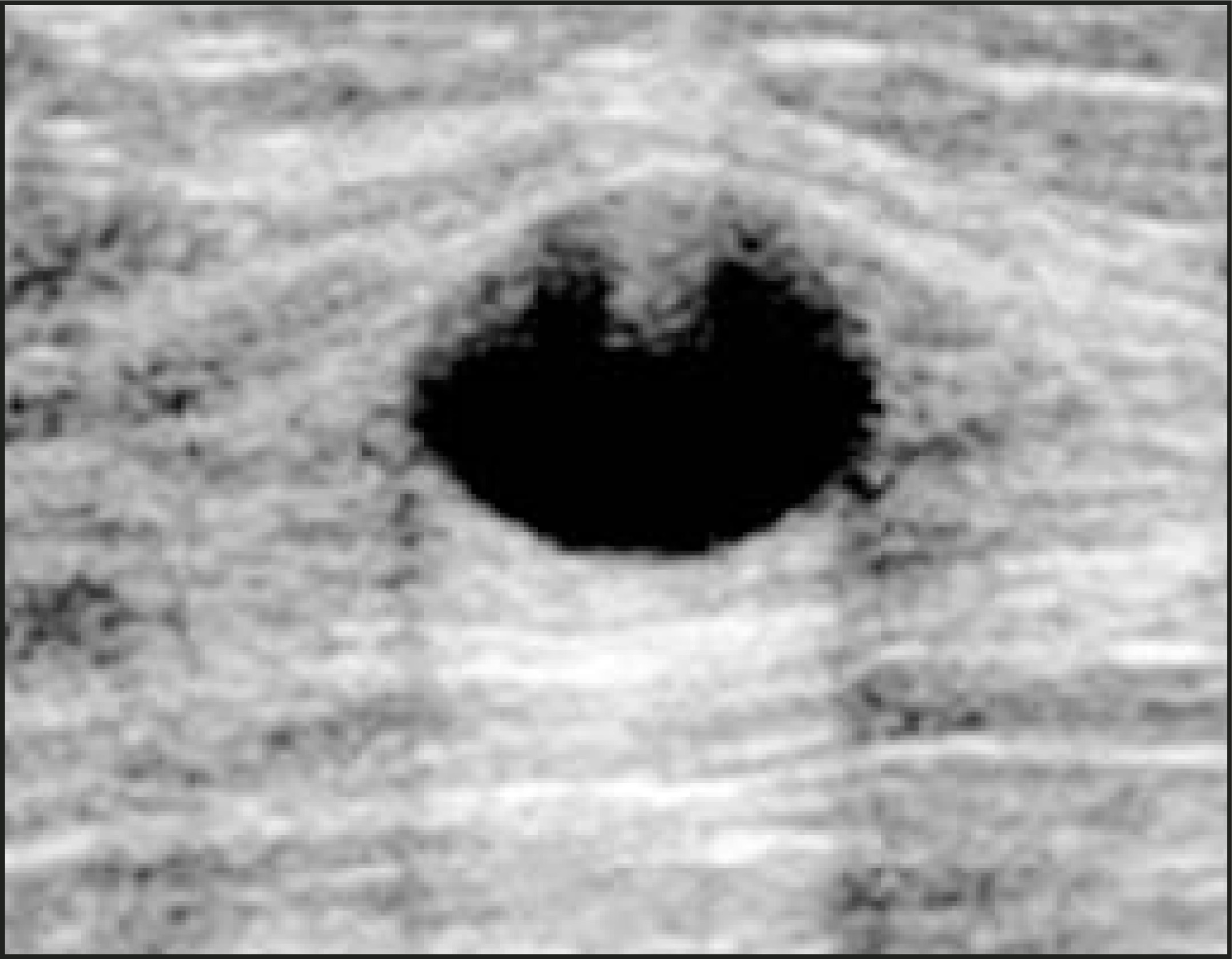
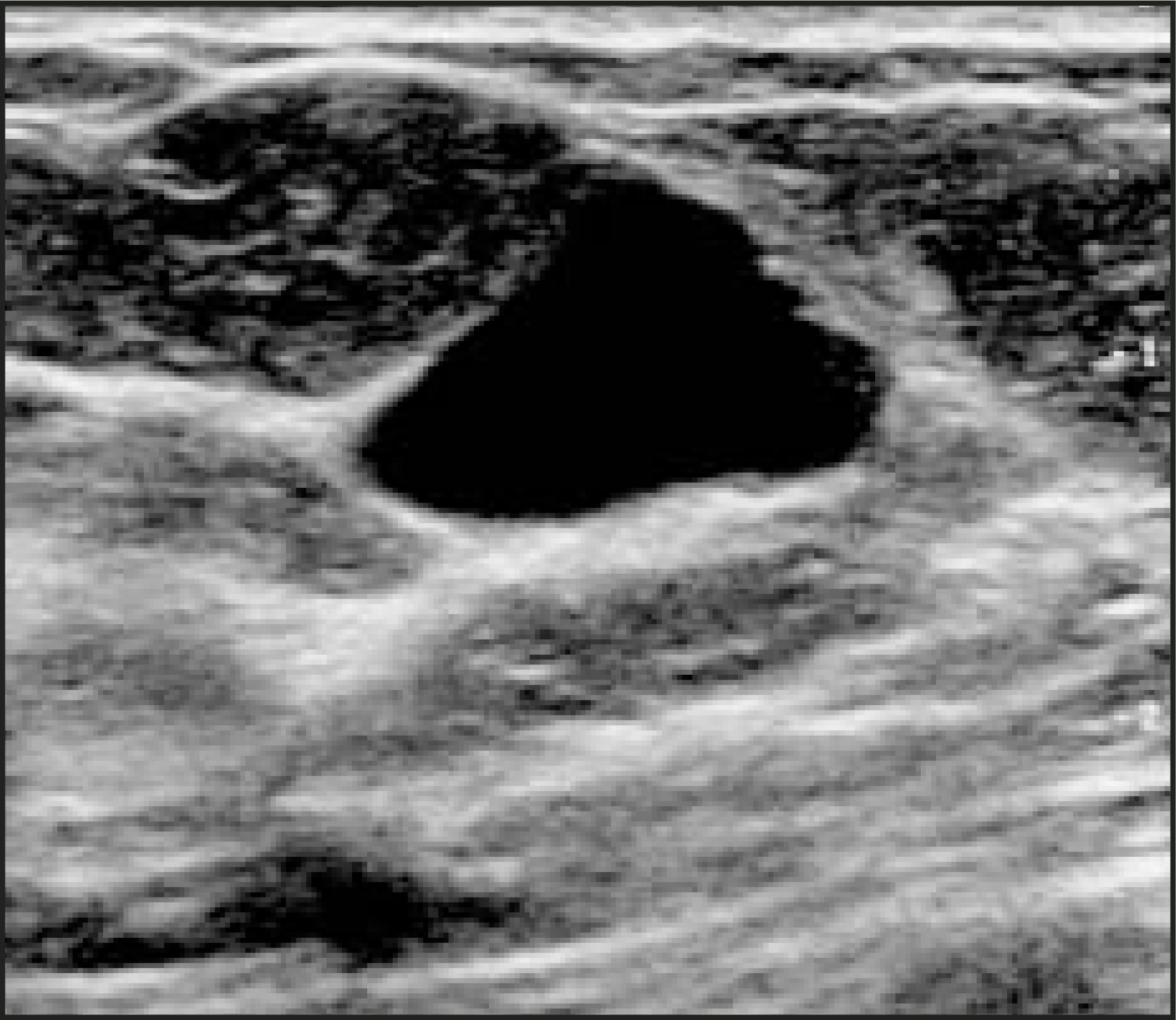
In the mid-1980s, the seminal article on simple cysts, on which current practice of breast ultrasound is founded, was reported by Hilton.2 In this study, 300 consecutive patients were evaluated with real-time ultrasound to define the characteristics of simple cysts. These authors determined that ultrasound could reliably diagnose simple cysts, obviating the need for surgical or needle intervention. The criteria proposed in that article have withstood the test of time and include: 1) round or oval shape; 2) anechoic; 3) well-defined margins, particularly the posterior wall; and 4) posterior enhancement. Two important points were noted in Hilton’s work. First, reverberation echoes were occasionally present in the anterior field of a simple cyst. These echoes are artifactually produced due to the physics of sound and tissue interfaces and should not be confused with real echoes in complicated cysts (Figure 1). Second, in 25% of cases, posterior enhancement was not present on all images (Figure 2). Therefore, a simple cyst can still be diagnosed even if this feature is absent. Note that the remaining criteria should be strictly applied in order to establish the diagnosis of simple cyst.
Solid masses: Benign/malignant differentiation
At a time when the prevailing sentiment was that ultrasound could not distinguish between benign and malignant solid breast masses, Fornage3 was evaluating the fibroadenoma. He noted that the length to anteroposterior (AP) diameter was >1.4 in 86% of fibroadenomas, and <1.4 in 100% of 28 carcinomas. In 1995, Stavros4 evaluated 750 solid breast masses to attempt to differentiate benign from malignant or indeterminate lesions. The criteria for malignant masses were: angular margins, spiculation, marked hypoechogenicity, shadowing, microlobulation, duct extension, branch pattern, and calcification. The criteria for benignity were no malignant findings, and combinations of the following features: intense hyperechogenicity, ellipsoid in shape, up to 3 macrolobulations, and a thin pseudocapsule. For indeterminate lesions, the criteria were: isoechogenicity, maximum lesion diameter, mild hypoechogenicity, normal sound transmission, enhanced transmission, heterogeneous texture, and homogeneous texture. More specifically, if there were no malignant characteristics and none of the combinations of benign criteria was present, then the lesion was placed in the indeterminate category. Among 625 pathologically benign masses, this study correctly classified 424 as benign (68% true negative) and 201 as malignant or indeterminate (32% false positive). Among 125 pathologically malignant masses, 123 were correctly classified as malignant (98.4% true positive) and 2 were incorrectly classified as benign (1.6% false negative). The results indicated that their classification scheme could be accurately applied to distinguish between benign and malignant solid breast masses.
However, there were important limitations to the Stavros study.4 First, the readers of the ultrasound images were not blinded to the results of corresponding mammograms. Although one may argue that this methodology mirrors clinical practice, this method cannot adequately test a given classification scheme without introducing bias. Moreover, the data were not reported with consideration of age stratification. Mean age in this study was 47 years, with a range of 18 to 88 years. One must question whether the same classification scheme would prove as effective if it were applied to a postmenopausal age group, in which fibroadenomas are uncommon and the prevalence of malignancy is peaking. The results did not describe whether detected masses were new or if interval change could be documented. Lastly, one should question whether the criteria are equally accurate if applied to palpable lesions. Of the 125 breast cancers evaluated, 44 (35%) were palpable. One of the two false-negative cases was also palpable. One should keep these limitations in mind when applying the criteria proposed in this study.
There have been several corroborating studies, only two of which are highlighted in this review. In 1999, Buchberger5 reported the prospective evaluation of 687 symptomatic patients that were identified with either a palpable or a mammographically detected mass with exclusion of simple cysts. There were 353 masses remaining that were categorized into the 3-way classification scheme proposed by Stavros.4 There were no malignant lesions among the 109 lesions prospectively diagnosed as benign. There were 2 cancers among 141 masses called indeterminate and 26 cancers in 103 called malignant. Thus, there were no false-negative results, but again the readers were not blinded to corresponding mammograms.
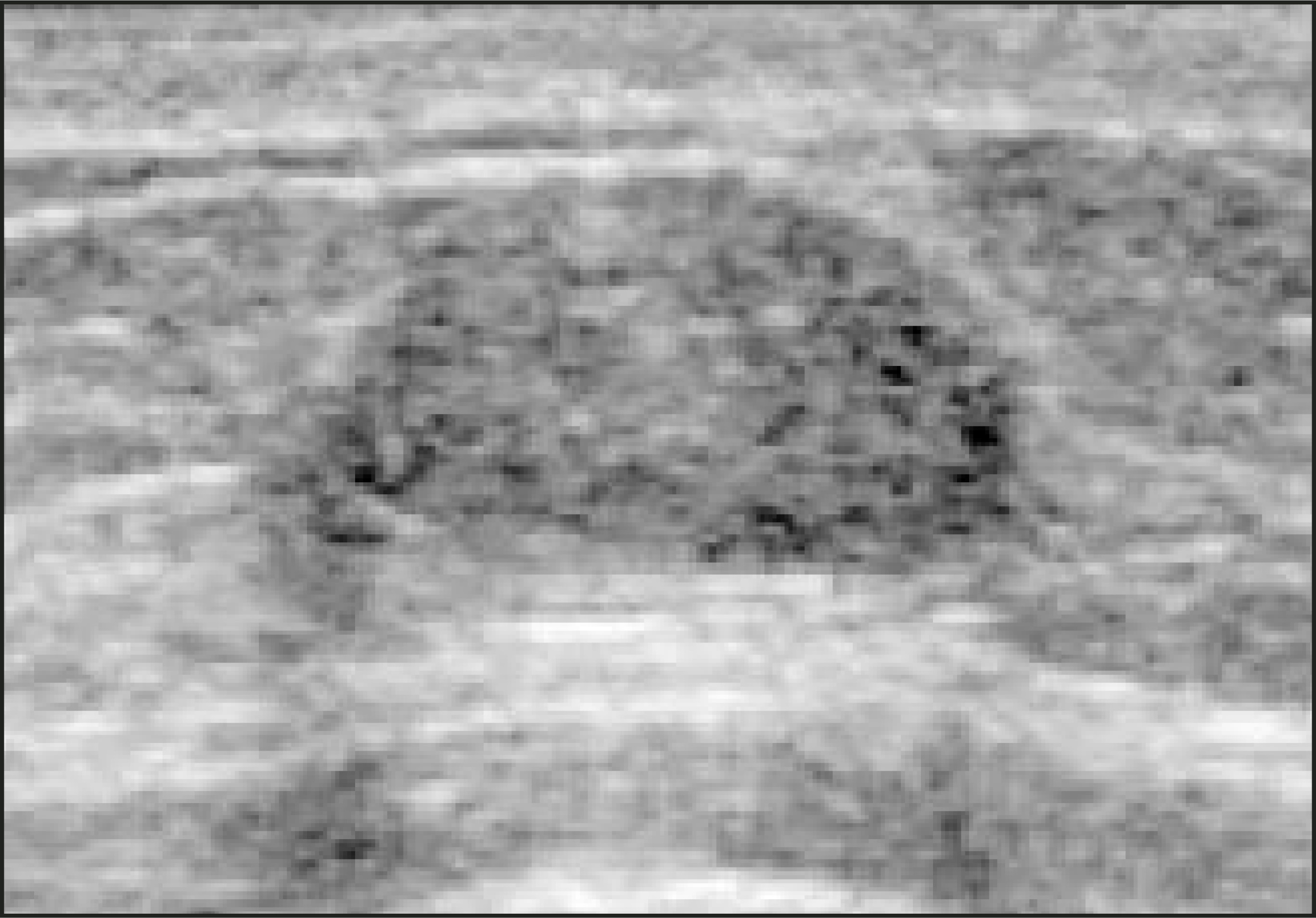
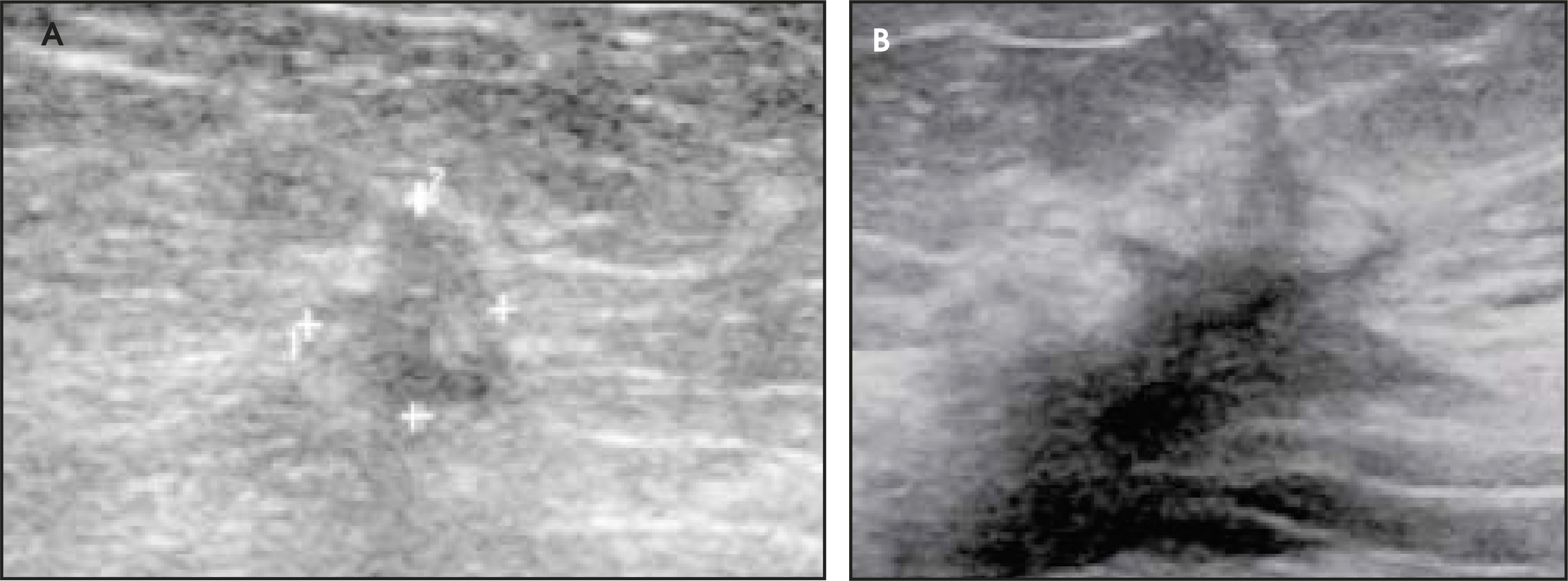
Another study by Rahbar,6 combining results from the laboratories of Lawrence Bassett at the University of California at Los Angeles and Valerie Jackson at the University of Indiana, reported on their experience attempting to differentiate benign from malignant solid masses. This was a retrospective review that evaluated the applicability of all literature-based benign/malignant imaging criteria and interobserver variability in sonographic differentiation of benign from malignant lesions. There were 162 masses reported in 161 consecutive patients who had undergone biopsy. A total of 118 (73%) of the masses were palpable, and 38 (23%) were malignant. Mammograms were available for 133 of the masses and for 32 of the cancers. The masses were evaluated in 3 phases: the first phase was with ultrasound only, the second phase was with mammography only, and the third phase, performed after time had passed, was with mammography and ultrasound results together. The specific sonographic features that were tested included shape, margins, length:AP ratio,3 echogenicity and echo texture, presence of calcifications, lateral edge shadowing, and presence of a pseudocapsule. The original report by Stavros et al4 stressed this latter feature as an important one for a benign lesion. The results showed that the features most predictive of benignity were an oval/round shape, circumscribed margins, edge refractory shadowing, and a length:AP ratio >1.4 (Figure 3). The hyperechoic, well-circumscribed lesion, which is most commonly a lipoma or fat necrosis, correlated well with benignity but occurred too infrequently to be generalized. The pseudocapsule also predicted benignity but was subject to a low interobserver agreement. Spiculation, microlobulation, ill-defined margins, irregular shape, and length:AP ratios <1.4 were the best predictors of malignant masses (Figure 4). If the 3 most reliable criteria had been applied to predict benignity, the malignant biopsy yield rate would have increased by 16% from 23% to 39%. The ultrasound contribution to mammography would have increased the biopsy yield rate by 2%, but this improvement was not statistically significant. The major limitation of this study was that the sonographic images were reviewed from hard copy and not in real time.
In summary, these studies support the use of ultrasound with state-of-the-art equipment to distinguish between benign and malignant solid breast masses. One should be aware of the limitations of the studies that propose these various classification schemes. If one chooses to practice based on the results of these studies, this author encourages each radiologist to read the original reports and to perform a self-audit of one’s practice to ensure that the criteria studied are being applied accurately.
Ultrasound “screening” for breast cancer: Review of the literature and controversies
The largest study to date reporting sonographic screening to detect malignancies is the 1995 study by Gordon and Goldberg7 that involved 12,706 patients. These patients were all symptomatic in that they either underwent scanning for a palpable lesion or for a mammographically detected mass. In addition to the focal area of interest, the investigators scanned the entire breast. Of the entire patient cohort there were 1575 (12.4%) solid masses in which 44 malignancies were detected by ultrasound among 30 patients. Half of these patients had primary malignant lesions. There was a 0.3% prevalence of sonographically detected malignancies among the 12,000 patients.
Reports of ultrasound screening for breast cancer are summarized below. The study of Buchberger,5 cited above, also reported on the prevalence of malignancies detected by ultrasound screening among 6113 asymptomatic patients. Pa-tients with only fat tissue density by mammography were excluded. There were 23 (0.3%) occult cancers among the 6113 patients. Kolb8 evaluated 3626 women with negative mammograms and normal physical examinations. The prevalence of breast cancer was also 0.3% (11 of 3626). Other “screening” ultrasound studies have shown similar results: Kaplan9: 0.3% (3 of 1862); Kolb et al10: 0.27% (37 of 13,547); Leconte et al11:0.5% (16 of 3084); and Crystal et al12: 0.46% (7 of 1517). All of these studies were performed with the investigator having initial knowledge of the appearance of the mammogram. Therefore, bias was introduced into all of these sonography-based studies.
In contrast, the AVON/ACRIN study, which opened in May 2004 with Berg13 as the primary investigator, is a multi-institutional study in which screening ultrasound will be performed and interpreted before screening mammography is obtained. The patient population will be asymptomatic women at high risk for breast cancer. These results will be important to understand the influence of ultrasound on management and outcome independent of mammographic results.
Do these studies “prove” benefit?
A significant question remaining is whether the detection of malignancies by sonography will lead to a reduction in breast-cancer–related mortality, as has been demonstrated for mammography on multiple occasions. A randomized, controlled trial comparing patient survival with and without screening sonography is the only scientific method that can provide evidence of mortality reduction.14,15 Such a study has never been performed with ultrasound and may be cost prohibitive. Discussions regarding “surrogate endpoints” have been debated for years.16 Feig16 defined these surrogate end points as “a factor or group of factors that accurately predict outcome that can be substituted for waiting for death, and used to determine efficacy more rapidly.” In other words, studies using mortality as the end point of measure are very long and very expensive. Surrogate end points may be alternative methods of measure.
The reason that detection of these incidentally found lesions may not affect mortality is that it is not known whether these lesions grow so slowly that they will never become clinically significant or whether the converse is true—that these lesions, while small, are already metastatic so that their detection is not life-saving. Additionally, the patient population being studied in a screening trial is healthy and asymptomatic. Screening for malignancies comes with additional radiation-associated risks, and any mortality benefit may not be great enough to add that risk to a healthy individual. General concepts concerning breast cancer screening are well described by Kopans in “Screening for Cancer—When is it Valid ? Lessons from the Mammography Experience.”14
Conclusion
This article has provided a historical perspective on breast ultrasound. The use of ultrasound is an important adjunctive tool to mammography and physical examination in the evaluation of breast cancer. One’s practice should be founded on combining both academic scientific results and personal experience. This article is intended to provide an objective perspective from which radiologists can draw to apply to their own practices.
References
- Texidor H. The use of ultrasonography in the management of masses of the breast. Surg Gynecol Obstet. 1980;150:486-490.
- Hilton S, Leopold G, Olson L, Willson S. Real-time breast sonography: Application in 300 consecutive patients. AJR Am J Roentgenol. 1986;147:479-486.
- Fornage B, Lorigan J, Andry E. Fibroadenoma of the breast: Sonographic appearance. Radiology. 1989;172:671-675.
- Stavros A, Thickman D, Rapp C. Solid breast nodules: Use of sonography to distinguish between be- nign and malignant lesions. Radiology. 1995;196:123-134.
- Buchberger W, DeKoekkoek-Doll P, Springer P. Incidental findings on sonography of the breast: Clinical significance and diagnostic workup. AJR Am J Roentgenol. 1999;173:921-927.
- Rahbar G, Sie A, Hansen G, Prince J. Benign versus malignant solid breast masses: US differentiation. Radiology. 1999;213:889-894.
- Gordon P, Goldenberg S. Malignant breast masses detected only by ultrasound, a retrospective review. Cancer. 1995;76:626-630.
- Kolb T, Lichy J, Newhouse J. Occult cancer in women with dense breasts: Detection with screening US—diagnostic yield and tumor characteristics. Radiology. 1998;207:191-199.
- Kaplan S. Clinical utility of bilateral whole-breast US in the evaluation of women with dense breast tissue. Radiology. 2003;227:607-608.
- Kolb T, Lichy J, Newhouse J. Comparison of the performance of screening mammography, physical examination, and breast US and evaluation of factors that influence them: An analysis of 27,825 patient evaluations. Radiology. 2002;225:165-175.
- Leconte I, Feger C, Galant C. Mammography and subsequent whole-breast sonography of nonpalpable breast cancers: The importance of radiologic breast density. AJR Am J Roentgenol. 2003;180:1675-1679.
- Crystal P, Strano S, Shcharynski S. Using sonography to screen women with mammographically dense breasts. AJR Am J Roentgenol. 2004;182:259-260.
- Berg W. Rationale for a trial of screening breast ultrasound: American College of Radiology Imaging Network (ACRIN) 6666. AJR Am J Roentgenol. 2003;180:1225-1228.
- Kopans D, Monsees B, Feig S. Screening for cancer—When is it valid? Lessons from the mammography experience. Radiology. 2003;229:319-327.
- Kopans D. Sonography should not be used for breast cancer screening until its efficacy has been proven scientifically. AJR Am J Roentgenol. 2004;182:489-491.
- Feig S. Determination of mammographic screening intervals with surrogate measures for women aged 40-49 years. Radiology. 1999;193:311-314.
Citation
. Current status of breast ultrasound. Applied Radiology. 2004;33(9):16-21. doi:10.37549/AR1278.