Cystic renal neoplasms in adults: Current imaging update
Applied Radiology — Vol. 44 , Issue 1 , pp. 23 -33
DOI: 10.37549/AR2149
Published: January 1, 2015
Categories
Cystic neoplasms of the adult kidney are a diverse group of pathologically distinct tumors with variable clinico-biological profiles. Cystic tumors may be histo-biologically benign or malignant. Benign tumors include lymphangioma, cystic nephroma, and mixed epithelial and stromal tumor. Cystic renal cell carcinoma (RCC), multilocular cystic RCC, and primary renal synovial sarcoma constitute malignant cystic neoplasms. Recent advances in pathology have expanded the spectrum of cystic renal neoplasms. Indeed, new tumors such as tubulocystic RCC, acquired cystic disease-associated RCC, and angiomyolipoma with epithelial cysts have been identified as ‘distinct tumor entities’ as they show characteristic pathological and imaging findings (Table 1).1,2 Radiologists play an important role in the detection, characterization, treatment follow-up, and long-term surveillance of cystic masses. The major responsibility of radiologists is to segregate renal cystic masses into three different groups: lesions that can be safely ignored, lesions that need regular follow-up, and masses that require surgical resection. In this article, we will describe the imaging findings of various cystic renal neoplasms and how we can differentiate these from benign cysts; additionally, current imaging and pathological update regarding select tumors will also be presented.
Bosniak classification of renal cysts: Impact on patient management
This is a classification system of renal cysts based on the specific imaging findings to separate surgical lesions from nonsurgical ones; proper understanding and application of Bosniak classification is the key step in the management of cystic renal masses.3 Contrast-enhanced multiphasic CT and/or MRI are the imaging modalities commonly used to characterize renal cysts. Based on morphology and enhancement patterns, renal cysts can be grouped into 5 categories that can assess natural history and guide management (Figure 1, Table 2).3,4 Category I and II cysts are benign and require no follow-up, except in some rare cases; category III and IV lesions carry high risk of malignancy and need surgical resection.5 Category IIF cysts are slightly more complicated than category II cysts and are indeterminate lesions that should undergo regular follow-up.5,6 Image-guided radiofrequency ablation of category III and IV cysts is a safe and effective management strategy in surgically high-risk patients.7 Novel imaging techniques such as contrast-enhanced ultrasound, diffusion-weighted and blood-oxygen level dependent (BOLD) MR imaging are extremely helpful in differentiating categories IIF and II masses in patients with compromised renal function as they are not suited for contrast-enhanced MRI or CT.8
Benign neoplasms
Lymphangioma
Renal lymphangioma is a rare, benign mesenchymal neoplasm that commonly presents as a cystic lesion in the renal sinus and perinephric space; about 50 cases have been described so far in the literature.9 Lymphangioma is considered as a developmental malformation of the perirenal lymphatic system rather than a true neoplasm; these lymphatics fail to communicate with retroperitoneal counterparts that result in lymphangioma formation.9,10 Adult patients are commonly affected without any sex predilection; many of them are asymptomatic. Histologically, these tumors show multiple cystic spaces containing clear fluid, lined by flattened endothelial cells.2
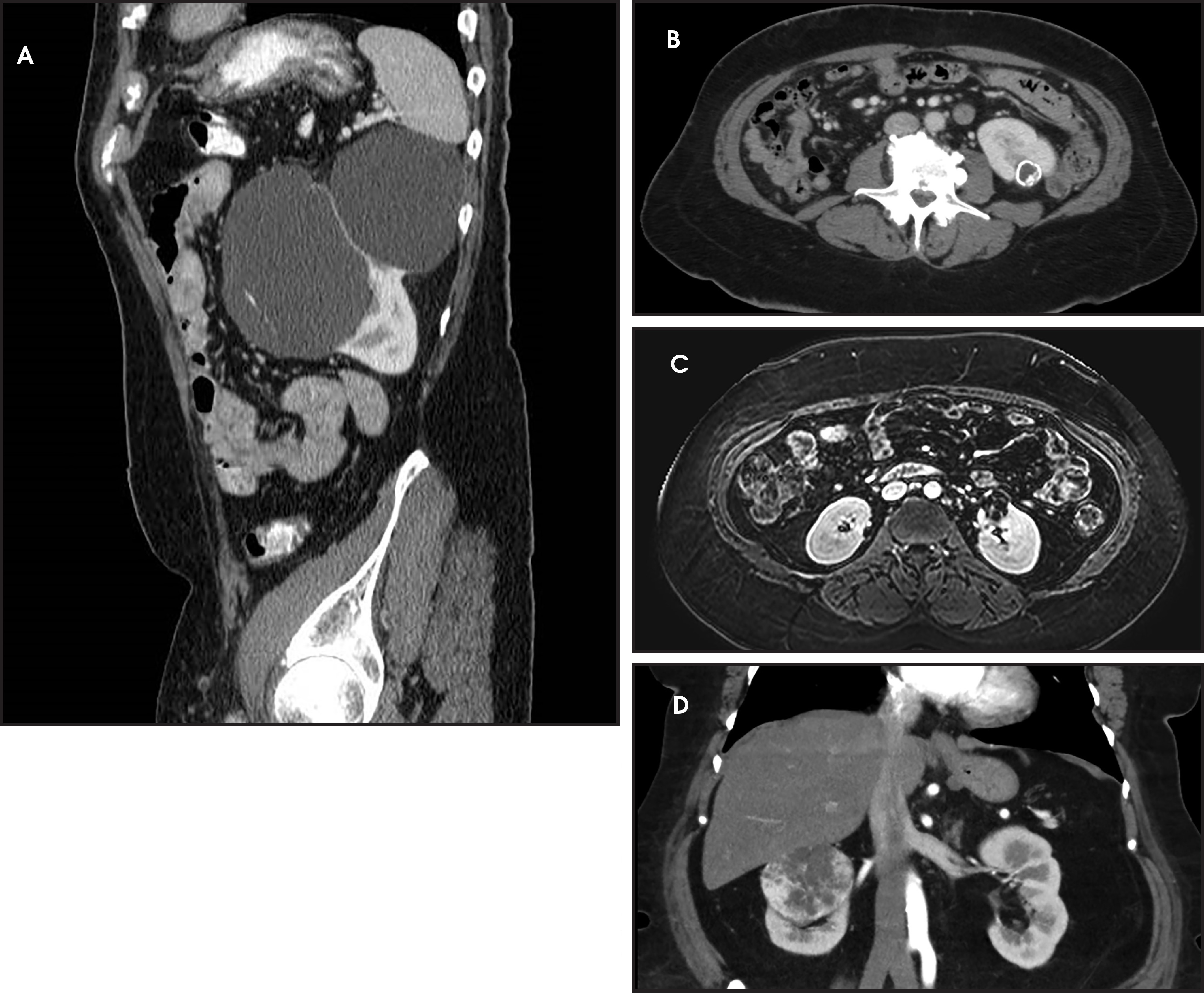
On imaging, renal lymphangioma appears as a uni- or multilocular cystic mass in the perinephric space and/or in the renal sinus (Figure 2).9 Rarely, tortuous, dilated lymphatic channels in the retroperitoneum can also be identified. The CT attenuation of the fluid can vary from water density to higher densities depending on fluid content; at MRI, fluid signal characteristics may be variable depending on the amount of protein or hemorrhage within it.10 Spontaneous regression of these lesions is not uncommon and no treatment is required in select patients without any symptoms. Cyst marsupialization, aspiration, and nephrectomy are treatment options in symptomatic patients.9
Cystic nephroma
Cystic nephroma (CN) is an uncommon, benign cystic neoplasm of the kidney, composed of epithelial and stromal elements; CN typically develops in adults older than 30 years with a strong female sex predilection.11 Currently, this tumor is considered as a distinct, unrelated entity from pediatric cystic nephroma. Painless abdominal mass and flank pain are the common presenting symptoms. At gross pathologic examination, CN is composed entirely of cysts and thin septae without any definite solid component; on histology, multiple interconnecting cystic spaces lined by cuboidal or hobnail epithelium are observed.12
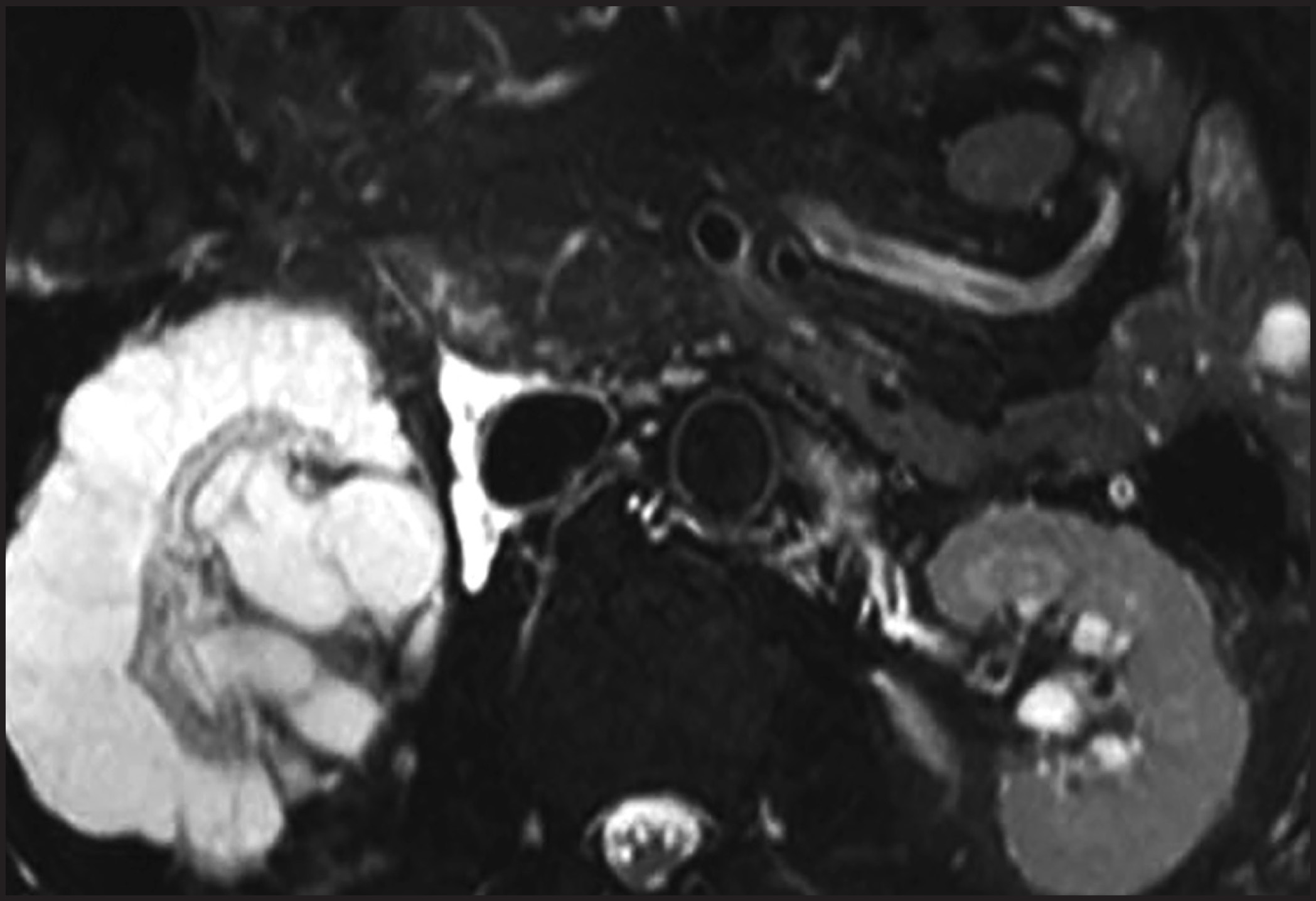
At CT/MRI, cystic nephroma presents as a well-circumscribed, multilocular cystic mass with multiple minimally thickened septations that enhance on contrast administration; cystic spaces may contain simple or hemorrhagic fluid. However, discernible solid, enhancing nodularity is absent (Bosniak category IIF and III) (Figure 3).12,13 Although rare, tumor herniation into the pelvicalyceal system and renal sinus is characteristic and may result in hydronephrosis and hemorrhage.14 Septal calcifications may be seen in up to 30% of cases. On MRI, septae are usually hypointense on T1- and T2-weighted images due to fibrous content, and cyst fluid may have varying appearances depending on protein and hemorrhagic contents.15 Nephron-sparing surgery is the treatment of choice and select nonsurgical patients can be followed up with continued surveillance.16 CNs show excellent prognosis after surgical resection with very minimal risk of local recurrence.2
Mixed epithelial and stromal tumor
Mixed epithelial and stromal tumor (MEST) is a rare neoplasm, composed of both epithelial and stromal elements and demonstrating solid and cystic architecture. MEST occurs predominantly in female patients with a mean age of 52 years, and especially in those with a history of estrogen therapy, which could indicate an underlying association between estrogen and MEST.17,18 Most patients are asymptomatic and tumors are incidentally detected on imaging done for unrelated reasons. Pathologically, tumor is composed of multiple cysts with thick septations and solid components; on microscopy, an admixture of ovarian-like stroma and epithelial-lined cysts and tubules is identified.1,19
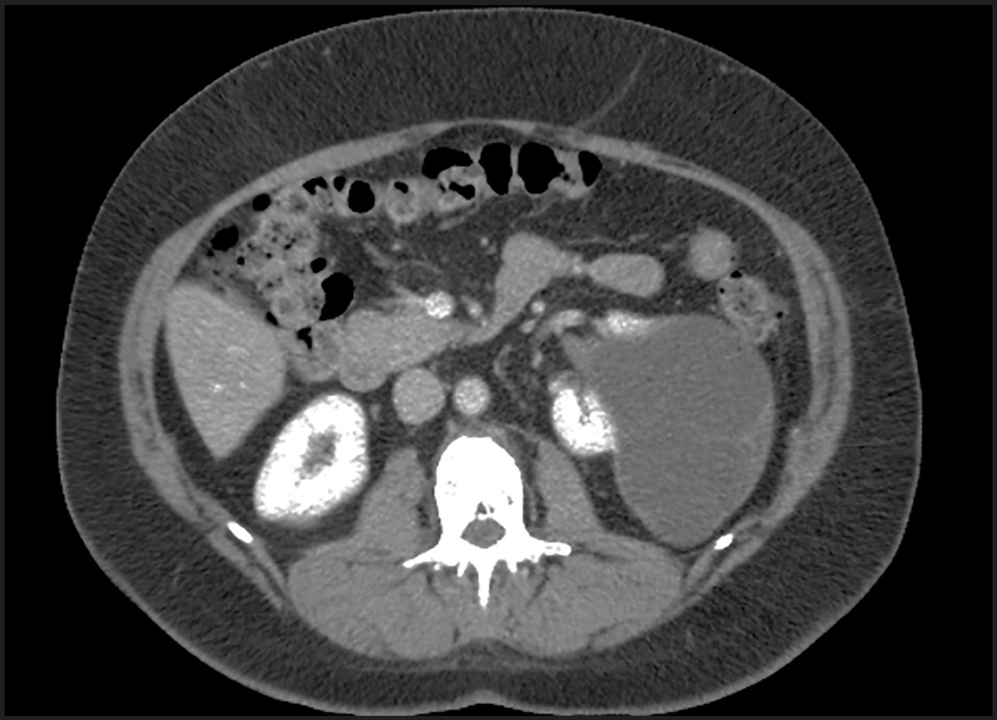
On imaging, MEST appears as a well-defined, multilocular cystic mass with a variable proportion of solid and cystic components; multiple thickened internal septae and solid nodules that demonstrate heterogeneous contrast material enhancement (Bosniak category III and IV) (Figure 4).20 Calcifications and hemorrhagic areas may be identified rarely. Partial nephrectomy is the treatment of choice and tumors show a good prognosis after surgical resection. As there are significant pathologic, genetic, and clinical similarities between CN and MEST, it has been proposed that these tumors are two ends of the spectrum of the same entity, which is best described as ‘Renal Epithelial and Stromal Tumor’ (REST).21 The major morphologic difference between adult CN and MEST is the amount of solid component; while CN is mostly cystic with thin septae, MEST demonstrates thicker septations and solid components.1
Malignant neoplasms
Cystic renal cell carcinoma
Cystic change in RCCs could be secondary to intrinsic growth pattern of the tumor or extensive tumor necrosis.2 Based on morphology, cystic RCCs can be subdivided into four types: multilocular cystic RCC, unilocular cystic RCC, cystic tumor with one or more tumor nodules, and RCC with extensive necrosis.2 About 15% of all RCCs are almost completely cystic; both clear cell and papillary types can present as cystic RCC.14
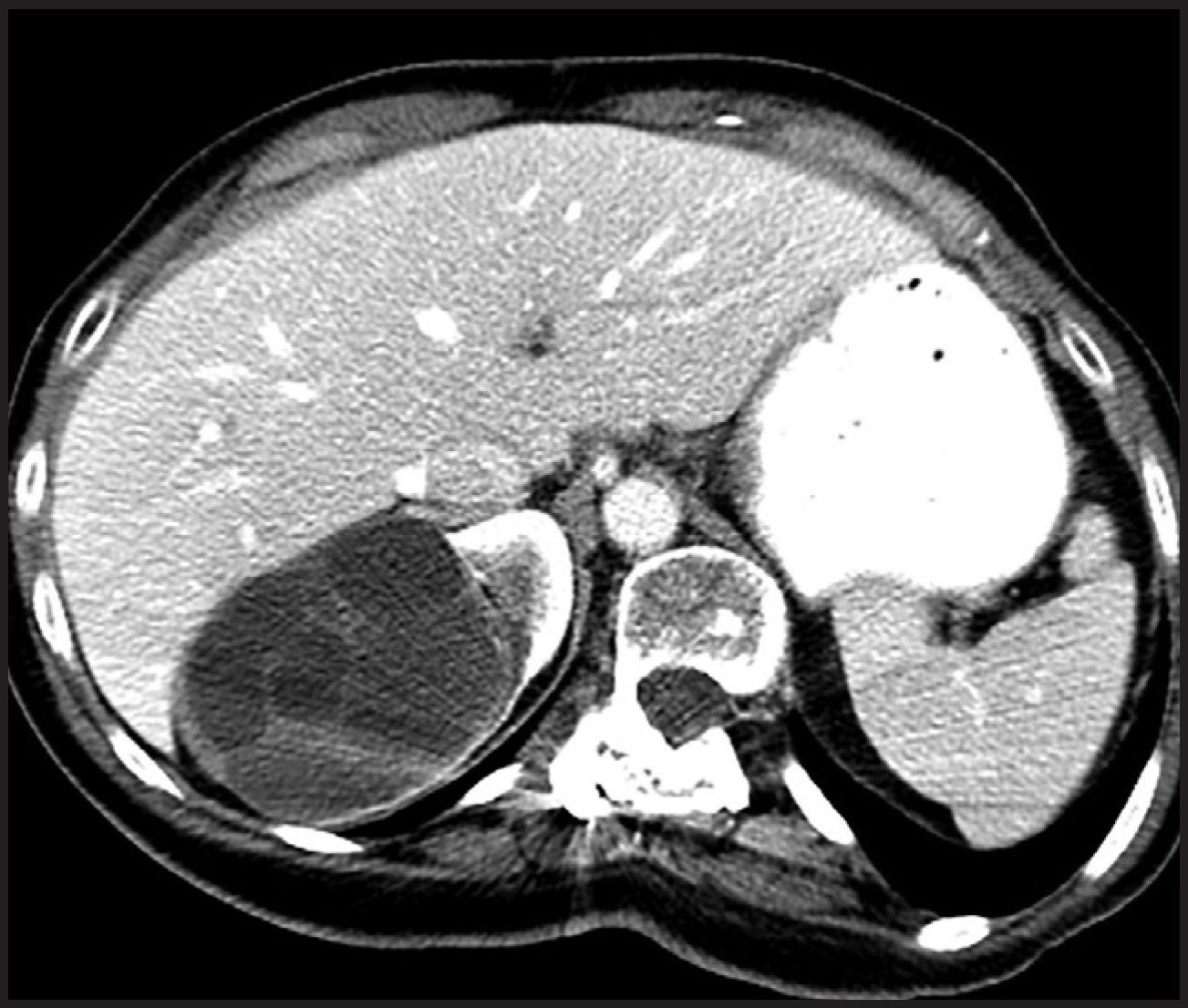
At CT/MR imaging, cystic RCCs appear as Bosniak category III/IV cysts with irregularly thickened, enhancing septations and solid components; cystic RCCs with extensive necrosis are mostly of clear cell type and demonstrates necrotic and hemorrhagic areas (Figure 5).18 Most cystic RCCs demonstrate indolent growth with no recurrent or metastatic potential and with excellent clinical outcomes; among all, cystic RCC with extensive necrosis is the most aggressive with metastases and death in up to 40% of cases.2,22
Multilocular cystic renal cell carcinoma
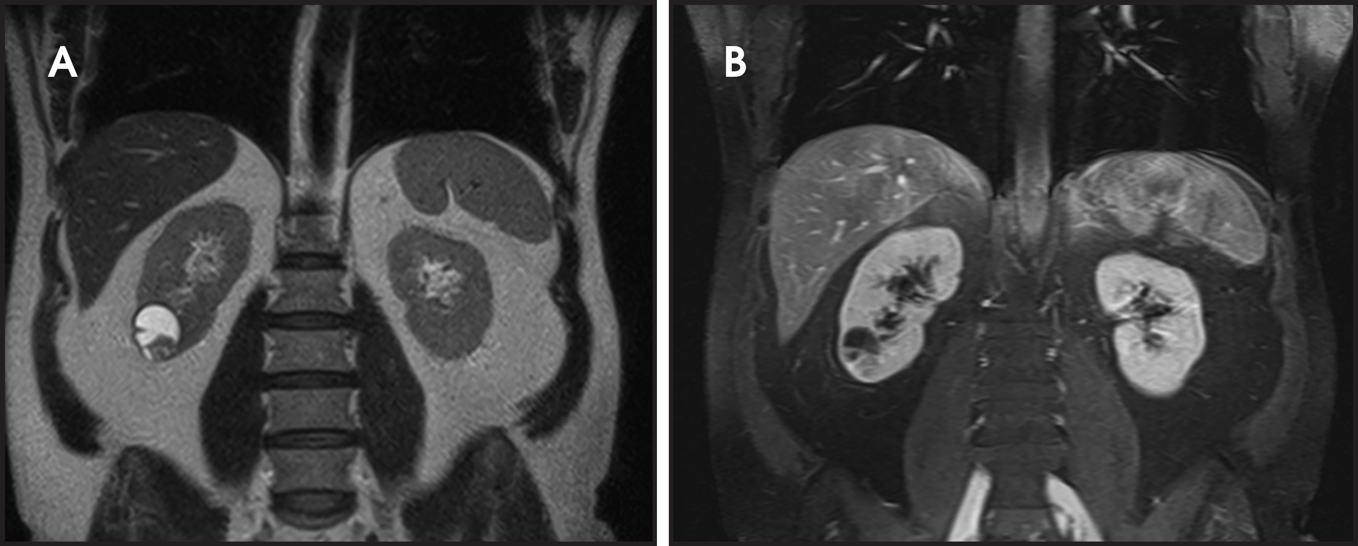
Multilocular cystic renal cell carcinoma (MCRCC) is a subtype of clear cell RCC and constitutes approximately 5% of all clear cell RCCs.23 Most of the patients with MCRCCs are asymptomatic and incidental detection is common. Pathologically, the tumor wall is lined by single or multiple layers of clear cells and septae consisting of dense collagenous stroma with epithelial cells.24 Solid nodules containing clear cells are very uncommon. On imaging, MCRCC demonstrates variable imaging appearances ranging from a Bosniak IIF to IV cystic lesion, although multilocular cystic masses containing thick, enhancing septations surrounded by a well-defined fibrous capsule (Bosniak III) is the commonest presentation (Figure 6).18,24 Intracystic fluid may show varied signal intensities depending on its contents and septal and/or wall calcification can be seen in up to 20% of cases.14 The important differential diagnosis is cystic RCC with extensive necrosis, which usually demonstrates thicker and nodular septations, decreased number of loculations, and presence of enhancing mural nodules, whereas these findings are typically absent in MCRCC.25 MCRCC shows excellent prognosis with no known evidence of recurrence or metastatic disease so far; considering this fact, MCRCC is being designated as a ‘tumor with low malignant potential’ and follow-up guidelines are changing accordingly.26
Primary renal synovial sarcoma
Primary renal synovial sarcoma (PRSS) of the kidney is an extremely rare, mesenchymal neoplasm of the kidney, first described by Argani and colleagues as a distinct subset of embryonal sarcomas of the kidney that shows characteristic cytogenetic and pathologic features.27 About 90 cases of PRSS have been described so far in the literature; young adults and adolescents are commonly affected with a mean age of 35 years with equal incidence in both sexes27. Abdominal pain and hematuria are the most common presenting symptoms. PRSS is characterized by a translocation, t(X; 18) (p11; q11) resulting in a fusion between the SYT gene on chromosome 18 and SSX family genes on chromosome X that produces a SYT-SSX gene transcript.28,29 A solid mass with multiple smooth walled cysts and/or scattered areas of necrosis and hemorrhage are the common gross pathologic appearances of PRSS. At histology, PRSS consists of monomorphic spindle cells interspersed with hobnail epithelium lined cysts.27
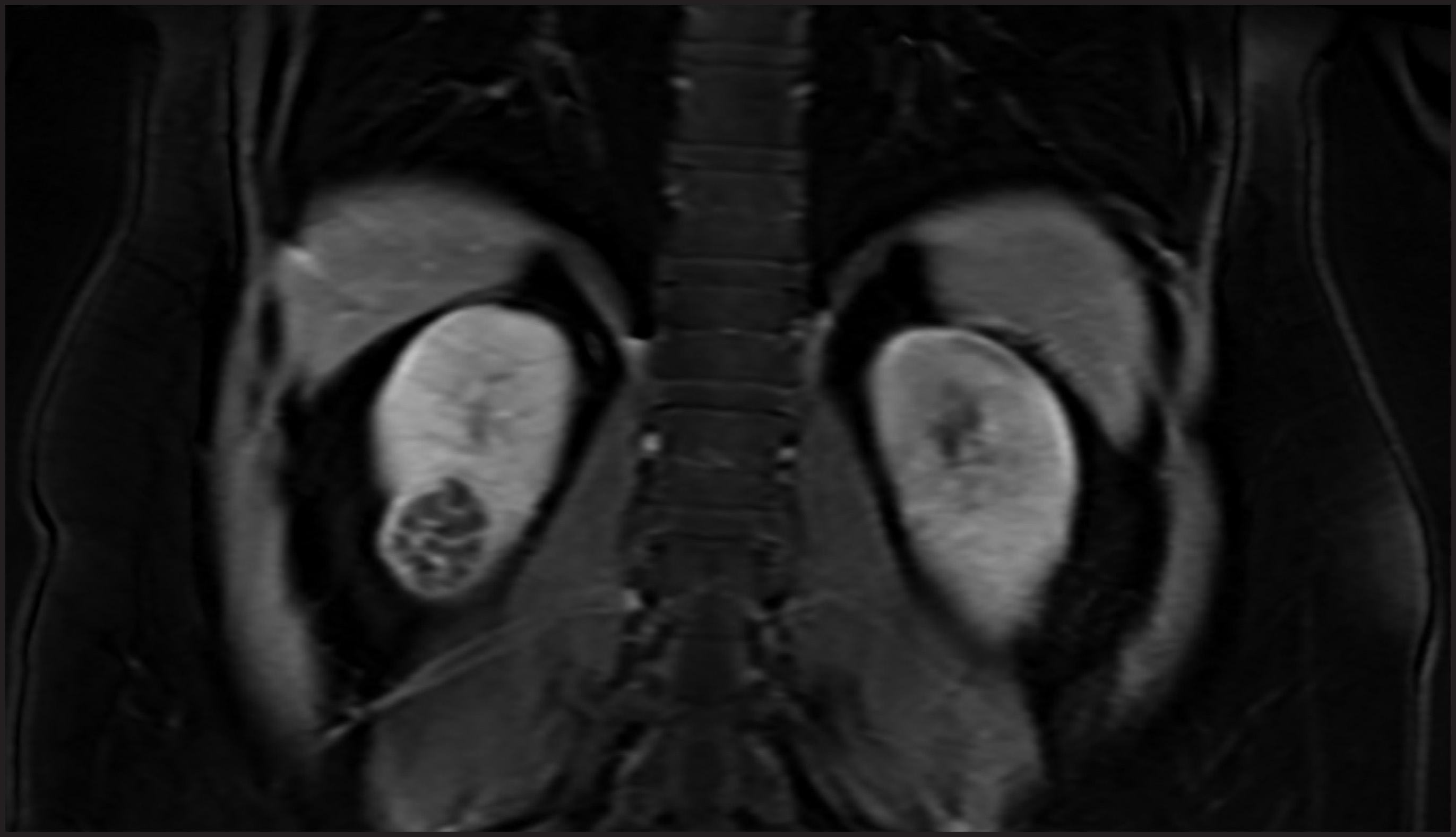
On imaging, PRSS presents as a large, well-circumscribed, solid-cystic mass with a pseudo-capsule and heterogeneous enhancement; solid components demonstrate a ‘rapid wash-in and slow wash-out’ pattern of enhancement; retroperitoneal lymphadenopathy is rare.30 In addition, this tumor may appear as a solid renal mass with multiple smooth walled cysts.31 On T2W MR imaging, PRSS may demonstrate marked heterogeneity with areas of high, intermediate, and low signal intensity, which has been described as ‘‘triple sign.”32 Surgical resection is the main stay of treatment; radiation and chemotherapy are beneficial in treating local spread and metastatic disease. PRSS is an aggressive tumor with poor patient outcome despite appropriate management.29 PET/CT is useful for surveillance and monitoring of treatment response.33
Emerging entities
Tubulocystic renal cell carcinoma
Previously referred to as ‘low-grade collecting duct carcinoma’ or ‘Bellinian epithelioma’, tubulocystic renal cell carcinoma (TC-RCC) is a unique, recently identified subtype of renal cell carcinoma. This tumor was variably thought to arise from collecting duct, proximal convoluted tubule or of intercalated cell origin.34,35 However, TC-RCC is currently considered as a distinct morphological entity that is separate from the biologically aggressive collecting duct carcinoma but is closely related to papillary RCC.1,36 About 70 cases have been reported so far in the English literature with a male predominance. Many of the previously identified cases were incidentally detected in patients in their 5th or 6th decade.36 On gross pathology, TC-RCC appears as a well-circumscribed, encapsulated, multicystic tumor with a characteristic “bubble wrap” or “sponge” appearance.36 On histology, small to medium-sized tubules combined with cystically dilated larger tubules are commonly identified.26,37
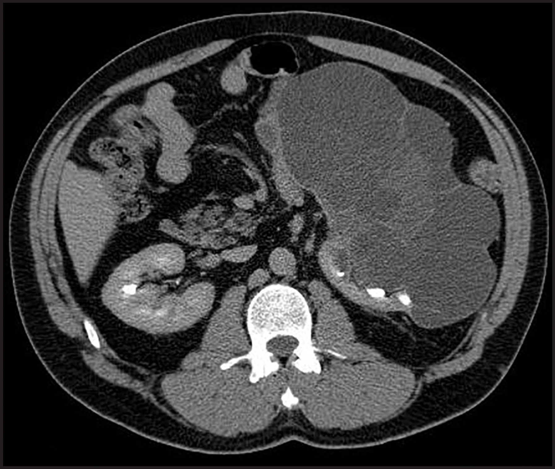
At CT/MR imaging, TC-RCC presents as a multiloculated cystic mass with ‘spider web’ enhancing septae and/or enhancing mural nodules suggestive of the Bosniak III/IV category. Hemorrhagic and necrotic areas are very uncommon (Figure 7).14 In addition, multiple small, uniform-sized cysts giving a “spongy appearance” is another imaging appearance. Although most of the TC-RCCs show an indolent course with excellent prognosis after resection, an exact biologic profile has not been fully characterized.38
Acquired cystic disease-associated renal cell carcinoma
Acquired cystic disease-associated RCC (ACD-RCC) is being identified as a distinct renal neoplasm and is the most common RCC subtype in end-stage kidneys, especially in patients with acquired cystic kidney disease (ACKD); clear cell papillary subtype is the second most common one in these patients.26 This tumor is strongly associated with longer duration of dialysis (>10 yrs.); multifocal and bilateral tumors are very frequent.39 At gross pathologic examination, ACD-RCC may appears as a small nodule attached to cyst wall or solid lesion filling a cyst, or solid mass separate from the underlying cysts. Histologically, cribriform/sieve-like growth pattern with intratumoral oxalate crystals and abundant eosinophilic cytoplasm is characteristic of ACD-RCC.26
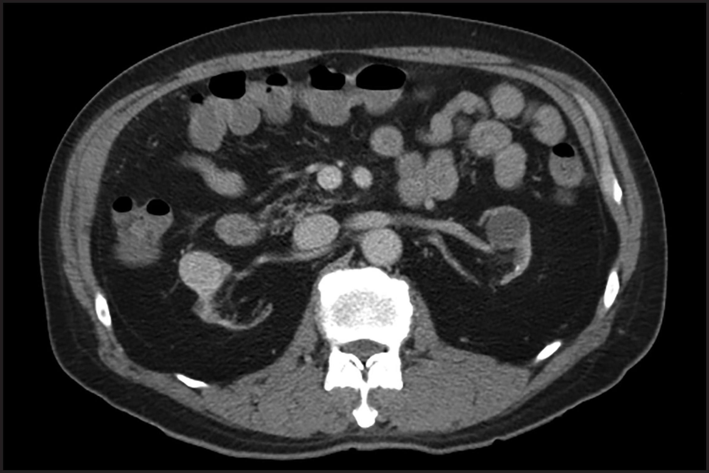
On imaging, ACD-RCC appears as a heterogeneously enhancing solid mass either within the atrophic kidneys or kidneys with multiple cysts secondary to acquired cystic kidney disease (Figure 8).14 Development of enhancing solid components, diffuse wall thickening, and multiple septations within the pre-existing cysts indicate development of RCC.14 However, no characteristic imaging findings have been described so far to differentiate ACD-RCC from clear cell papillary RCC. ACD-RCC demonstrates aggressive behavior than other RCC subtypes in end-stage kidneys. However, the prognosis is relatively good because of early detection secondary to continued surveillance.26
Angiomyolipoma with epithelial cysts
Angiomyolipoma with epithelial cysts (AMLEC) is a rare, distinct cystic variant of angiomyolipoma, characterized by multiple cysts and minimal or no macroscopic fat; AMLEC was first described by Davis et al. as cystic AML and renamed as AMLEC by Fine and colleagues.40,41 Fewer than 20 cases have been reported in the literature so far with slight female predominance.40 Although etiopathogenesis of AMLEC is unclear, histogenesis of the Müllerian-like stroma with subsequent cyst development has been proposed due to the embryological proximity between the urinary and genital systems.41 On imaging, AMLEC may appear as a cystic lesion with enhancing solid components (Bosniak 4) or as a solid mass with scattered cystic foci.42 As the presence of macroscopic fat is rare, it is difficult to differentiate AMLEC from more common RCCs and other cystic neoplasms. Biopsy could be considered in these cases to potentially confirm this benign lesion and to avoid unnecessary surgery.42
Conclusion
Complex cystic renal masses may be benign or malignant. Diagnosis and management of these masses predominantly is based on cross-sectional imaging findings. The Bosniak classification system is the most commonly used to separate cystic lesions requiring surgery from those that can be safely ignored or followed-up. Contrast-enhanced CT and MRI are the imaging modalities of choice in characterizing cystic kidney masses. On imaging, it is very important to identify thick, enhancing septations and/or solid components within the cystic mass that differentiate Bosniak category III/IV lesions from the remaining non-surgical masses. In addition to existing neoplasms, novel cystic renal tumors such as tubulocystic RCC and ACKD-associated RCC have been identified and likely to be added to World Health organization classification in the near future. Awareness of the wide spectrum of benign and malignant cystic renal neoplasms and non-neoplastic cystic masses and familiarity with their characteristic imaging findings should afford appropriate patient management.
References
- Moch H. Cystic renal tumors: New entities and novel concepts.. Adv Anat Path.. 2010;17:209-214.
- Truong L, Choi Y, Shen S, Ayala G, Amato R, Krishnan B. Renal cystic neoplasms and renal neoplasms associated with cystic renal diseases: Pathogenetic and molecular links.. Adv Anat Path.. 2003;10:135-159.
- Bosniak M. The use of the Bosniak classification system for renal cysts and cystic tumors.. J Urol.. 1997;157:1853-1852.
- Hartman D, Choyke P, Hartman M. From the RSNA refresher courses: a practical approach to the cystic renal mass.. Radiographics.. 2004(1):S101-115.
- Israel G, Silverman S. The incidental renal mass.. Radiol Clin North Am. 2011;49:369-383.
- Gabr A, Gdor Y, Roberts W, Wolf J. Radiographic surveillance of minimally and moderately complex renal cysts.. BJU Intl.. 2009;103:1116-1119.
- Felker E, Lee-Felker S, Alpern L, Lu D, Raman S. Efficacy of imaging-guided percutaneous radiofrequency ablation for the treatment of biopsy-proven malignant cystic renal masses.. AJR Am J Roentgen.. 2013;201:1029-1035.
- Park B, Kim C, Kim E. Differentiation of Bosniak categories IIF and III cystic masses: what radiologists should know.. J Comp Asst Tomo.. 2010;34:847-854.
- Katabathina V, Vikram R, Nagar A, Tamboli P, Menias C, Prasad S. Mesenchymal neoplasms of the kidney in adults: imaging spectrum with radiologic-pathologic correlation.. Radiographics.. 2010;30:1525-1540.
- Upreti L, Dev A, Kumar P. Imaging in renal lymphangiectasia: report of two cases and review of literature.. Clin Radiol.. 2008;63:1057-1062.
- Eble J, Bonsib S. Extensively cystic renal neoplasms: cystic nephroma, cystic partially differentiated nephroblastoma, multilocular cystic renal cell carcinoma, and cystic hamartoma of renal pelvis.. Semin Diag Pathol.. 1998;15:2-20.
- Wilkinson C, Palit V, Bardapure M. Adult multilocular cystic nephroma: Report of six cases with clinical, radio-pathologic correlation and review of literature.. Urology annals. 2013;5:13-17.
- Raman S, Hruban R, Fishman E. Beyond renal cell carcinoma: rare and unusual renal masses.. Abdom Imaging.. 2012;37:873-884.
- Katabathina V, Garg D, Prasad S, Vikram R. Cystic renal neoplasms and renal neoplasms associated with cystic renal diseases in adults: cross-sectional imaging findings.. J Comp Asst Tomo.. 2012;36:659-668.
- Silver I, Boag A, Soboleski D. Best cases from the AFIP: Multilocular cystic renal tumor: cystic nephroma.. Radiographics.. 2008;28.
- Lane B, Campbell S, Remer E. Adult cystic nephroma and mixed epithelial and stromal tumor of the kidney: clinical, radiographic, and pathologic characteristics.. Urology. 2008;71:1142-1148.
- Adsay N, Eble J, Srigley J, Jones E, Grignon D. Mixed epithelial and stromal tumor of the kidney.. Am J Surg Pathol.. 2000;24:958-970.
- Freire M, Remer E. Clinical and radiologic features of cystic renal masses.. AJR Am J Roentgen.. 2009;192:1367-1372.
- Antic T, Perry K, Harrison K. Mixed epithelial and stromal tumor of the kidney and cystic nephroma share overlapping features: reappraisal of 15 lesions.. Arch Path Lab Med.. 2006;130:80-85.
- Chu L, Hruban R, Horton K, Fishman E. Mixed epithelial and stromal tumor of the kidney: radiologic-pathologic correlation.. Radiographics.. 2010;30:1541-1551.
- Turbiner J, Amin M, Humphrey P. Cystic nephroma and mixed epithelial and stromal tumor of kidney: a detailed clinicopathologic analysis of 34 cases and proposal for renal epithelial and stromal tumor (REST) as a unifying term.. Am J Surg Pathol.. 2007;31:489-500.
- Tan P, Cheng L, Rioux L. Renal tumors: Diagnostic and prognostic biomarkers.. Am J Surg Pathol.. 2013;37:1518-1531.
- Han K, Janzen N, McWhorter V. Cystic renal cell carcinoma: Biology and clinical behavior.. Uro Oncol. 2004;22:410-414.
- Hindman N, Bosniak M, Rosenkrantz A, Lee-Felker S, Melamed J. Multilocular cystic renal cell carcinoma: comparison of imaging and pathologic findings.. AJR Am J Roent gen.. 2012;198:W20-26.
- Aubert S, Zini L, Delomez J, Biserte J, Lemaitre L, Leroy X. Cystic renal cell carcinomas in adults. Is preoperative recognition of multilocular cystic renal cell carcinoma possible?. J Urol.. 2005;174:2115-2119.
- Srigley J, Delahunt B, Eble J. The International Society of Urological Pathology (ISUP) Vancouver Classification of Renal Neoplasia.. Am J Surg Pathol.. 2013;37:1469-1489.
- Argani P, Faria P, Epstein J. Primary renal synovial sarcoma: molecular and morphologic delineation of an entity previously included among embryonal sarcomas of the kidney.. Am J Surg Pathol.. 2000;24:1087-1096.
- Schoolmeester J, Cheville J, Folpe A. Synovial sarcoma of the kidney: a clinicopathologic, immunohistochemical, and molecular genetic study of 16 cases.. Am J Surg Pathol.. 2014;38:60-65.
- Iacovelli R, Altavilla A, Ciardi A. Clinical and pathological features of primary renal synovial sarcoma: analysis of 64 cases from 11 years of medical literature.. BJU Intl.. 2012;110:1449-1454.
- Lv X, Qiu Y, Han L. Primary renal synovial sarcoma: computed tomography imaging findings.. Acta Radiol. 2014.
- Gong J, Kang W, Li S, Yang Z, Xu J. CT findings of synovial sarcomas of the kidney with pathological correlation.. Clinical imaging.. 2013;37:1033-1036.
- Zakhary M, Elsayes K, Platt J, Francis I. Magnetic resonance imaging features of renal synovial sarcoma: a case report.. Cancer Imaging.. 2008;8:45-47.
- Erturhan S, Seckiner I, Zincirkeser S. Primary synovial sarcoma of the kidney: use of PET/CT in diagnosis and follow-up.. Ann Nuc Med. 2008;22:225-229.
- MacLennan G, Bostwick D. Tubulocystic carcinoma, mucinous tubular and spindle cell carcinoma, and other recently described rare renal tumors.. Clin Lab Med.. 2005;25:393-416.
- Osunkoya A, Young A, Wang W, Netto G, Epstein J. Comparison of gene expression profiles in tubulocystic carcinoma and collecting duct carcinoma of the kidney.. Am J Surg Pathol.. 2009;33:1103-1106.
- Zhou M, Yang X, Lopez J. Renal tubulocystic carcinoma is closely related to papillary renal cell carcinoma: implications for pathologic classification.. Am J Surg Pathol.. 2009;33:1849-1840.
- Yang X, Zhou M, Hes O. Tubulocystic carcinoma of the kidney: clinicopathologic and molecular characterization.. Am J Surg Pathol.. 2008;32:177-187.
- Bhullar J, Varshney N, Bhullar A, Mittal V. A New Type of Renal Cancer-Tubulocystic Carcinoma of the Kidney: A Review of the Literature.. Intl J Surg Pathol.;22:297-302.
- Tickoo S, dePeralta-Venturina M, Harik L. Spectrum of epithelial neoplasms in end-stage renal disease: an experience from 66 tumor-bearing kidneys with emphasis on histologic patterns distinct from those in sporadic adult renal neoplasia.. Am J Surg Pathol.. 2006;30:141-153.
- Davis C, Barton J, Sesterhenn I. Cystic angiomyolipoma of the kidney: a clinicopathologic description of 11 cases.. Modern Pathol.. 2006;19:669-674.
- Fine S, Reuter V, Epstein J, Argani P. Angiomyolipoma with epithelial cysts (AMLEC): a distinct cystic variant of angiomyolipoma.. Am J Surg Pathol.. 2006;30:593-599.
- Rosenkrantz A, Hecht E, Taneja S, Melamed J. Angiomyolipoma with epithelial cysts: mimic of renal cell carcinoma.. Clin Imaging.. 2010;34:65-68.
Citation
. Cystic renal neoplasms in adults: Current imaging update. Applied Radiology. 2015;44(1):23-33. doi:10.37549/AR2149.