Differential diagnosis of esophageal disease on esophagography
Applied Radiology — Vol. 30 , Issue 10 , pp. 11 -21
DOI: 10.37549/AR1048
Published: October 1, 2001
Categories
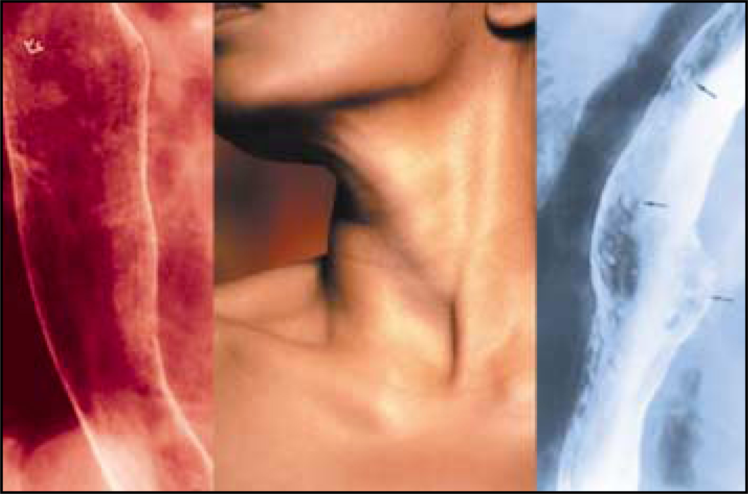
This review article will present a gamut or pattern approach to the diagnosis of esophageal diseases.1,2 For most patients, analysis of the radiographic findings in combination with consideration of the clinical history leads to the diagnosis or a graded differential diagnosis.3
Normal esophagus
The normal esophagus is a muscular tube covered by nonkeratinized squamous epithelium. The esophagus lies in the neck and mediastinum. The aorta, left mainstem bronchus, and posterior border of the heart impress upon the wall of the esophagus. During double-contrast esophagography, the barium-etched contour is visible as a smooth, white line (figure 1). En face, the barium-coated esophageal mucosa is smooth and featureless, fading to gray.1 The squamocolumnar junction with the stomach may be seen as a zigzag line, also known as the Z line.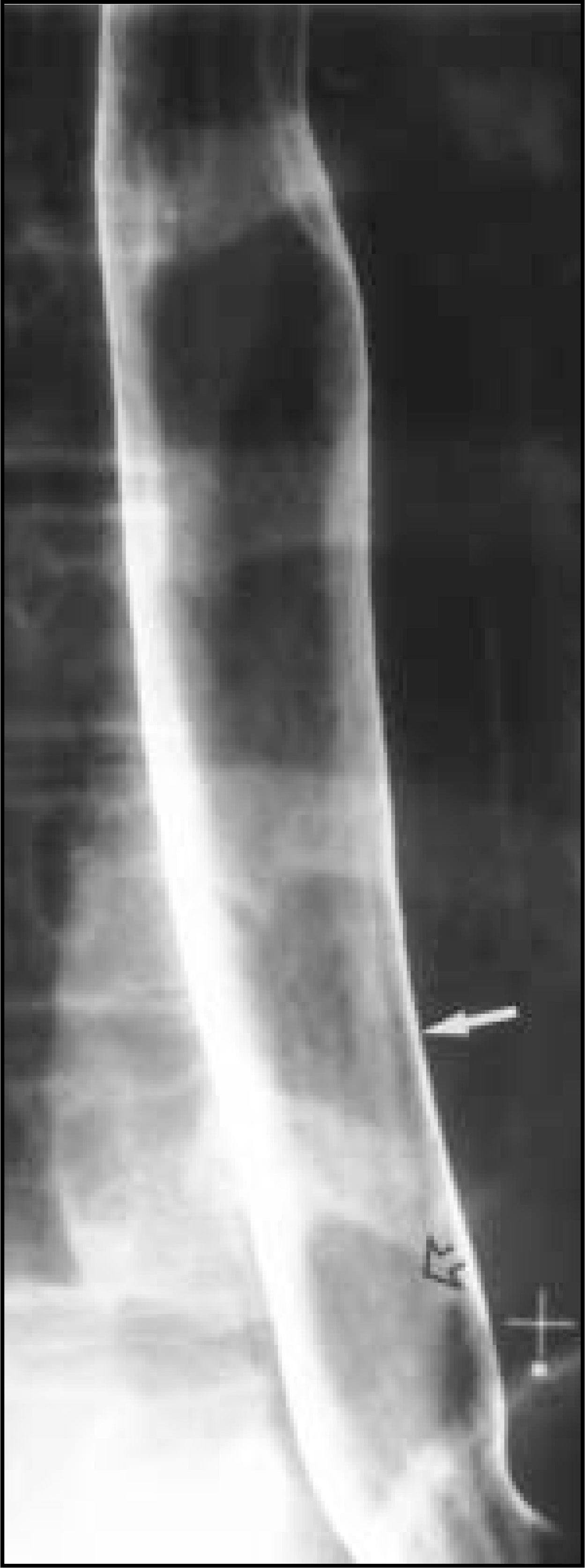
Small esophageal ulcers
A wide variety of diseases are associated with small esophageal ulcers <1 cm in diameter (Table 1).4-12 The most common causes of radiographically diagnosed small esophageal ulcers are viral-induced esophagitis, drug-induced esophagitis, and reflux esophagitis.
Herpes simplex virus type I most frequently causes esophagitis in immunosuppressed patients, although immunocompetent patients are affected occasionally. On barium studies, small, discrete superficial ulcers are seen on a background of normal mucosa (figure 2).4-6 The ulcers may have a round, stellate, linear, or serpentine configuration. Rarely, herpes esophagitis may develop in immunocompetent patients. In this self-limited form of herpes esophagitis, the ulcers manifest as smaller, punctate collections of barium.7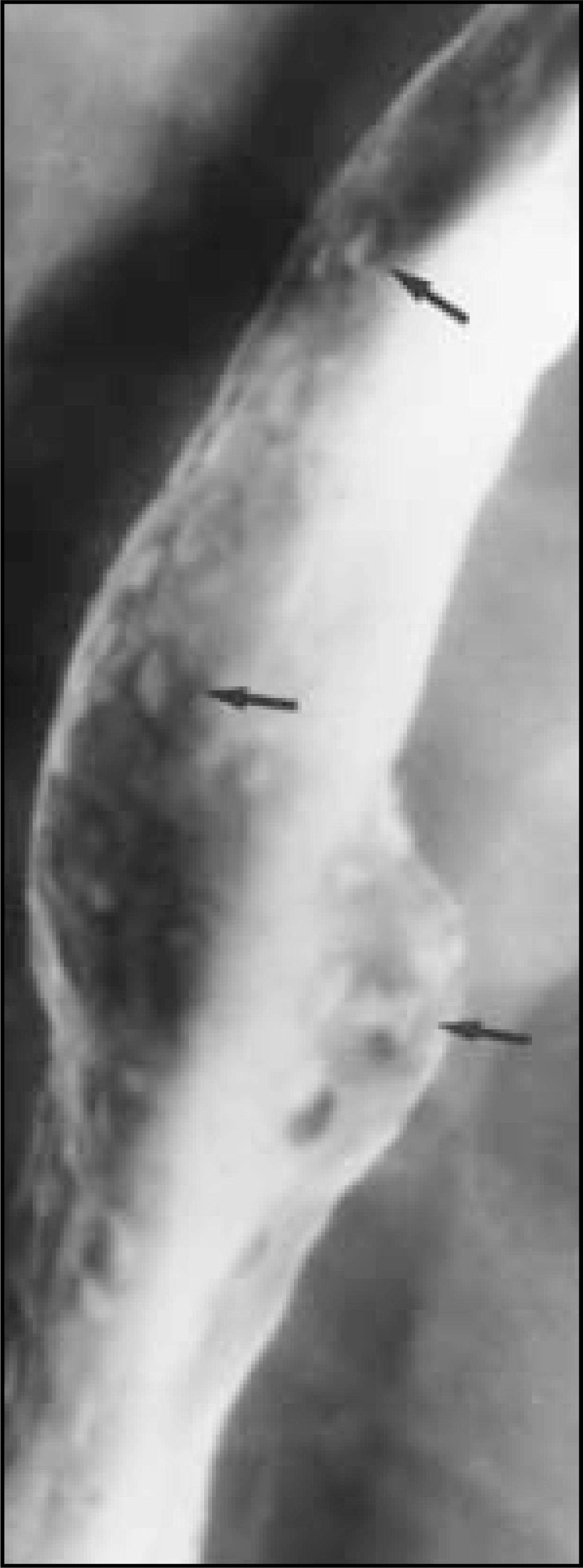
Drug-induced esophagitis is primarily a contact esophagitis caused by a variety of medications, including tetracycline or its derivatives, quinidine, potassium chloride, non-steroidal anti-inflammatory agents, and alendronate sodium. If little or no water is used during ingestion of these medications, pills may transiently lodge in the esophagus at the level of normal extrinsic impressions, including the aortic arch, the left mainstem bronchus, and the left atrium. Typically, small, shallow ulcers will be clustered together in the mid-esophagus.8,9 These ulcers usually heal within 7 to 10 days after cessation of the offending medication.
In patients with small esophageal ulcers, the clinical history provides the key to the diagnosis. Patients with herpes esophagitis are usually immunosuppressed. If drug ingestion is the cause of the ulcer, a clinical history of using the offending medication can usually be elicited. Finally, patients with reflux-induced ulcers usually have a history of heartburn, and the ulcers are usually located in the distal esophagus near the esophagogastric junction. These patients also usually have other radiographic findings of reflux esophagitis.2
Large esophageal ulcers
The most common causes of large esophageal ulcers, those >1 cm in diameter, are human immunodeficiency virus (HIV) and cytomegalovirus (CMV) in immunocompromised patients, primarily patients with acquired immunodeficiency syndrome (AIDS).13-16 HIV ulcers sometimes occur near the time of clinical presentation and seroconversion. The ulcers in CMV and HIV esophagitis (figure 3) tend to be large, flat, ovoid-shaped barium collections or barium-etched craters.14,16 Endoscopic biopsy specimens are necessary to distinguish CMV or HIV ulcers, as the treatment for each of these infections is different. Esophageal ulceration due to HIV is a diagnosis of exclusion, so this diagnosis can be made only when endoscopic biopsies, brushings, and cultures are negative for CMV.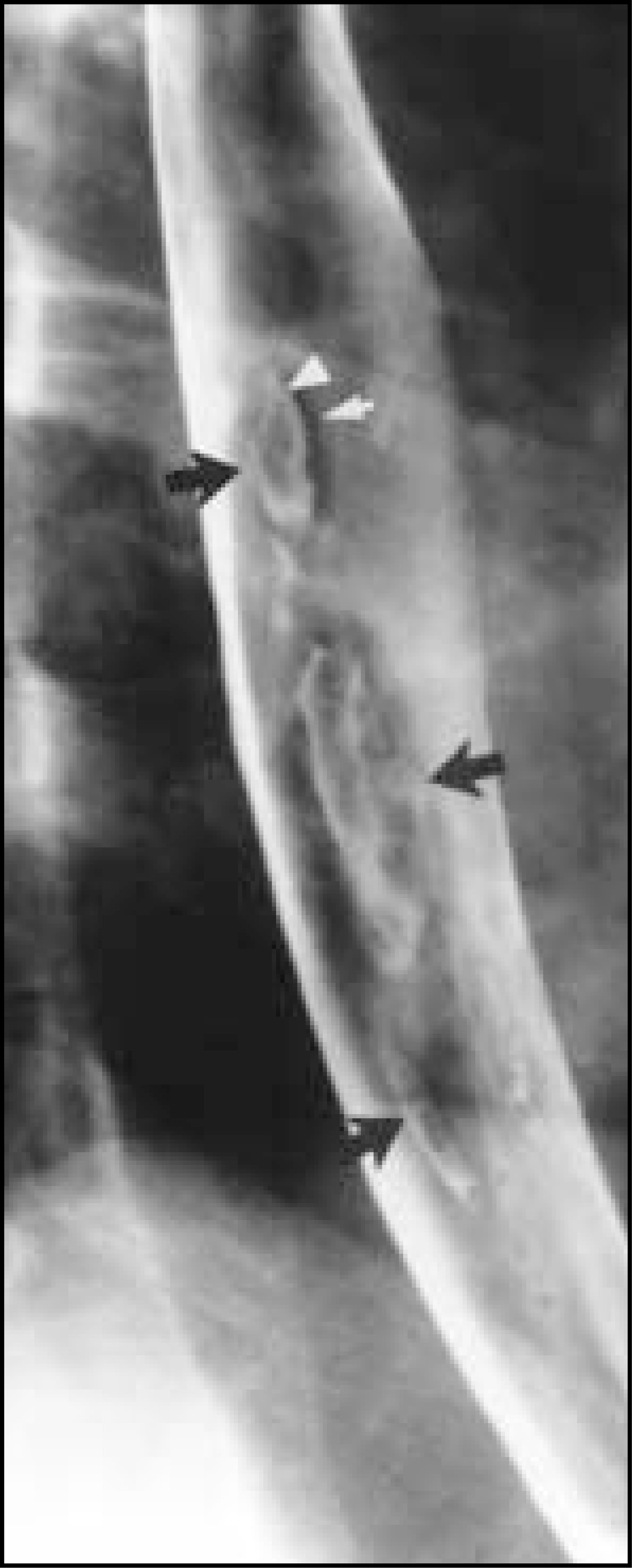
Other common causes of large esophageal ulcers include carcinoma (figure 4), drug-induced ulcers, or Barrett’s esophagus3,17,18 (Table 1). However, these ulcers tend to be deeper than viral-induced ulcers, and their edges are frequently thickened and lobulated. In such cases, biopsy specimens may be necessary to exclude neoplasia.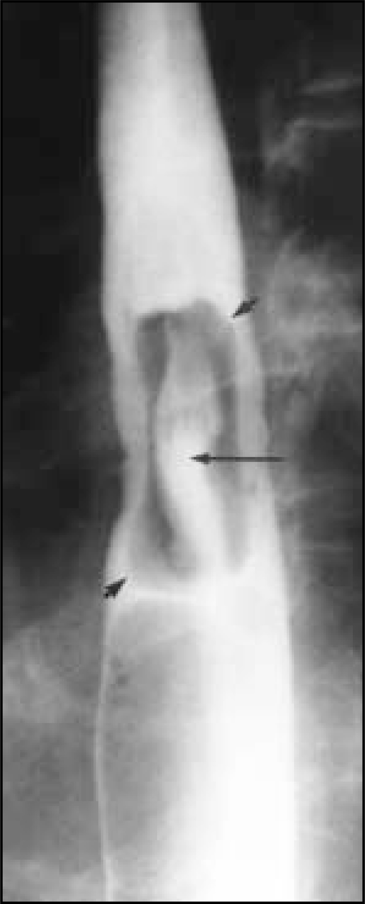
Mucosal nodules and plaques
Mucosal nodules and plaques are elevated lesions of varying size. Plaques are usually discrete, irregular or ovoid elevations that barely protrude above the mucosal surface. Nodules are smaller elevations and are more rounded than plaques. The morphology of the elevations in combination with the clinical history allows a specific diagnosis to be made in most patients (Table 2).
Candida esophagitis most frequently manifests as numerous small, discrete, ovoid or linear plaque-like elevations aligned parallel to the longitudinal folds of the esophagus (figure 5).19 In mild-to-moderate Candida esophagitis, the plaques are separated by intervening segments of normal mucosa. In more severe cases, the plaques may carpet the esophagus. In even more severe cases, confluent plaques, pseudomembranes, and barium burrowing beneath the inflammatory detritus may produce a grossly irregular appearance of the contour in profile and the mucosal surface en face, the so-called “shaggy esophagus.” When plaques and pseudomembranes slough, large ulcers may form, but these are almost always present on a background of diffuse plaque formation.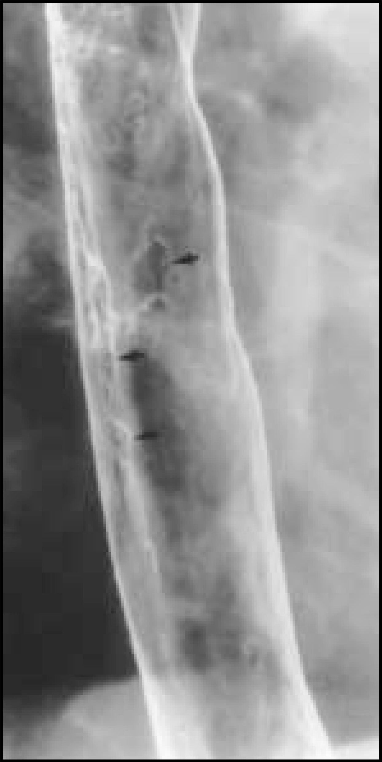
Candida albicans is the most common cause of infectious esophagitis. Immunosuppression is the most frequent predisposing factor. Patients usually complain of dysphagia or odynophagia. Thrush in the oral cavity or pharynx is seen in about one-half of patients. Candida esophagitis may also develop in patients with severe esophageal motility disorders, such as scleroderma, or in patients with esophageal obstruction and stasis due to achalasia or carcinoma.
Small nodules and plaques of varying sizes may also be seen in the esophagus in patients with glycogenic acanthosis, a common degenerative condition.20,21 However, the plaques of glycogenic acanthosis are usually seen in the upper or midesophagus in a random distribution. In this disorder, glycogen is accumulated in the cytoplasm of cells in the upper portion of the squamous epithelium. Glycogenic acanthosis typically occurs in elderly individuals who have no esophageal symptoms and who do not have a history of immunosuppression or a condition predisposing to stasis. In contrast, patients with Candida esophagitis are usually symptomatic, and the plaques tend to be more linear in shape and aligned longitudinally along the folds.
In some patients with reflux esophagitis, tiny mucosal nodules are seen (figure 6).2 However, these nodules are more ill-defined and less discrete than the plaques in Candida esophagitis. The nodules of reflux esophagitis also are more confluent and are located in the distal esophagus, usually in patients with gastro-esophageal reflux and hiatal hernias. Reflux esophagitis is more frequently characterized by poorly defined tiny mucosal elevations, termed mucosal “granularity” (figure 6B).1,2,22,23 This granularity may be associated with tiny or linear ulcers and thickened, nodular folds. These changes are also usually associated with a hiatal hernia and fluoroscopically detected gastroesophageal reflux. Some patients with reflux esophagitis have such severe inflammation that plaque-like pseudomembranes may eventually form.24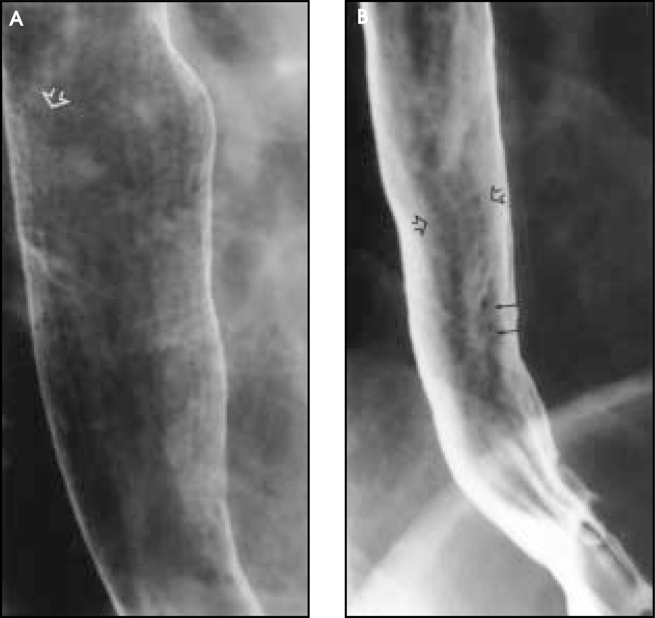
A focal area of confluent mucosal nodularity may be worrisome for superficial spreading carcinoma, a cancer confined to the mucosa and submucosa (figure 7).25,26 However, the nodules are not as discrete as those in Candida esophagitis, nor are they separated by normal intervening mucosa. Although most focal areas of mucosal irregularity will probably be caused by glycogenic acanthosis, an area of focal mucosal nodularity should be biopsied to exclude superficial spreading carcinoma.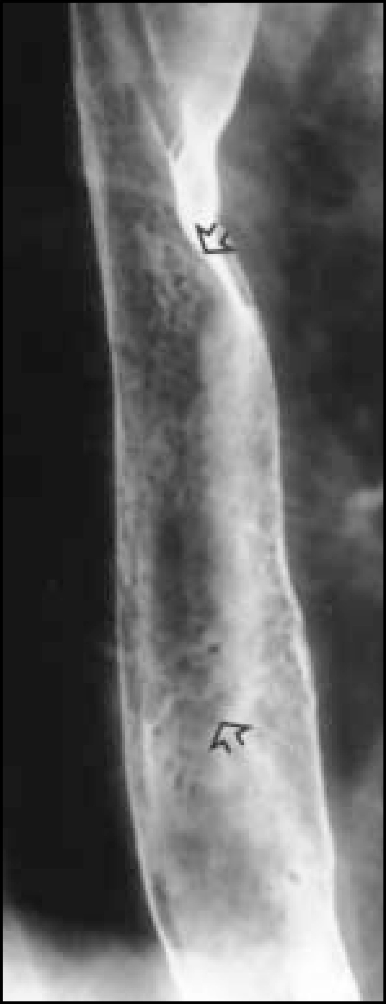
It is also difficult to distinguish a focal area of mucosal nodularity from the surface pattern termed “reticular mucosa,” which is seen in Barrett’s esophagus.27 Barrett’s esophagus is an acquired condition in which there is progressive columnar metaplasia of the esophagus due to long-standing gastroesophageal reflux disease. The reticular mucosal pattern resembles the areae gastricae of the stomach, with a fine, net-like web of barium-filled grooves surrounding small tufts of mucosa (figure 8).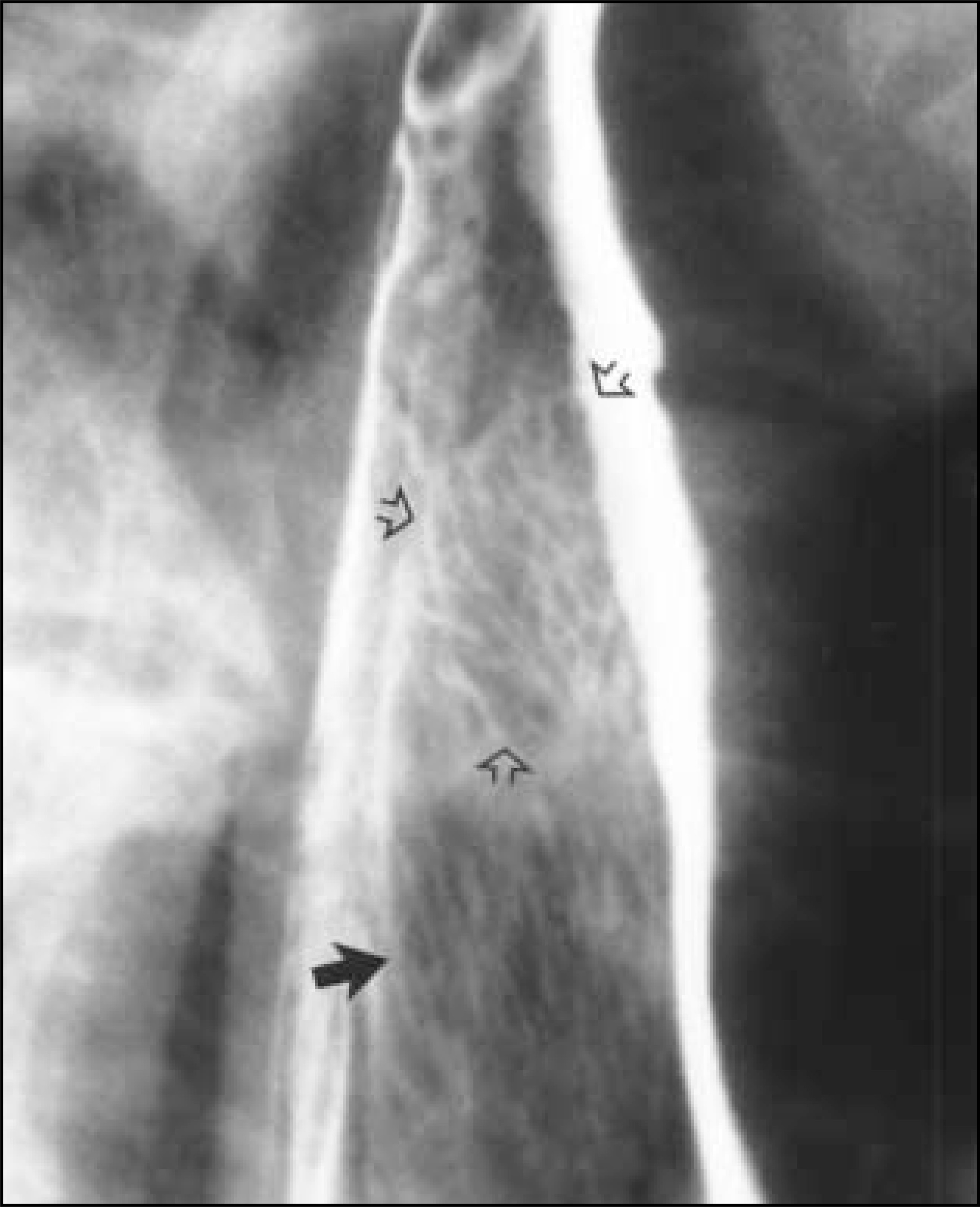
Abnormal esophageal folds
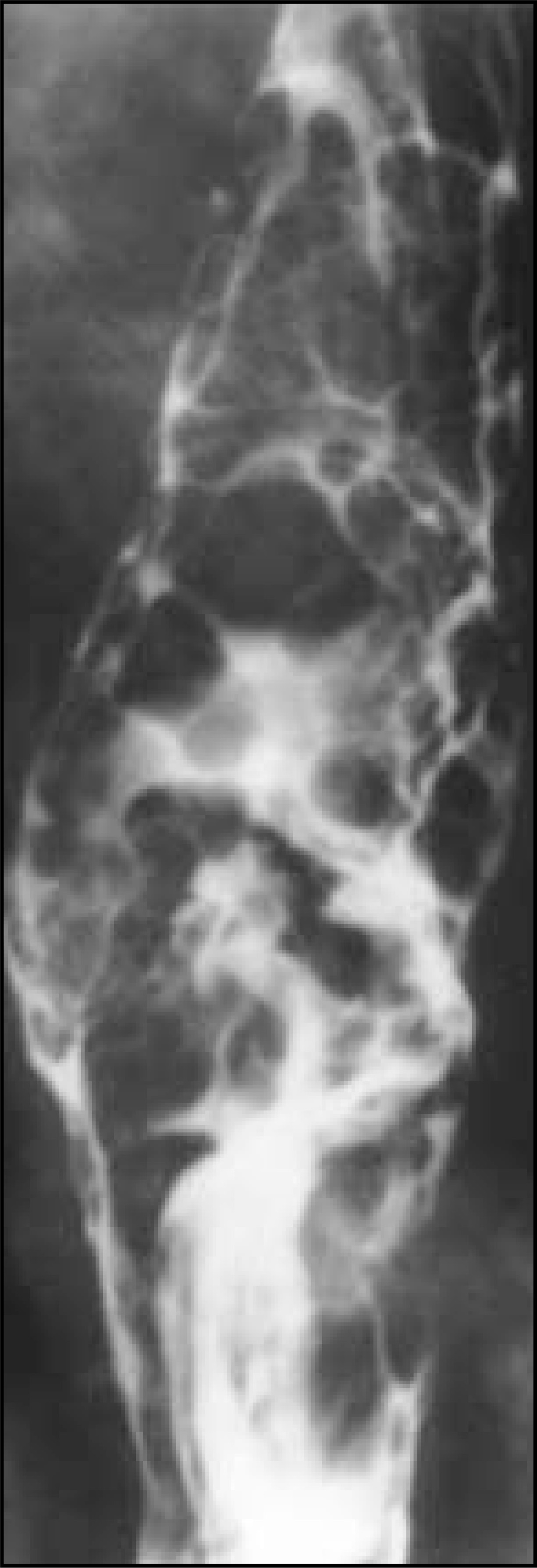
The longitudinal folds of the esophagus are composed of mucosa and submucosa and are best seen when the esophagus is underdistended. Therefore, abnormalities of folds reflect disease in the mucosa and submucosa (Table 3). In patients with reflux esophagitis, thickened esophageal folds are frequently seen when the esophagus is collapsed.1,2 These patients usually have other findings of reflux disease, including gastro-esophageal reflux, a granular mucosa, and hiatal hernia. In contrast, esophageal varices are serpentine, with a smooth surface (figure 9). Varices may change in size with varying degrees of esophageal distention and patient position. If the folds are rigid, fixed, or irregular, however, the varicoid form of squamous cell carcinoma must be excluded (figure 10).28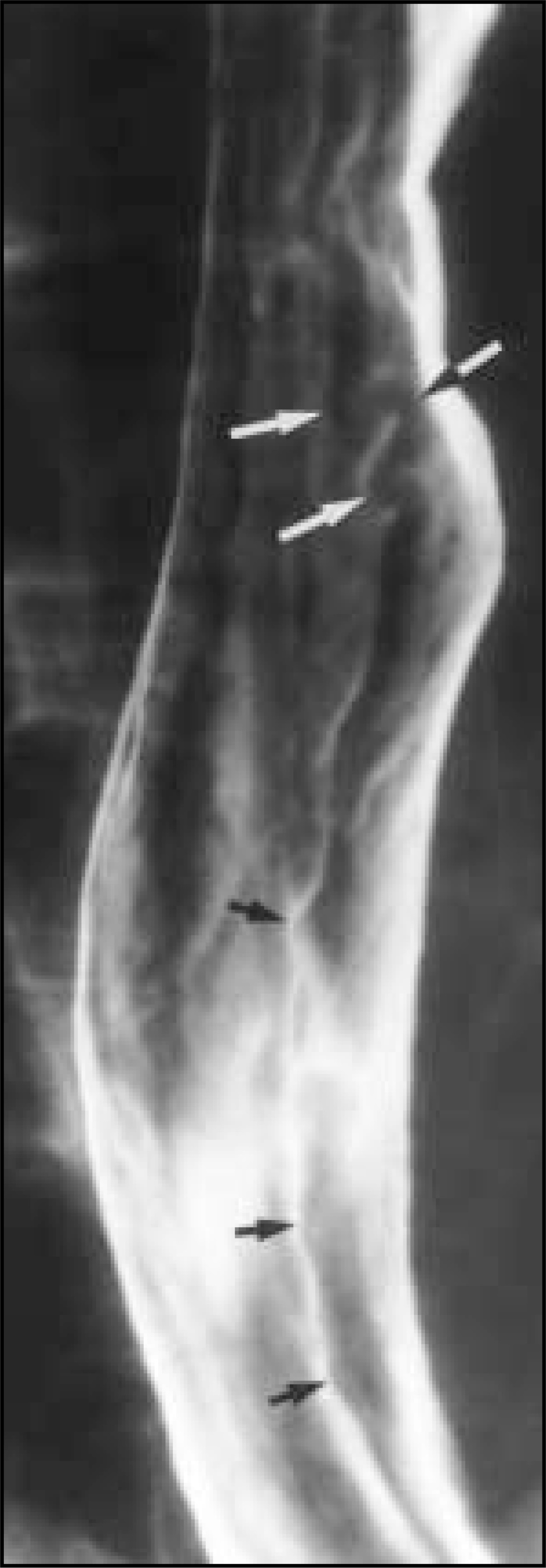
Esophageal strictures
The differential diagnosis of an esophageal stricture depends on the morphology and location of the stricture as well as on the clinical history. Benign strictures typically manifest as smooth, tapered areas of concentric narrowing (figure 11).1 Asymmetric scarring may result in sacculation, flattening, or other deformity of the wall. In contrast, malignant strictures are manifest as eccentric narrowings, thicker on the side where the tumor originated.23 The mucosal surface is irregular, with nodules of varying size disrupting the surface and barium being trapped in areas of ulceration. The margins of malignant strictures often appear abrupt and shelf-like (figure 12). Unlike malignant lesions in the colon, however, malignant esophageal lesions may have sloped or tapered margins, as the soft esophageal submucosa and muscularis propria provide little resistance to the longitudinal spread of tumor. In some patients, a plaque-like indentation of the lumen is seen (figure 13). If any mucosal irregularity or plaque-like flattening is identified in the region of an esophageal stricture, endoscopy and biopsy are required to exclude carcinoma.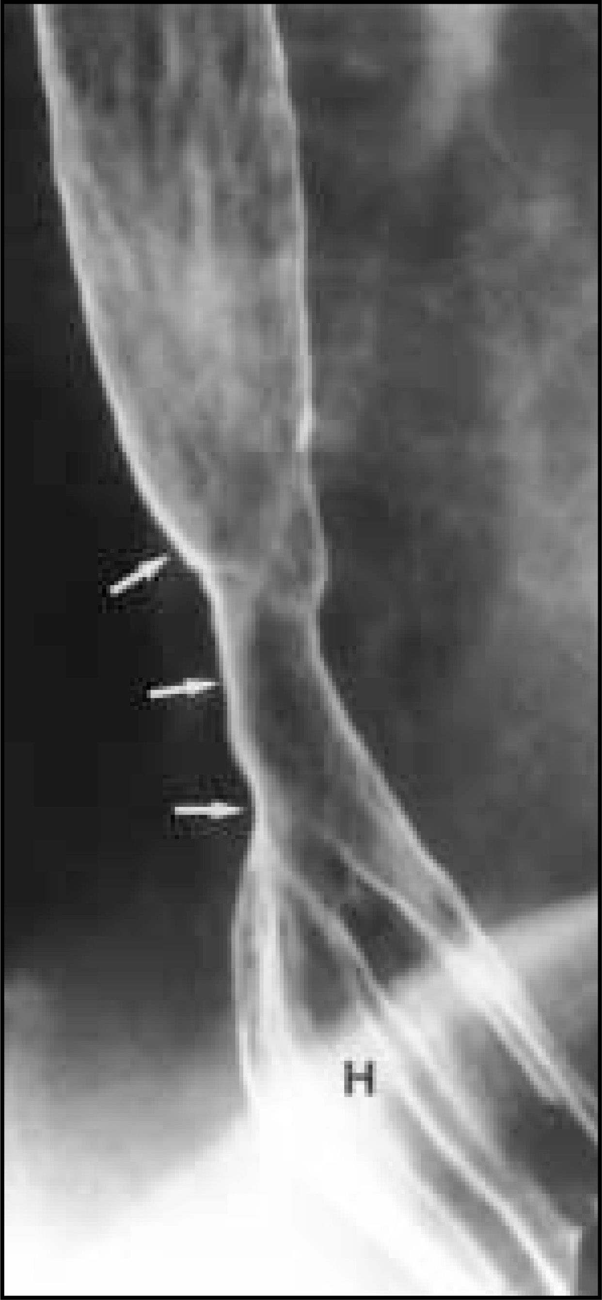
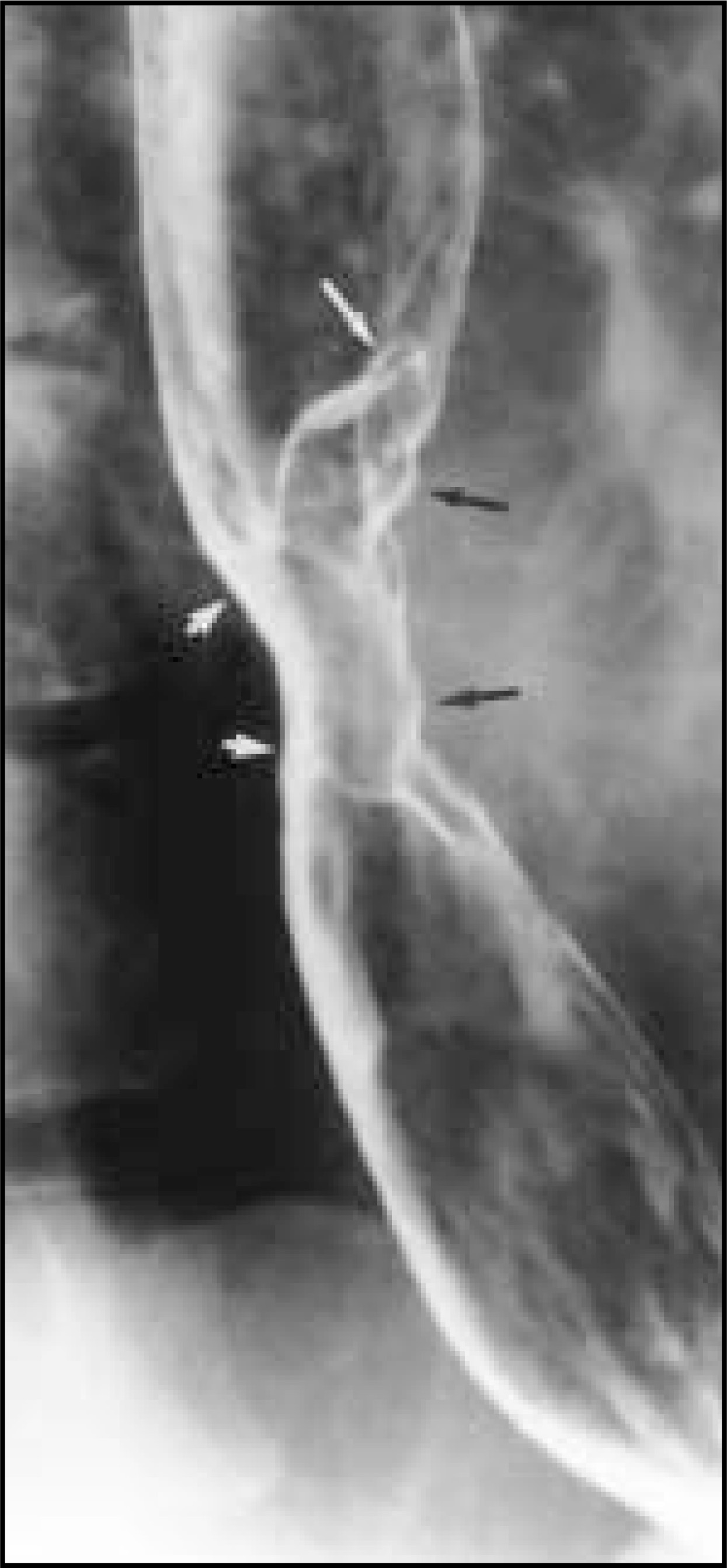
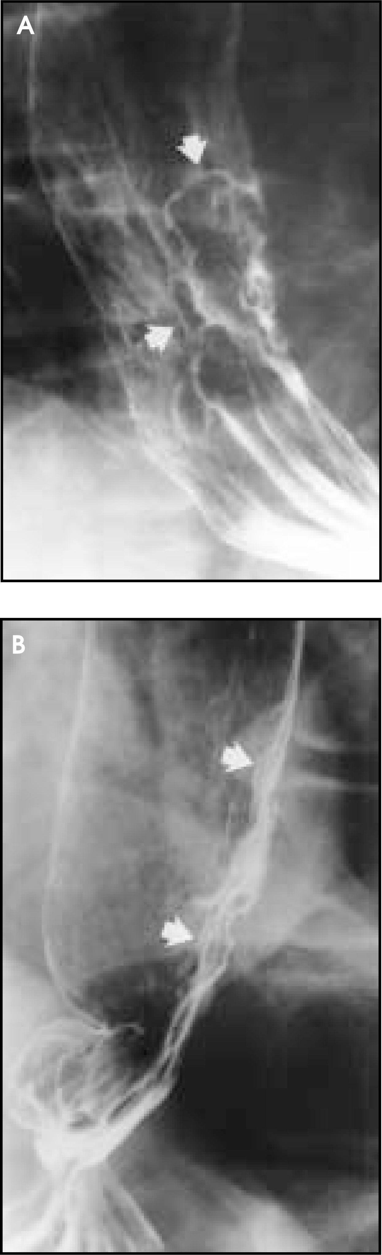
The most common causes of short distal esophageal strictures are gastroesophageal reflux and carcinoma (Table 4; figures 11 and 14). Long distal esophageal strictures are often due to severe acid exposure related to Zollinger-Ellison syndrome, prolonged nasogastric intubation, or alkaline reflux esophagitis (Table 4). Some patients with Crohn’s disease may also develop long distal esophageal strictures. A wide variety of conditions cause midesophageal strictures (Table 4).29-34 The combination of the clinical history, physical examination findings, and radiographic appearance of the strictures often enables a specific diagnosis.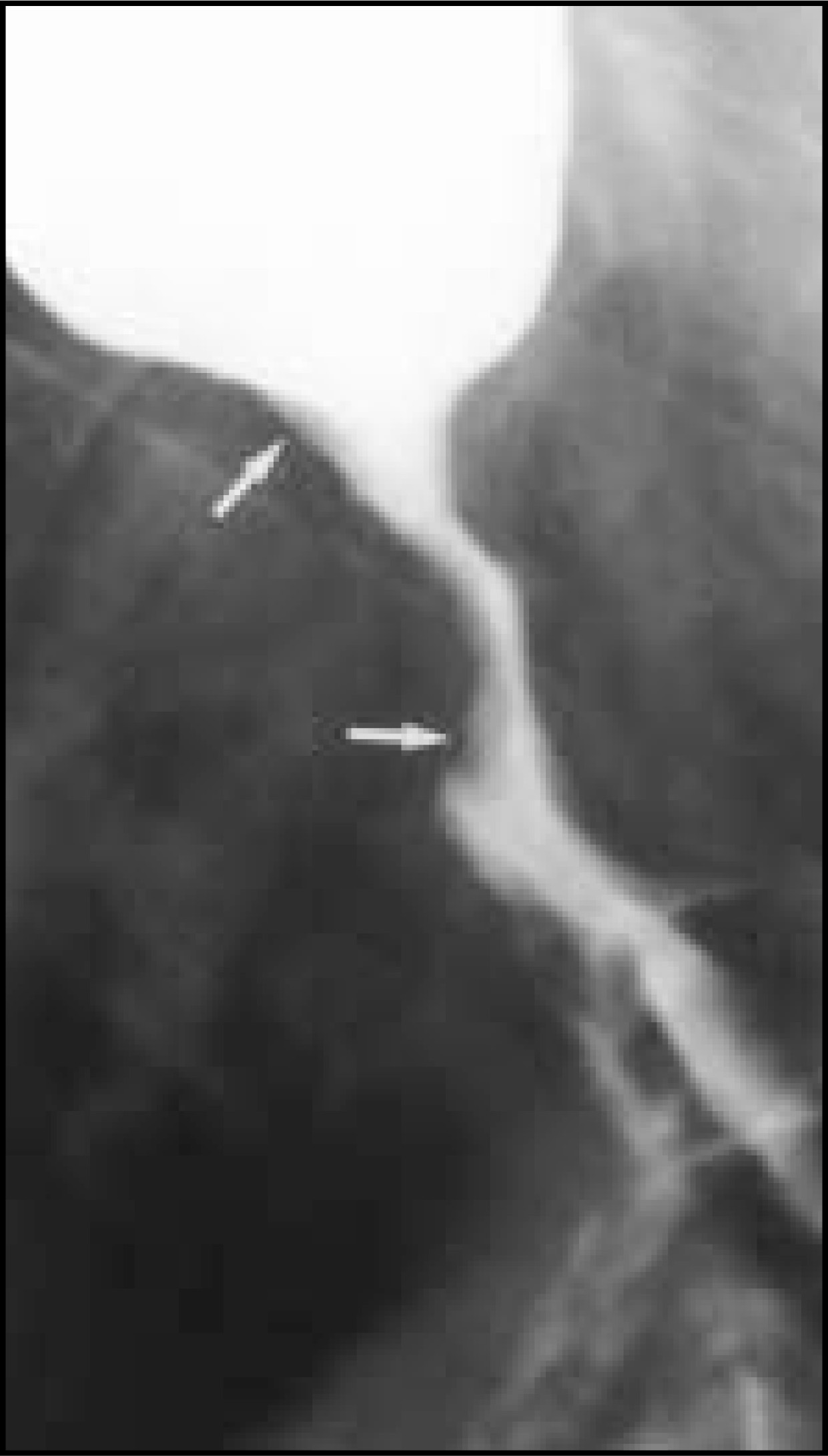
Strictures related to reflux esophagitis are usually seen in the distal esophagus. Some reflux-induced strictures are smooth and tapered (figure 11). However, other reflux-induced strictures are associated with enough asymmetric scarring to cause sacculation in the area of tapering. These sacculations due to scarring should not be confused with ulcers. Other reflux-induced strictures may be associated with such severe scarring and esophageal shortening that a hiatal hernia is even present in the erect position.
In the presence of a hiatal hernia and gastroesophageal reflux, a benign-appearing midesophageal stricture or reticular pattern should be strongly suggestive of Barrett’s esophagus.30 Strictures associated with Barrett’s esophagus are more frequently seen in the distal esophagus, however. Patients with distal esophageal strictures and reflux changes have a moderate risk of Barrett’s esophagus, between 20% and 40%.30 Patients with reflux esophagitis alone have about a 10% risk of Barrett’s esophagus.30 Conversely, a very low risk of Barrett’s esophagus is present if the esophageal mucosa is smooth, if a stricture is not seen, and if only gastroesophageal reflux or a hiatal hernia is present.
Schatzki rings are of unknown etiology, possibly related to gastro-esophageal reflux. They are thin (1 to 3 mm in thickness), symmetric rings at the esophagogastric junction, frequently seen above a small hiatal hernia (figure 15).31 Schatzki rings are best demonstrated in the prone position and are sometimes detected only with a solid bolus.32 Rings <13 mm in luminal diameter invariably cause dysphagia, whereas rings 13 to 20 mm in luminal diameter may or may not cause dysphagia.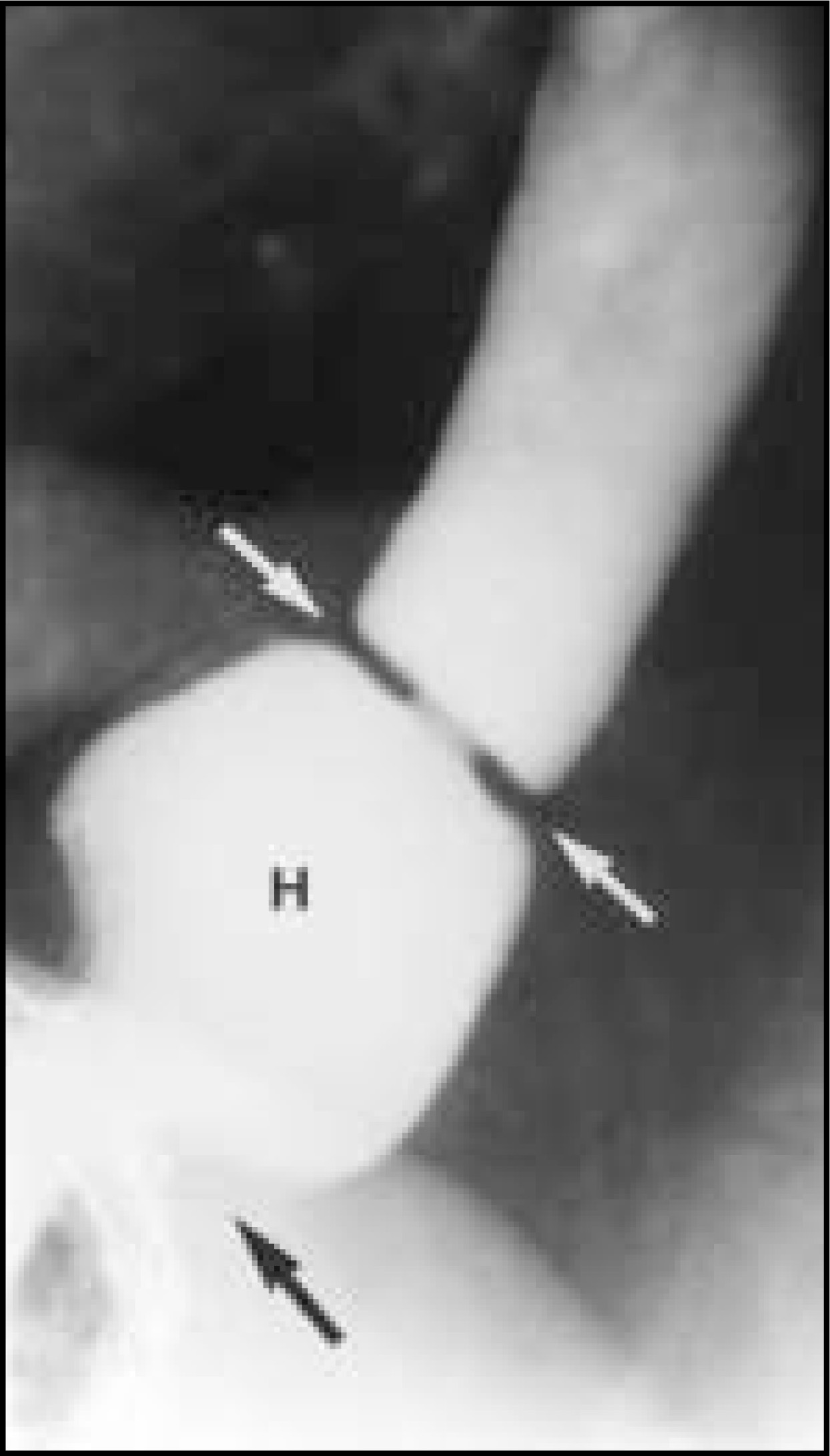
Acute radiation damage causes small ulcers, mucosal granularity, and abnormal peristalsis.11 Chronic radiation strictures are usually smooth and tapered.
Caustic ingestion can result in one or more long strictures in the mid-esophagus or diffuse esophageal narrowing. Stricture formation occurs as early as 4 to 6 weeks after ingestion of a caustic agent. Any irregularity in a long-standing lye stricture should be suspicious for esophageal carcinoma until proven otherwise.
In the classic form of esophageal intramural pseudodiverticulosis, numerous 1- to-3-mm flask-shaped outpouchings are associated with a long cervical or upper thoracic esophageal stricture (figure 16).33 In the more common form of esophageal intramural pseudodiverticulosis, however, the outpouchings are associated with a short, distal, reflux-induced stricture.34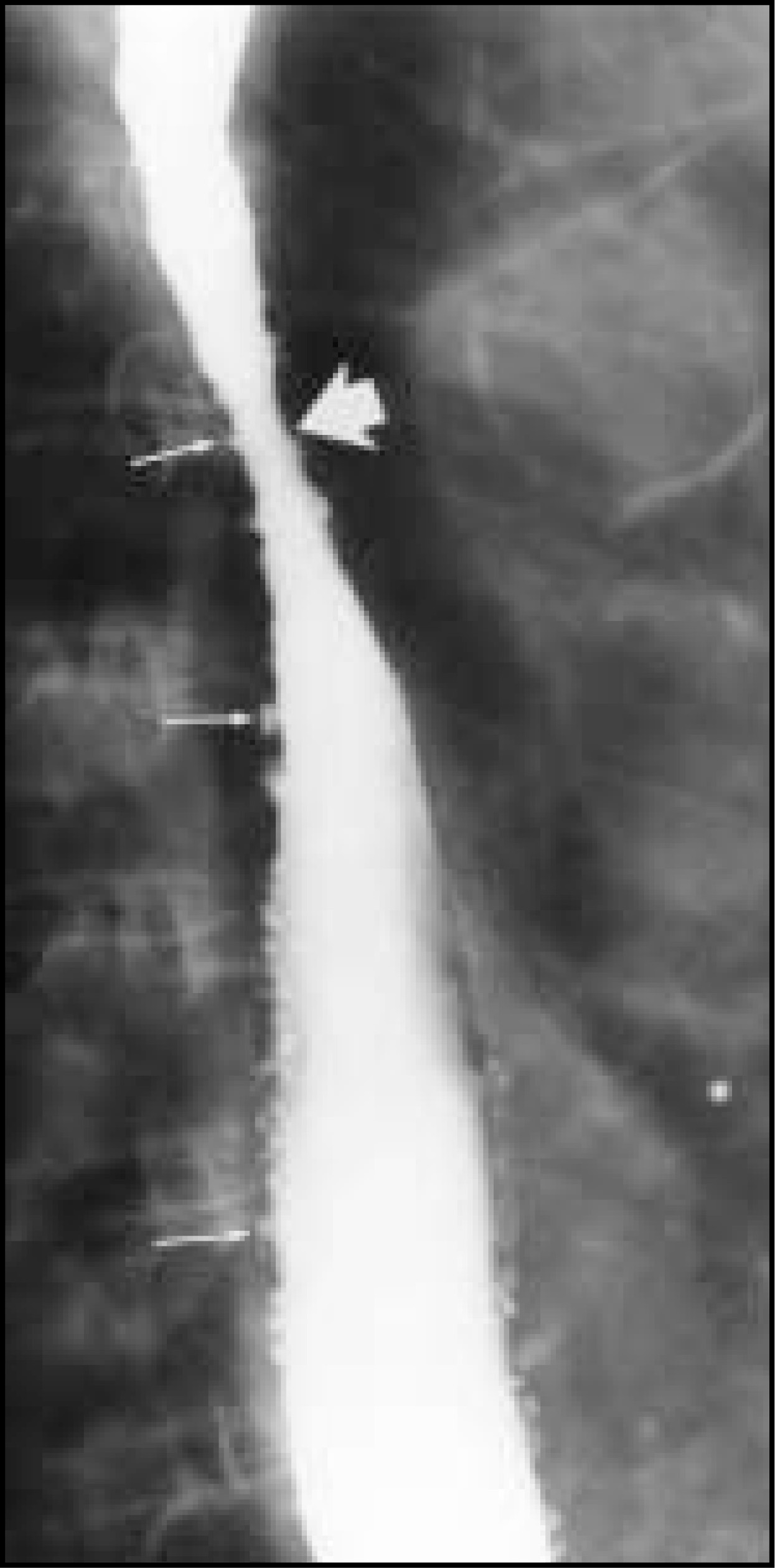
Primary or metastatic cancers are frequent causes of midesophageal strictures. Some squamous cell carcinomas have an elongated, circumferentially infiltrating appearance (figure 12). An eccentric, annular lesion may be seen with a coarsely lobulated contour and barium trapped within tumor nodules. Adenocarcinomas of the esophagus arise in dysplastic columnar epithelium within Barrett’s mucosa. Adenocarcinomas are found most frequently in the distal esophagus. Adenocarcinomas can have an infiltrative (figure 14), plaque-like (figure 13), ulcerative, or polypoid appearance. Unlike squamous cell carcinomas, adenocarcinomas have a marked tendency to invade the gastric cardia and fundus. Metastases to the esophagus most frequently involve the subcarinal region due to direct invasion by tumor from subcarinal lymph nodes (figure 17) or from the left mainstem bronchus.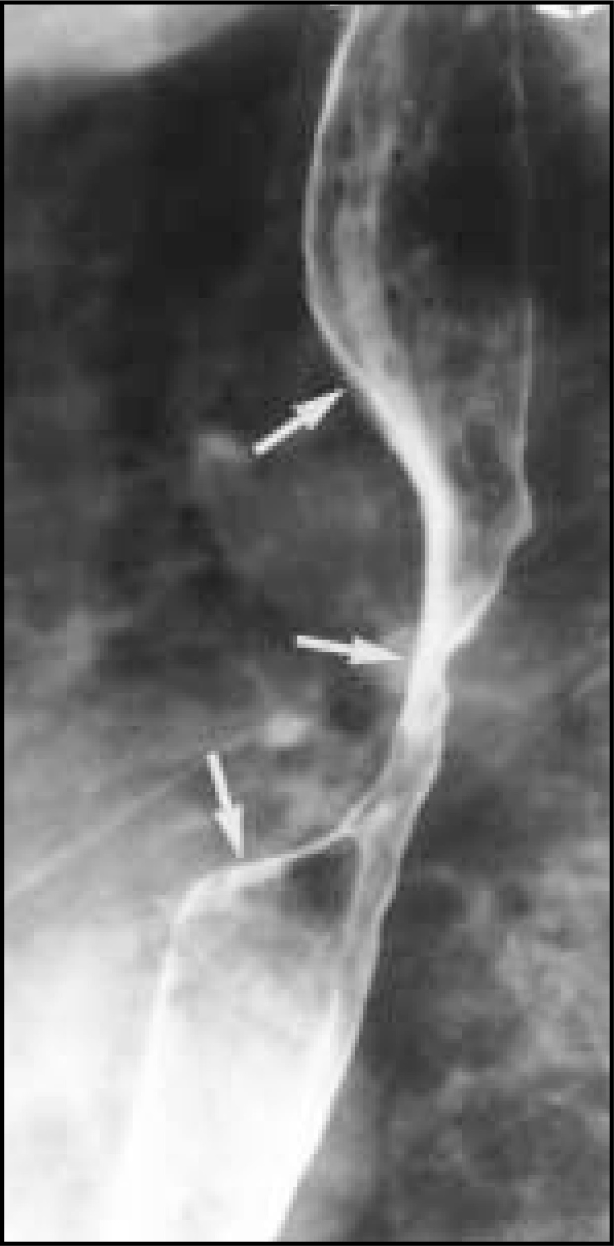
Polypoid intraluminal masses
A wide variety of polypoid masses are seen in the esophagus (Table 5).35-41 Polypoid masses are demonstrated radiographically as radiolucent filling defects in the barium pool (figure 18) or as barium-etched lines within the esophageal lumen. Polypoid masses must first be distinguished from foreign bodies (figure 19). The clinical history is crucial for the diagnosis of foreign bodies. These patients typically complain of abrupt-onset dysphagia or odynophagia during eating and the sensation that food is stuck in the substernal region. The polypoid filling defect of the foreign body may be associated with an irregular meniscus of barium superiorly. Perforation is uncommon, usually occurring after the impaction has been present longer than 24 hours. A repeat esophagram should be performed after the foreign body has been removed to exclude an esophageal stricture or motor disorder as the cause of the food impaction.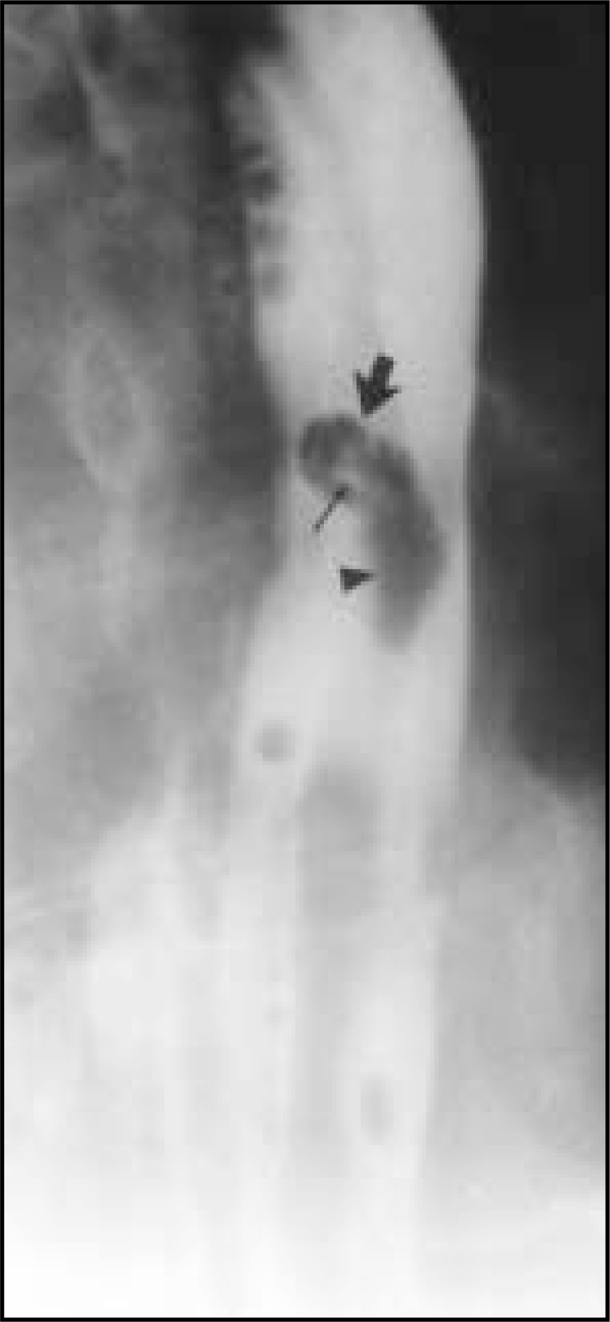
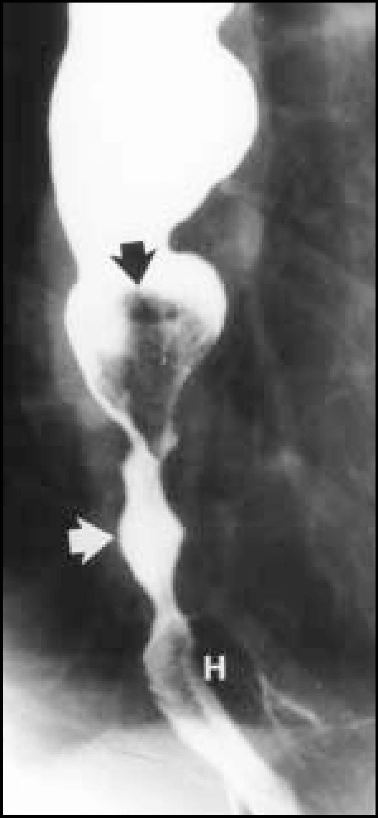
Squamous papillomas are the most common benign mucosal tumor of the esophagus (figure 18), appearing as small, sessile, slightly lobulated polyps. The esophagus is one organ of the gastrointestinal tract in which true leiomyomas form. Most tumors arising in the mesenchyme are undifferentiated gastrointestinal stromal tumors of unknown malignant potential. Leiomyomas, however, are true proliferations of smooth muscle and are the most common submucosal mass in the esophagus. Granular cell tumors are another rare cause of submucosal masses in the esophagus.36
Polyps at the esophagogastric junction are frequently related to chronic gastroesophageal reflux disease, termed “inflammatory esophagogastric” or “sentinel” polyps.40 These polyps are smooth-surfaced enlargements atop a thickened rugal fold at the gastric cardia. If any surface irregularity is seen, however, endoscopy must be performed to exclude a malignant tumor at the cardia or in Barrett’s esophagus.
Some squamous cell carcinomas have a polypoid (figure 20) rather than infiltrating appearance.38,39 Small cell carcinomas are rare tumors that typically manifest as small, centrally ulcerated masses in the midesophagus.37 Spindle cell carcinomas are usually large, polypoid masses that expand the esophageal lumen without causing significant obstruction.39 Despite the relatively noninfiltrating appearance of spindle cell carcinomas, 5-year survival rates in these patients are as dismal as in those patients with squamous cell carcinomas. Rarely, primary malignant melanoma of the esophagus may be manifest as a bulky, polypoid intraluminal mass indistinguishable from spindle cell carcinoma.41 AR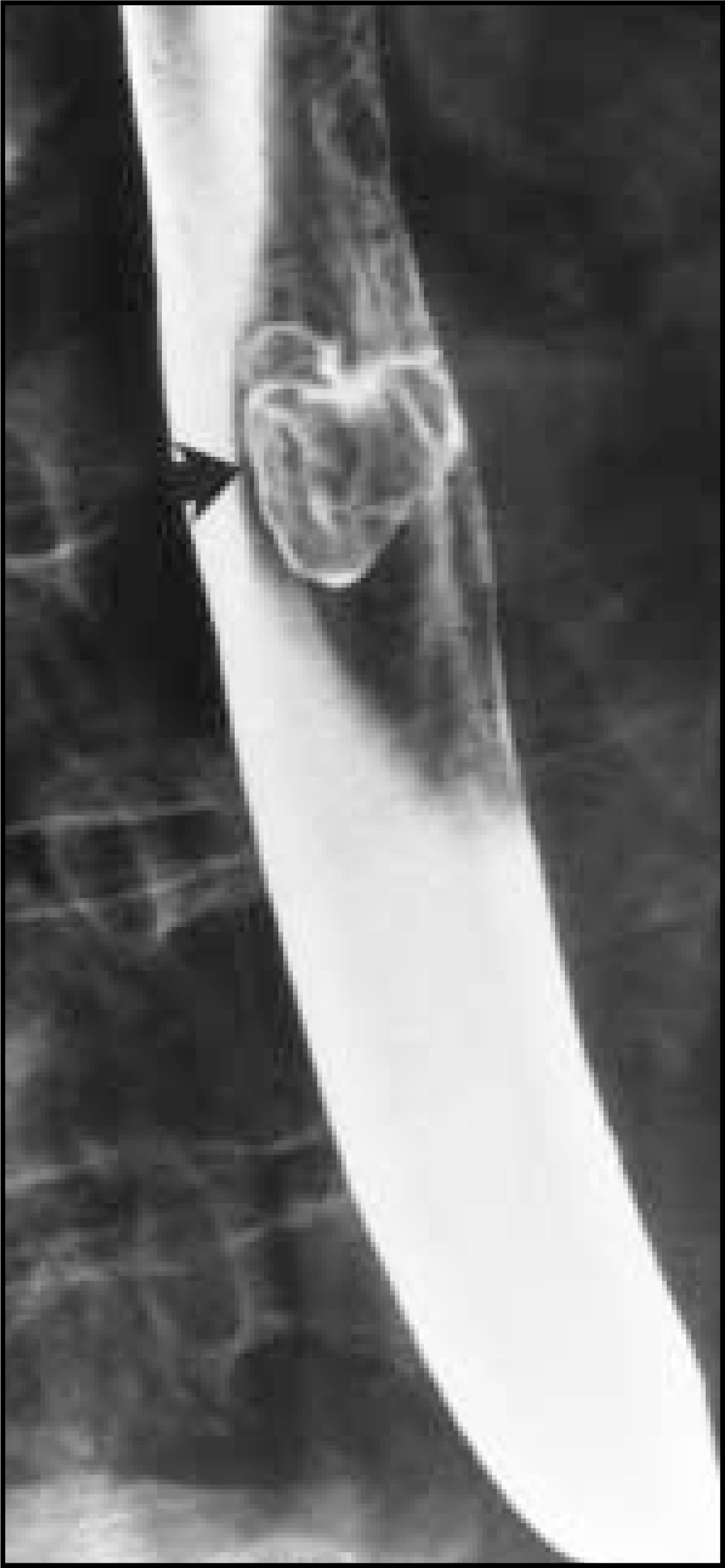
References
- Levine M, Laufer I, Levine M, Rubesin S, Laufer I. Double Contrast Gastrointestinal Radiology. 2000:90-126.
- Levine M. Radiology of esophagitis: A pattern approach. Radiology. 1991;179:1-7.
- Rubesin S, Laufer I, Gore R, Levine M. Textbook of Gastrointestinal Radiology. 2000:44-66.
- Levine M, Laufer I, Kressel H, Friedman H. Herpes esophagitis. AJR Am J Roentgenol. 1981;136:863-866.
- Levine M, Loevner L, Saul S. Herpes esophagitis: Sensitivity of double-contrast esophagography. AJR Am J Roentgenol. 1988;151:57-62.
- Rubesin S, Levine M, Laufer I, Thompson W. Common Problems in Gastrointestinal Radiology. 1989:108-117.
- Shortsleeve M, Levine M. Herpes esophagitis in otherwise healthy patients: Clinical and radiographic findings. Radiology. 1992;182:859-861.
- Creteur V, Laufer I, Kressel H. Drug-induced esophagitis detected by double-contrast radiography. Radiology. 1983;147:365-368.
- Bova J, Dutton N, Goldstein H, Hoberman L. Medication-induced esophagitis: Diagnosis by double contrast esophagography. AJR Am J Roentgenol. 1987;148:731-732.
- Gohel V, Long B, Richter G. Aphthous ulcers in the esophagus with Crohn colitis. AJR Am J Roentgenol. 1981;137:872-873.
- Collazzo L, Levine M, Rubesin S, Laufer I. Acute radiation esophagitis: Radiographic findings. AJR Am J Roentgenol. 1997;169:1067-1070.
- Levine M. Drug-induced disorders of the esophagus. Abdom Imaging.. 1999;24:3-8.
- Levine M, Loercher G, Katzka D. Giant HIV-related ulcers in the esophagus. Radiology. 1991;180:323-326.
- Sor S, Levine M, Kowalski T. Giant esophageal ulcers in HIV-positive patients: Clinical, radiographic and pathologic findings. Radiology.. 1995;194:447-451.
- Levine M, Woldenberg R, Herlinger H, Laufer I. Opportunistic esophagitis in AIDS: Radiographic diagnosis. Radiology. 1987;165:815-820.
- Balthazar E, Megibow A, Hulnick D. Cytomegalovirus esophagitis in AIDS: Radiographic features in 16 patients. AJR Am J Roentgenol.. 1987;149:919-923.
- Gloyna R, Zornoza J, Goldstein H. Primary ulcerative carcinoma of the esophagus. AJR Am J Roentgenol.. 1977;129:599-600.
- Levine M, Rothstein R, Laufer I. Giant esophageal ulcer due to Clinoril. AJR Am J Roentgenol. 1991;156:955-956.
- Levine M, Macones A, Laufer I. Candida esophagitis: Accuracy of radiographic diagnosis. Radiology. 1985;154:581-587.
- Berliner L, Redmond P, Horowitz L, Ruoff M. Glycogen plaques (glycogenic acanthosis) of the esophagus. Radiology. 1981;141:607-610.
- Glick S, Teplick S, Goldstein J. Glycogenic acanthosis of the esophagus. AJR Am J Roentgenol.. 1982;139:683-688.
- Graziani L, Bearzi I, Romagnoli A. Significance of diffuse granularity and nodularity of the esophageal mucosa at double-contrast radiography. Gastrointest Radiol.. 1985;10:1-6.
- Rubesin S. Gallery of double contrast terminology. Gastro Clin North Am.. 1995;24:259-288.
- Levine M, Cajade A, Herlinger H, Laufer I. Pseudomembranes in reflux esophagitis. Radiology.. 1986;159:43-45.
- Itai Y, Kogure T, Okuyama Y, Akiyama H. Superficial esophageal carcinoma: Radiological findings in double-contrast studies. Radiology.. 1978;126:597-601.
- Levine M, Dillon E, Saul S, Laufer I. Early esophageal cancer. AJR Am J Roentgenol.. 1986;146:507-512.
- Levine M, Kressel H, Caroline D. Barrett esophagus: Reticular pattern of the mucosa. Radiology. 1983;147:663-667.
- Yates C, LeVine M, Jensen K. Varicoid carcinoma of the esophagus. Radiology.. 1977;122:605-608.
- Levine M, Borislow S, Rubesin S, O’Brien C. Proximal esophageal stricture caused by Motrin. Abdom Imaging.. 1994;19:6-7.
- Gilchrist A, Levine M, Carr R. Barrett’s esophagus: Diagnosis by double-contrast esophagography. AJR Am J Roentgenol.. 1988;150:97-102.
- Levine M, Rubesin S. Radiologic investigation of dysphagia. AJR Am J Roentgenol.. 1990;154:1157-1163.
- Low V, Rubesin S. Contrast evaluation of the pharynx and esophagus. Rad Clin North Am.. 1993;31:1265-1291.
- Cho S, Sanders M, Turner M. Esophageal intramural pseudodiverticulosis. Gastrointest Radiol. 1981;5:9-16.
- Levine M, Moolten D, Herlinger H, Laufer I. Esophageal intramural pseudodiverticulosis: A reevaluation. AJR Am J Roentgenol.. 1986;147:1165-1170.
- Olmsted W, Lichtenstein J, Hyams V. Polypoid epithelial malignancies of the esophagus. AJR Am J Roentgenol.. 1983;140:921-925.
- Rubesin S, Herlinger H, Sigal H. Granular cell tumors of the esophagus. Gastrointest Radiology.. 1985;10:11-15.
- Levine M, Pantongrag-Brown L, Buck J. Small-cell carcinoma of the esophagus: Radiographic findings. Radiology.. 1996;199:703-705.
- Levine M, Laufer I, Yamada A, Levine M, Rubesin S, Laufer I. Double Contrast Gastrointestinal Radiology.. 2000:126-148.
- Levine M, Gore R, Levine M. Textbook of Gastrointestinal Radiology.. 2000:435-451.
- Bleshman M, Banner M, Johnson R. The inflammatory esophagogastric junction polyp and fold. Radiology.. 1978;128:589-593.
- Yoo C, Levine M, McLarney J, Lowry M. Primary malignant melanoma of the esophagus: Radiographic findings in seven patients. Radiology.. 1998;209:455-459.
Citation
. Differential diagnosis of esophageal disease on esophagography. Applied Radiology. 2001;30(10):11-21. doi:10.37549/AR1048.