Ectopic ureteral insertion into the seminal vesicle associated with ipsilateral renal dysplasia
Applied Radiology — Vol. 45 , Issue 9 , pp. 46 -47
DOI: 10.37549/AR2305
Published: September 1, 2016
Categories
CASE SUMMARY
A 34-year-old man presented with perineal and pelvic pain, urinary intermittency and dysuria of 2 months’ duration. He reported a previous episode of prostatitis.
IMAGING FINDINGS
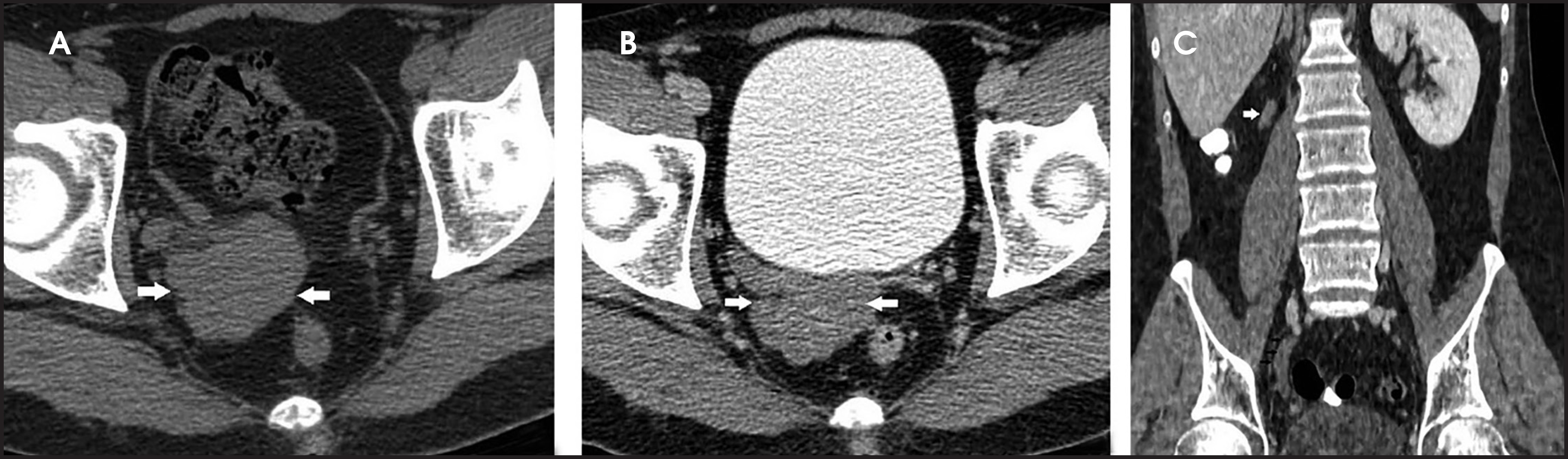
Computed tomography of the abdomen and pelvis revealed a non-enhancing cystic mass of spontaneously high density in the right paravesical area (Figures 1A and 1B). The right seminal vesicle was not identified. The right ureter was dilated, filled with the same density material and appeared blunted distally. An ipsilateral renal remnant was also noted (Figure 1C).
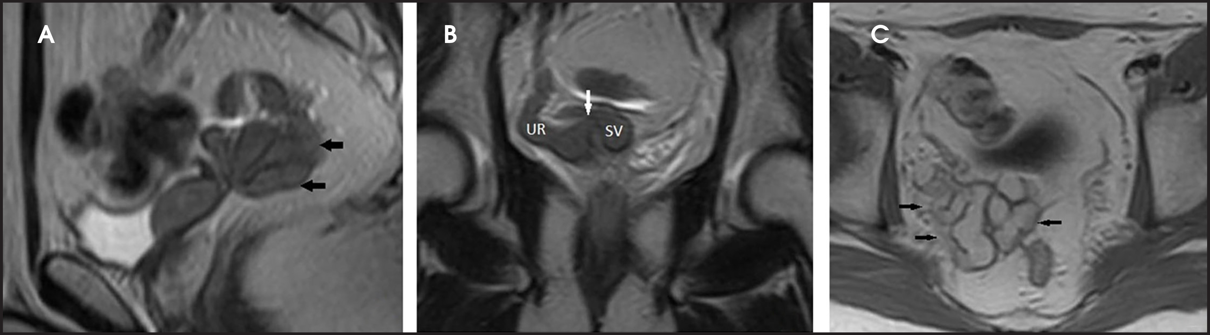
Magnetic resonance imaging of the pelvis demonstrated a dilated right ureter ectopically draining into a multiloculated cystic mass representing an ectatic right seminal vesicle (Figure 2). The cystic and ureteral content showed high signal on T1-weighted images (Figure 2C) and low signal on T2-weighted images (Figures 2A and 2B) suggestive of rich proteinaceous material.
DIAGNOSIS
Ectopic ureteral insertion into the seminal vesicle associated with ipsilateral renal dysplasia
DISCUSSION
There is an association between congenital renal abnormalities and internal genital malformations related to their close embryologic relationship.1 Ectopic ureter draining into the genital system is a rare entity and results from a more cranial origin of the ureteral bud from the mesonephric duct with resultant ureteral stump opening in the mesonephric duct derivatives (seminal vesicles, ejaculatory ducts, or vas deferens). The inadequate stimulation of metanephrogenic blastema by the ureteric bud results in renal maldevelopment.2
Ectopic ureters are more common in females and present early with incontinence.3 The presentation is often delayed in males to early adulthood.4 The commonly reported symptoms are perineal or pelvic pain, urinary tract infection, prostatitis, epididymitis, hematuria, hemospermia and infertility.4,5
Several imaging techniques have been used in the evaluation of these anomalies. Intravenous urography may show a smooth indentation on the urinary bladder from mass effect of the enlarged seminal vesicle. It can also demonstrate the abnormal excretion of the ipsilateral kidney.5,6
Pelvic or transrectal ultrasound can detect the enlarged seminal vesicle, confirm its cystic nature and delineate the dilated ureter.5,6
A computed tomography (CT) scan can demonstrate the cystic pelvic mass and reveal the associated renal anomalies. However, CT scans sometimes cannot show the ureteral opening into the seminal vesicle.4,6
Magnetic resonance imaging (MRI) has a superior soft tissue contrast and shows better the connection between the ectopic ureter and the seminal vesicle.4,6 The seminal vesicle appears as an elongated cystic structure with thin septa and of variable signal depending on protein content.6
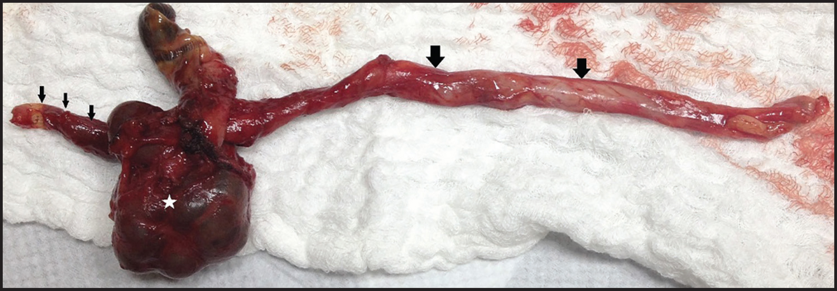
Our patient underwent surgical excision of the right seminal vesicle and the ipsilateral ureter. Histopathological analysis proved our radiological diagnosis.
CONCLUSION
There is an association between genital tract malformation and renal maldevelopment related to their close embryologic origin.
We report a rare case of right ectopic ureteral insertion into the seminal vesicle associated with ipsilateral renal agenesis. The diagnosis was suggested by computed tomography and confirmed on magnetic resonance imaging and pathologically.
References
- Narlawar R, Hanchate V, Raut A, Hira P, Nagar A, Chaubal N. Renal agenesis and seminal vesicle cyst. J Ultrasound Med. 2003;22:225-228.
- Buogo G, Rodrigues E, Rodriguez P. Laparoscopic removal of seminal vesicle cyst with ectopic ureteral insertion and renal remnant. International Braz J Urol. 2002;20:335-337.
- Berrocal T, Lopez-Pereira P, Arjonilla A, Gutierrez J. Anomalies of the distal ureter, bladder, and urethra in children: embryologic, radiologic, and pathologic features. Radiographics. 2002;22:1139-1164.
- Arora S, Breiman R, Webb E, Westphalen A, Yeh B, Coakley F. CT and MRI of congenital anomalies of the seminal vesicles. AJR. 2007;189:130-135.
- Livingston L, Larsen C. Seminal vesicle cyst with ipsilateral renal agenesis. AJR. 2000;175:177-180.
- Abou El-Ghar M, El D. Ectopic insertion of the ureter into the seminal vesicle. World J Radiol. 2013;5((9)):349-351.
Citation
. Ectopic ureteral insertion into the seminal vesicle associated with ipsilateral renal dysplasia. Applied Radiology. 2016;45(9):46-47. doi:10.37549/AR2305.