Endogenous lipoid pneumonia
Applied Radiology — Vol. 37 , Issue 5 , pp. 32 -34
DOI: 10.37549/AR1612
Published: May 1, 2008
Categories
CASE SUMMARY
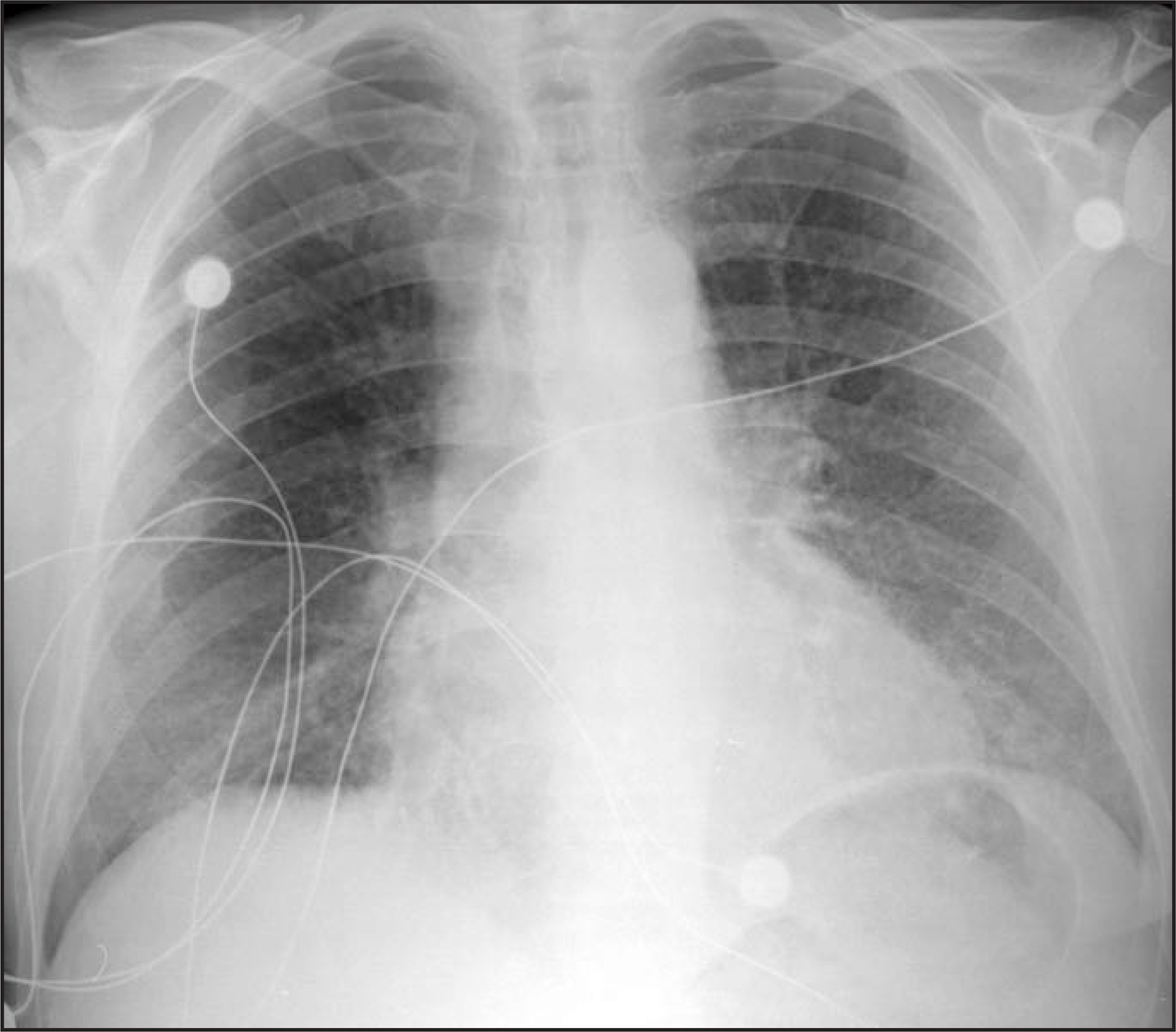
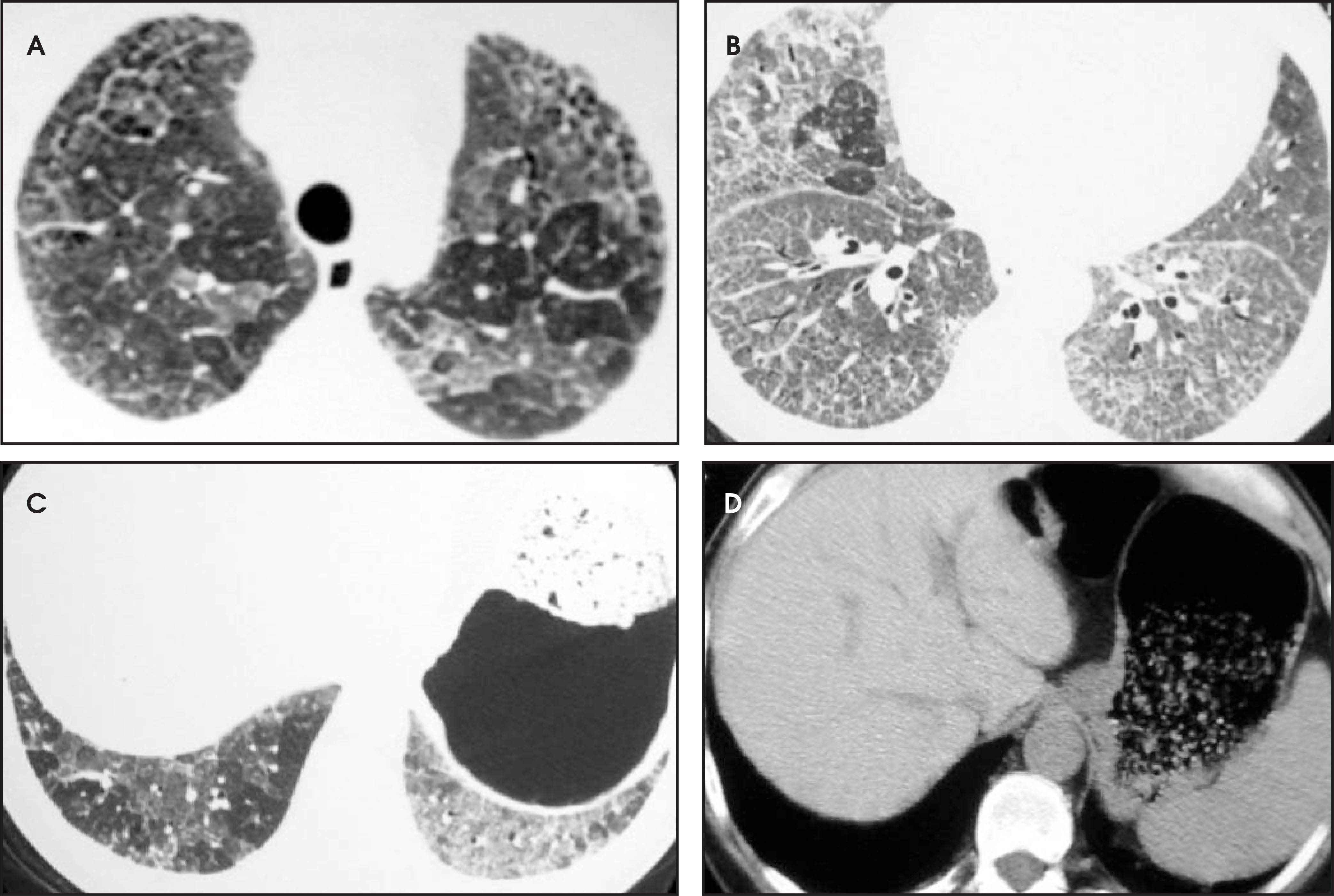
A 50-year-old man who was receiving amiodarone therapy for ventricular tachycardia presented to the emergency department following 2 syncopal episodes. A chest radiograph was obtained (Figure 1), and based on these findings, high-resolution CT (HRCT) of the chest was performed (Figure 2).
IMAGING FINDINGS
The chest radiograph showed nonspecific bibasilar reticular opacities (Figure 1), which were new since the previous radiograph 1 year earlier (not shown). High-resolution CT showed extensive interlobular and intralobular interstitial thickening superimposed on diffuse ground-glass opacities (the “crazy-paving” pattern), primarily involving the middle and lower lungs (Figure 2, A through C). There was also relatively high attenuation of the hepatic parenchyma as compared with the spleen (Figure 2D).
Transbronchial biopsy of the right lower lobe revealed pulmonary fibrosis, hemosiderin-laden macrophages, and scattered “foamy” macrophages with clear cytoplasm. No granulomatous in flam mation was identified. A clinical diag nosis of amiodarone pulmonary toxicity was made, and the drug was discontinued after a cardiac defibrillator was implanted.
DIAGNOSIS
Endogenous lipoid pneumonia resulting from amiodarone toxicity
DISCUSSION
Amiodarone has been used for >30 years for various cardiac disorders, particularly in treating life-threatening cardiac dysrhythmias.1-4 Various adverse effects involving the nervous system, gastrointestinal system, eyes, skin, thyroid, and liver are well known.1 Pulmonary toxicity, first recognized in 1980,5 is the most severe complication. It is estimated that 5% to 10% of patients treated with amiodarone will develop amioda rone pulmonary toxicity, and 5% to 10% of those affected will die from subsequent respiratory complications.2,4,6 Pulmonary toxicity may be mild and reversible or may progress to fibrosis and, ultimately, may be fatal. In most cases, it occurs months after therapy is begun, typically with doses of ≥400 mg per day, although several cases of toxicity developing after 48 hours of intravenous therapy have been reported in patients with acute respiratory distress syndrome who were treated for tachydysrhythmias. Presumably, the alveolar damage leads to im paired surfactant metabolism and makes amiodorone clearance more difficult.7,8
Pharmacologically, amiodarone has high lipid solubility, a large volume of distribution, and an elimination half-life ranging from 40 to 60 days. Thus, not only does the drug have the potential to accumulate in the lung in large quantities, but also, because of extremely slow elimination, lung injury may persist or progress despite cessation of therapy.4
The insidious onset of dry cough, dyspnea, pleuritic pain, weight loss, and sometimes fever comprise the most common symptoms of amiodarone pulmonary toxicity. Physical examination often reveals bilateral crackles and an occasional pleural rub. Although nonspecific, laboratory abnormalities common in amiodarone toxicity include leukocytosis and elevated lactate dehydrogenase level and erythrocyte sedimentation rate. Pulmonary function tests typically show a restrictive pattern and decreased diffusion capacity.3
The initial underlying pathologic process in the lung from amiodarone toxicity is postulated to be a drug-induced phospholipidosis,9 and the mechanism by which phospholipids accumulate in the cell is believed to be by inhibition of intracellular phospholipase when the drug and its metabolites are trapped in alveolar macrophages and type II pneumocyte lysosomes.4 This is speculated to lead to inhibition of lipid degradation and surfactant turnover, a mechanism similarly implicated in pulmonary alveolar proteinosis.10 Alternatively, a hypersensitivity response to the drug may develop, resulting in indirect lung injury.3
On light microscopy, characteristic macrophages with foamy and pale cytoplasm are seen. When interstitial pneumonia is present, nonspecific interstitial pneumonia is the most common pattern identified. Diffuse alveolar damage and bronchiolitis obliterans organizing pneumonia have also been described.1,9 Areas of fibrosis and hemorrhage can occur as well. In a patient developing signs and symptoms of drug toxicity, the presence of these various histologic abnormalities is highly suggestive of drug toxicity.9
Radiographic findings of amiodarone pulmonary toxicity typically include bilateral, sometimes asymmetric, patchy opacities.1,3 CT may show homogeneous peripheral and high-attenuation opacities due to incorporation of iodine-rich amiodarone (37% iodine by weight) and its metabolite desethylamiodarone in type II pneumocytes and alveolar macrophages.6 Only a limited number of pathologic processes are known to result in high-attenuation pulmonary parenchymal abnormalities on CT, including metastatic pulmonary calcifications from secondary hyperparathyroidism in the setting of renal failure, amyloid deposition, and diffuse pulmonary ossification. Besides in the lung, amiodarone can also accumulate in the liver, spleen, and myocardium, causing increased attenuation in these tissues on unenhanced CT. Patients who have pulmonary toxicity almost always have increased liver attenuation, a finding that can help clarify the diagnosis given that the pattern of pulmonary parenchymal abnormalities can vary.
In some cases of amiodarone pulmonary toxicity, the accumulation of phospholipids in alveolar macrophages and in other parenchymal cells leads to lipoid pneumonia.7 On HRCT, lipoid pneumonia may show crazy-paving, a colorful description of patchy interlobular and intralobular septal thickening and ground-glass opacity that may also be seen with other diseases, including pulmonary alveolar proteinosis, lung edema, Pneumocystis jiroveci pneumonia, and bronchioloalveolar carcinoma.
The diagnosis of drug-related pulmonary toxicity is often made based on a combination of clinical and imaging findings with little or no pathologic confirmation given that empirically discontinuing a particular drug may be safer than open or transbronchial lung biopsy. The radio graphic and histologic patterns of drug-induced lung injury are nonspecific, so these findings must be interpreted in the context of detailed clinical information.
CONCLUSION
Amiodarone, while a highly effective antidysrhythmic, has the potential to cause severe pulmonary toxicity, ranging from mild, reversible injury to pulmonary fibrosis. The underlying mechanism is believed to be related to both amiodarone’s large volume of distribution, long tissue half-life, and its interference with the normal lipid degradation pathways in the alveolar macrophages and other pulmonary parenchymal cells. Although the imaging findings may be nonspecific, the constellation of clinical history with the described imaging characteristics and amiodarone deposition in other tissues should alert the radiologist that toxicity from this drug may likely be the source of the patient’s respiratory dysfunction.
References
- Kennedy J, Myers J, Plumb V, Fulmer J. Amiodarone pulmonary toxicity. Clinical, radiologic, and pathologic correlations. Arch Intern Med. 1987;147:50-55.
- Martin W, Rosenow E. Amiodarone pulmonary toxicity. Recognition and pathogenesis (Part 2). Chest. 1988;93:1242-1248.
- Martin W, Rosenow E. Amiodarone pulmonary toxicity. Recognition and pathogenesis (Part I). Chest. 1988;93:1067-1075.
- Martin W. Mechanisms of amiodarone pulmonary toxicity. Clin Chest Med. 1990;11:131-138.
- Rotmensch H, Liron M, Tupilski M, Laniado S. Possible association of pneumonitis with amiodarone therapy. Am Heart J. 1980;100:412-413.
- Rossi S, Erasmus J, McAdams H. Pulmonary drug toxicity: Radiologic and pathologic manifestations. RadioGraphics. 2000;20:1245-1259.
- Donaldson L, Grant I, Naysmith M, Thomas J. Acute amiodarone-induced lung toxicity. Intensive Care Med Intensive Care Med.
- Lapinsky S, Mullen J, Balter M. Rapid pulmonary phospholipid accumulation induced by intravenous amiodarone. Can J Cardiol. 1993;9:322-324.
- Smith G. The histopathology of pulmonary reactions to drugs. Clin Chest Med. 1990;11:95-117.
- Kuhlman J, Teigen C, Ren H. Amiodarone pulmonary toxicity: CT findings in symptomatic patients. Radiology Radiology.
Citation
. Endogenous lipoid pneumonia. Applied Radiology. 2008;37(5):32-34. doi:10.37549/AR1612.