Epidermoid inclusion cyst of the scrotum
Applied Radiology — Vol. 37 , Issue 6 , pp. 40C -40E
DOI: 10.37549/AR1621
Published: June 1, 2008
Categories
CASE SUMMARY
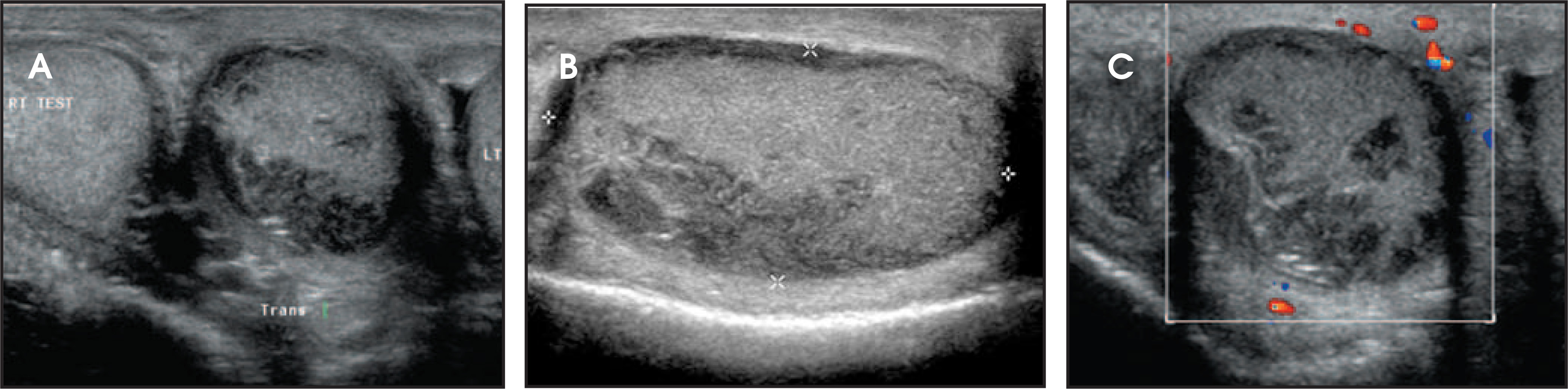
A 62-year-old white man presented to the urology clinic with a painless midline scrotal mass that had reportedly undergone recent growthafter nearly 30 years of stability. On physical examination, the scrotum exhibited moderate enlargement and tethered midline cutaneous thickening. Additionally, there was a palpable midline scrotal mass from which the testicles could not be clearly differentiated. An ultrasound examination was performed (Figure 1).
IMAGING FINDINGS
Sonographic evaluation revealed a midline solid scrotal mass that measured 3.0 × 3.0 × 2.5 cm, with dominant echogenicity and echotexture similar to that of normal testicle but with large heter o geneously hypoechoic regions throughout (Figure 1, A and B). Color Doppler imaging depicted no internal or capsular flow (Figure 1C). The left and right testicles were sonographically normal and entirely separate from the mass.
SURGICAL FINDINGS
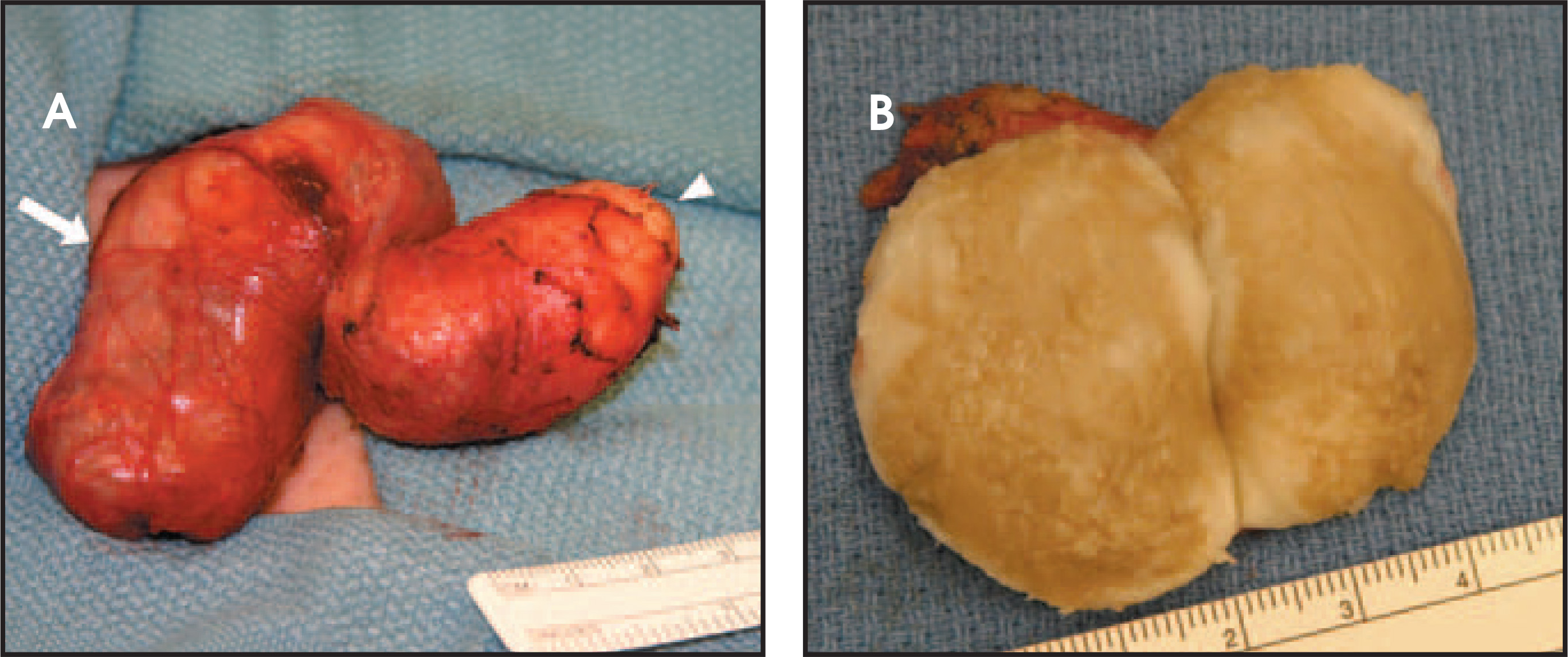
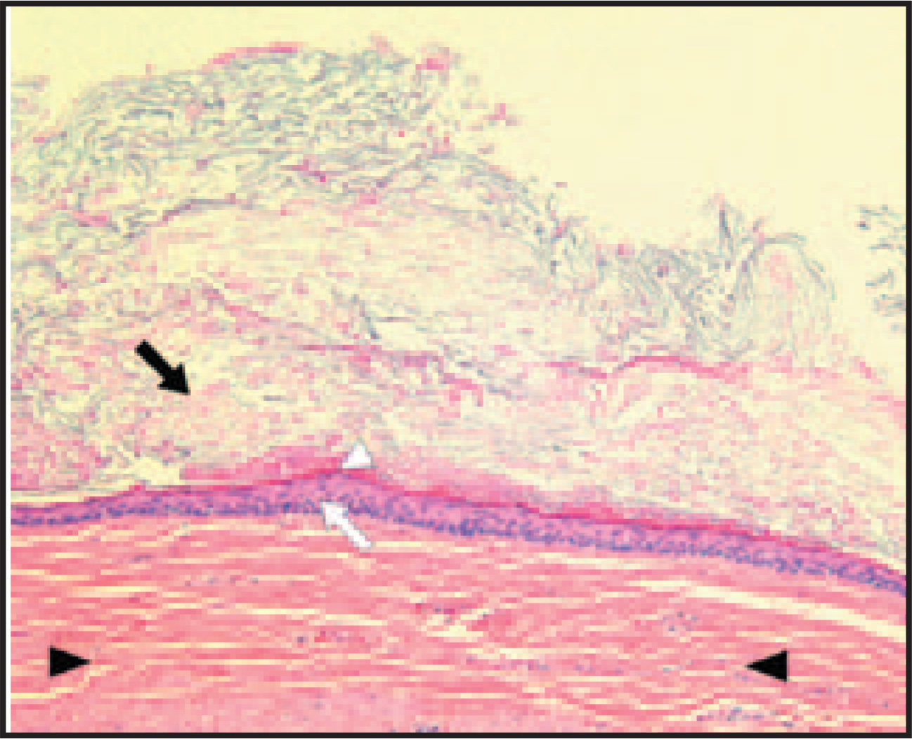
The patient underwent surgical excision; the intrascrotal extratesticular nature of this lesion was confirmed (Figure 2A). Sectioningrevealed a solid ovoid structure containing a soft tan-white to tan-brown cheesy material (Figure 2B). The histopathologic examination confirmed the lesion to be an epidermoid inclusion cyst (EIC) and noted evidence of prior rupture and giant cell granulomatousreaction (Figure 3).
DIAGNOSIS
Epidermoid inclusion cyst of the scrotum
DISCUSSION
Epidermoid inclusion cysts are benign tumors that occur after implantation of epidermal tissue into the dermis or subcutis, which may result eitherfrom trauma or abnormal embryologic closure of the median raphe and the urethral groove.1The slowly enlarging mass usually contains a combination of keratin and cholesterol, often in a laminated configuration arising from a stratified squamous epithelial wall.1While patients usually presentasymptomatically as young or middle-aged adults, cases complicated by rupture and bacterial infection,2extension into the pelvis,3and even extension to the rectal wall4have been reported. Epidermoid inclusion cysts appear solid on all imaging modalities and are often found to contain a laminated cheesy material on gross examination.
When evaluating the scrotum, ultrasound is an extremely effective imaging modality, detecting intrascrotal masses with a sensitivity ofnearly 100%.5Further, ultrasound has been reported to be 98% to 100% accurate in differentiating intratesticular lesions (which are more commonly malignant) from extratesticular lesions (which are usually benign).5Benign intrascrotal extratesticular lesions include EICs, adenomatoid tumor of the epididymis, fibroma, lip oma, and cholesterol granuloma. Granulomatous infectious lesions such as tuberculosis and coccidioidomycosis have also been described as producing masslike lesions of the epididymis.6Malignant lesions are far more rare but may includelymphoma, liposarcoma, and fibrosarcoma.
Sonographically, an EIC commonly appears as a well-circumscribed round or oval mass that is most often hypoechoic but ranges fromanechoic to hyperechoic depending on its content.1Lee et al7described 5 sonographic patterns found in 24 patients with EICs locatedthroughout the body; 71% of the lesions were either hypoechoic with scattered internal echoes or inhomogeneously hypoechoic.7Only 13%of cases showed the more specific “onion skin” appearance that has been described in intratesticular epidermoid cysts. Lesions with varyingechogenicity similar to those in our case were found in only 8% of cases and were noted to be associated with cyst rupture and granulomatous reaction. Of the 9 cases found to have ruptured, 4 demonstrated internal blood flow on color Doppler. In fact, these were the only lesionsreported the series by Lee et al7to exhibit internal flow by color Doppler.
Magnetic resonance imaging can be particularly helpful in scrotal mass characterization. An EIC typically is seen as a well-demarcated solid mass of characteristically high T2-weighted signal intensity, often surrounded by a low signal capsule.8T1-weightedimaging often reveals homogenous or heterogeneous low signal intensity, and gadolinium administration results in no significant enhancement.8Irregularity of the cyst capsule may indicate past rupture. However, while these imaging features are characteristic of epidermoid cysts,they do not exclude malignant neoplasm, and thus surgical excision is often performed.
CONCLUSION
As this case illustrates, epidermoid inclusion cysts do not always present with classic sonographic findings. This case, with the cyst’s varying echotexture, exhibits the least common sonographic features reported in the series presented by Lee et al.7These unusual imaging features led those involved to consider rarer extratesticular lesions such as polyorchism with malignant degeneration, posttraumatic extra-abdominal splenule implantation, and lymphoma.
Retrospectively, the most helpful feature in making the correct diagnosis in this case was the lesion’s lack of internal or capsular flow oncolor Doppler imaging, a characteristic feature of epidermoid inclusion cysts. It is important to consider both typical and atypical presentations of epidermoid cysts in the differential diagnosis of intrascrotal extra-testicular masses found on sonography. Because epidermoid inclusion cysts can mimic rare malignant tumors such as liposarcoma, fibrosarcoma and even metastatic disease, surgical excision remains thepreferred treatment.9
References
- Yang W, Whitman G, Tse G. Extratesticular epidermal cyst of the scrotum. AJR Am J Roentgenol. 2004;183:1084.
- Nezasa S, Ehara H, Deguchi T. Rupture of intra-scrotal epidermoid cyst complicated by bacterial infection: A case report [in Japanese]. Hinyokika Kiyo. 2001;47:441-443.
- Sadler B, Greenfield S, Wan J, Glick P. Intrascrotal epidermoid cyst with extension into the pelvis. J Urol. 1995;153:1265-1266.
- Picanco-Neto J, Lipay M, D’Avila C. Intrascrotal epidermoid cyst with extension to the rectum wall: A case report. J Pediatr Surg. 1997;32:766-767.
- Dambro T, Stewart R, Carroll B, Rumack C, Wilson S, Charboneau J. Diagnostic Ultrasound. 1997:791-821.
- Dykes T, Stone A, Canby-Hagino E. Coccidioidomyocosis of the epididymis and testis. AJR Am J Roentgenol. 2005;184:552-553.
- Lee H, Joo K, Song H. Relationship between sonographic and pathologic findings in epidermal inclusion cysts. J Clin Ultrasound. 2001;29:374-383.
- Cho J, Chang J, Park B. Sonographic and MR imaging findings of testicular epidermoid cysts. AJR Am J Roentgenol AJR Am J Roentgenol.
- Frates M, Benson C, DiSalvo D. Solid extratesticular masses evaluated with sonography: pathologic correlation. Radiology Radiology.
Citation
. Epidermoid inclusion cyst of the scrotum. Applied Radiology. 2008;37(6):40C-40E. doi:10.37549/AR1621.