1 University of Arizona College of Medicine, Tucson, Arizona
2 Department of Radiology, Phoenix Children’s Hospital, Phoenix, Arizona
3 Department of Radiology, Children’s Mercy Hospital, Kansas City, Missouri
4 Department of Radiology, Cincinnati Children’s Hospital, University of Cincinnati College of Medicine, Cincinnati, Ohio
* Corresponding author: Richard B. Towbin (rtowbin@gmail.com)
Abstract
Epiglottitis is a bacterial infection of the epiglottis and/or adjacent structures that causes the supraglottic region to become inflamed. The supraglottitis that results from the infection leads to a partial or near-total airway obstruction at this level. Supraglottic structures include the arytenoids, aryepiglottic folds, and vallecula. If untreated, the inflammation can result in asphyxia and respiratory arrest, making this potentially a life-threatening condition. Recognition of the clinical and imaging findings is essential for prompt diagnosis as this condition is a medical emergency.
Keywords
infection, head and neck, epiglottis, supraglottitis, inflammation
Categories
Case Summary
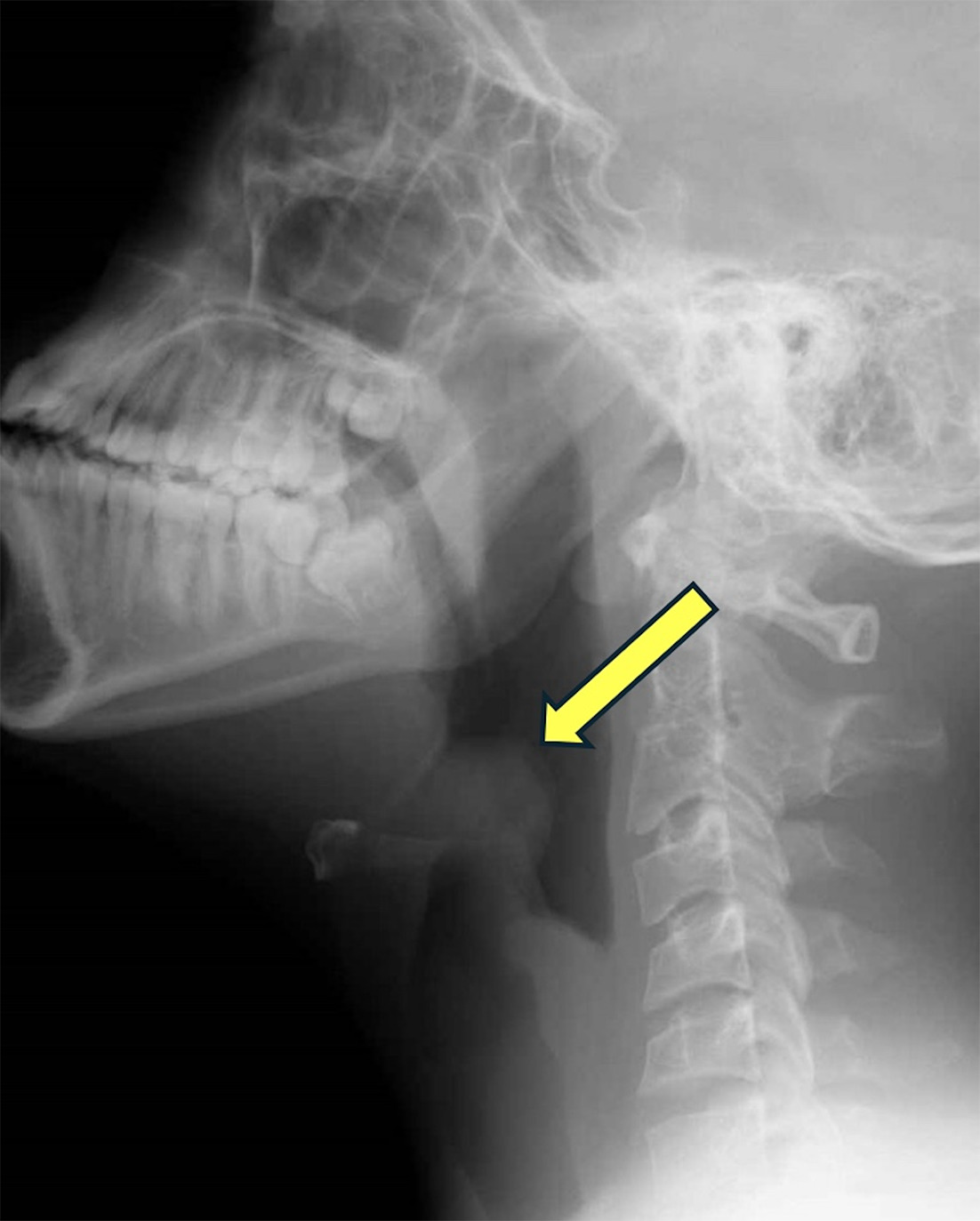
A school-aged child presented to the emergency department with high fever, stridor, throat pain, drooling, and difficulty swallowing. When sitting, the patient leaned forward. Sitting lateral neck radiographs were acquired, demonstrating an enlarged epiglottis. No radiographic signs of abscess were seen.
Imaging Findings
Lateral neck radiograph (Figure 1) showed enlarged epiglottis (arrow) consistent with epiglottitis.
Diagnosis
Epiglottitis.
The clinical differential diagnosis includes croup, pharyngitis, peritonsillar or retrotonsillar abscess, bacterial tracheitis, airway foreign body, and diphtheria.
Discussion
Epiglottitis is a bacterial infection of the epiglottis and/or adjacent structures that causes the supraglottic region to become inflamed. 1 The supraglottitis that results from the infection leads to a partial or near-total airway obstruction at this level. Supraglottic structures include the arytenoids, aryepiglottic folds, and vallecula. 1 If untreated, the inflammation can result in asphyxia and respiratory arrest, making this potentially a life-threatening condition. Recognition of the clinical and imaging findings is essential for prompt diagnosis as this condition is a medical emergency.
Infection most commonly affects children ages 2-6. 1 Haemophilus influenzae is the most common cause of epiglottitis infection. 1 H. influenzae is a gram-negative coccobacillus that causes several pediatric and adult infections. Its virulence is primarily determined by its polysaccharide polyribosyl ribitol phosphate capsule. 2 The first H. influenzae B vaccine was first introduced in 1985; however, it was not very effective in children 18 months old or younger. 3 A conjugate H. influenzae vaccine was introduced in 1990, which was effective in all age ranges. 3 H. influenzae B vaccine has decreased infection in the pediatric age group significantly. 1 The incidence of acute epiglottitis has decreased from 4.9 cases/100,000/year in children to 0.02/100,000/year since the introduction of the vaccine. 4 On the other hand, the incidence in adults has remained constant, 4 and adults are now more likely to die of acute epiglottitis than children. 1
A lateral neck radiograph in the upright position is the primary imaging modality (Figure 1). 5 On a normal lateral neck radiograph, the epiglottis, which is a flap of cartilage that covers the airway during swallowing, has well-defined thin margins. 6 The aryepiglottic folds are thin and convex inferiorly. In a patient with epiglottitis, there is a characteristic “thumb sign” because of swelling and submucosal edema of the epiglottitis. 6 There are also thickened aryepiglottic folds that become convex superiorly. Swelling of the aryepiglottic folds significantly narrows the upper airway that already exists because of the enlargement of the epiglottis and is a serious prognostic sign. Lastly, aryepiglottic folds add to the difficulty of intubation. 6 The pediatric and adult airway differs in anatomy. In children, the narrowest segment of the airway is the subglottis; any edema in these structures can cause airway obstruction by reducing the diameter of the airway. 7 Furthermore, this edema can force the epiglottis posteriorly, which also exacerbates airway obstruction. 7
A physical examination is also important for diagnosis. Clinical symptoms include a sudden onset of pain with swallowing, severe sore throat, high fever (>100.4°F), difficulty swallowing, change in voice quality, rapid breathing, a high-pitched respiratory sound caused by turbulent airflow (stridor), drooling, and sitting in a position leaning forward and upright. 5,6 Children assume this position unconsciously in order to increase the diameter of the airway, maximize laryngeal opening, and improve breathing. 8 It is imperative to not disturb the children and acquire the lateral neck radiograph in the position. Moving the child into a supine position could lead to a respiratory arrest. Symptoms typically appear rapidly, within hours in children. 7 To confirm bacterial infection, blood cultures can also be useful. 5
The treatment of epiglottitis is with empiric intravenous antibacterial therapy with third-generation cephalosporin and an anti-staphylococcal agent. 1 In the case of respiratory distress, children should be intubated or a tracheostomy should be placed. 1 Airway management in patients with epiglottitis is difficult due to the severe edema and obstruction, increased secretions, and distorted anatomy and requires a person skilled and experienced in intubation to perform the procedure. Monitoring the airway for obstruction is imperative. Administration of supplemental humidified oxygen and intravenous hydration can also be considered. 1
Complications of epiglottitis that may occur can be noninfectious or infectious. Some of these noninfectious complications include respiratory arrest, cardiac arrest, anoxic encephalopathy, and pulmonary edema. Examples of infectious complications are meningitis, pneumonia, cervical adenitis, and septic shock, with the most common being pneumonia. 9 Difficulty with intubation due to edema and sudden airway obstruction is associated with mortality in patients with epiglottitis. Another complication associated with mortality is the development of epiglottic abscess. 1 However, with prompt diagnosis and treatment, outcomes for children with epiglottitis are excellent, with mortality rates of about 0.5 per million patients. 10
Conclusion
Epiglottitis is a bacterial infection of the epiglottis and/or adjacent structures that causes the supraglottic region to become inflamed. The supraglottitis that results from the infection leads to a partial or near-total airway obstruction at this level. Supraglottic structures include the arytenoids, aryepiglottic folds, and vallecula. 1 If untreated, the inflammation can result in asphyxia and respiratory arrest, making this potentially a life-threatening condition. Recognition of the clinical and imaging findings is essential for prompt diagnosis as this condition is a medical emergency.
References
- Dowdy R, Cornelius B. Medical management of epiglottitis. Anesth Prog. 2020;67(2):90-97. doi:10.2344/anpr-66-04-08.
- Moxon E. The molecular basis of haemophilus influenzae virulence. J R Coll Physicians Lond. 1985;19(3):174-178.
- Broadhurst L, Erickson R, Kelley P. Decreases in invasive haemophilus influenzae diseases in US army children, 1984 through 1991. JAMA. 1993;269(2):227-231. doi:10.1001/jama.1993.03500020061032.
- Guldfred L, Lyhne D, Becker B. Acute epiglottitis: epidemiology, clinical presentation, management and outcome. J Laryngol Otol. 2008;122(8):818-823. doi:10.1017/S0022215107000473.
- Baiu I, Melendez E. Epiglottitis. JAMA. 2019;321(19):1946. doi:10.1001/jama.2019.3468.
- Darras K, Roston A, Yewchuk L. Imaging acute airway obstruction in infants and children. Radiographics. 2015;35(7):2064-2079. doi:10.1148/rg.2015150096.
- Stroud R, Friedman N. An update on inflammatory disorders of the pediatric airway: epiglottitis, croup, and tracheitis. Am J Otolaryngol. 2001;22(4):268-275. doi:10.1053/ajot.2001.24825.
- Rudner H. Differential diagnosis of upper airway disease in children. Can Fam Physician. 1985;31:1051-1053.
- Gonzalez C, Gartner J, Casselbrant M, Kenna M. Complication of acute epiglottitis. Int J Pediatr Otorhinolaryngol. 1986;11(1):67-71. doi:10.1016/s0165-5876(86)80029-5.
- Carenfelt C, Sobin A. Acute infectious epiglottitis in children and adults: annual incidence and mortality. Clin Otolaryngol Allied Sci. 1989;14(6):489-493. doi:10.1111/j.1365-2273.1989.tb00410.x.
Disclosures
The authors have no conflicts of interest to disclose. None of the authors received outside funding for the production of this original manuscript and no part of this article has been previously published elsewhere.
Citation
. Epiglottitis. Applied Radiology. 2025. doi:10.37549/JPCR-25-0070.