Erdheim-Chester Disease
Applied Radiology — Vol. 55 , Issue 2
Published: January 29, 2026
1 Department of Radiology, University of California-San Diego Medical Center, San Diego, California
* Corresponding author: Gary G. Ghahremani (gghahremani@ucsd.edu)
Categories
Case Summary
A middle-aged patient with diabetes presented with complaints of general malaise, dull abdominal pain, and weight loss of 12 pounds during the prior 3 months. Physical examination revealed periumbilical tenderness without a palpable mass, mild splenomegaly, and bilateral exophthalmos. Laboratory tests showed unremarkable hematologic and metabolic data, but abnormal renal function with an elevated creatinine level. The patient was referred to our institution for contrast-enhanced CT of the abdomen and MRI of the brain and orbits.
Imaging Findings
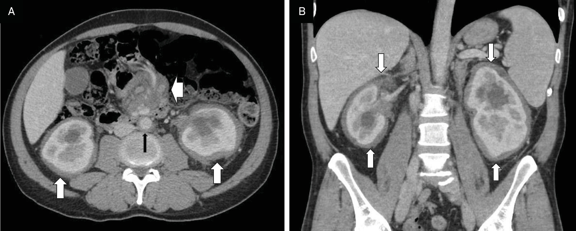
Contrast-enhanced CT of the abdomen demonstrated a thick rim of infiltrated subcapsular fat surrounding both kidneys. An ill-defined, 5 cm soft-tissue mass was present at the root of the small bowel mesentery, encasing the visceral vessels, extending into the retroperitoneal area, and surrounding the aorta, renal hilus, and proximal ureters, causing moderate hydronephrosis (Figure 1).
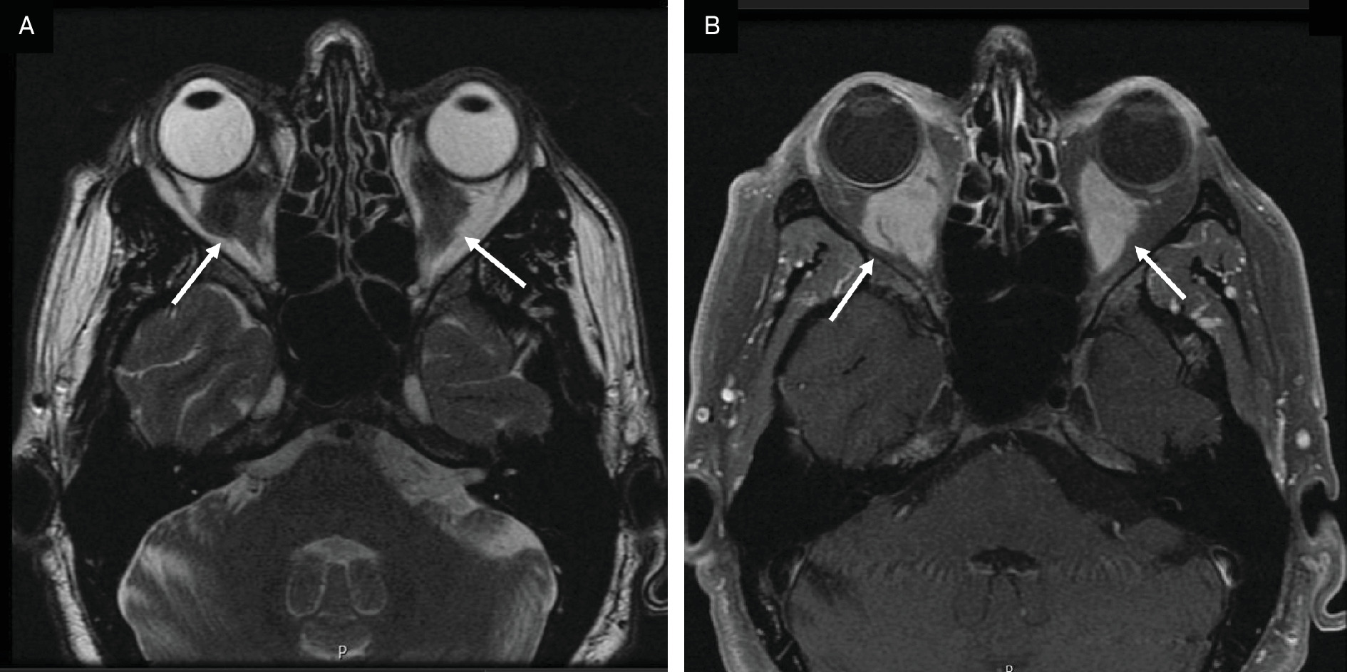
Contrast-enhanced MRI of the orbits demonstrated bilateral, cone-shaped, soft-tissue masses in the retrobulbar regions, resulting in a mild degree of exophthalmos. These lesions were hypointense and homogeneous on T1 and T2 sequences but appeared hyperintense on T1 with fat suppression (Figure 2).
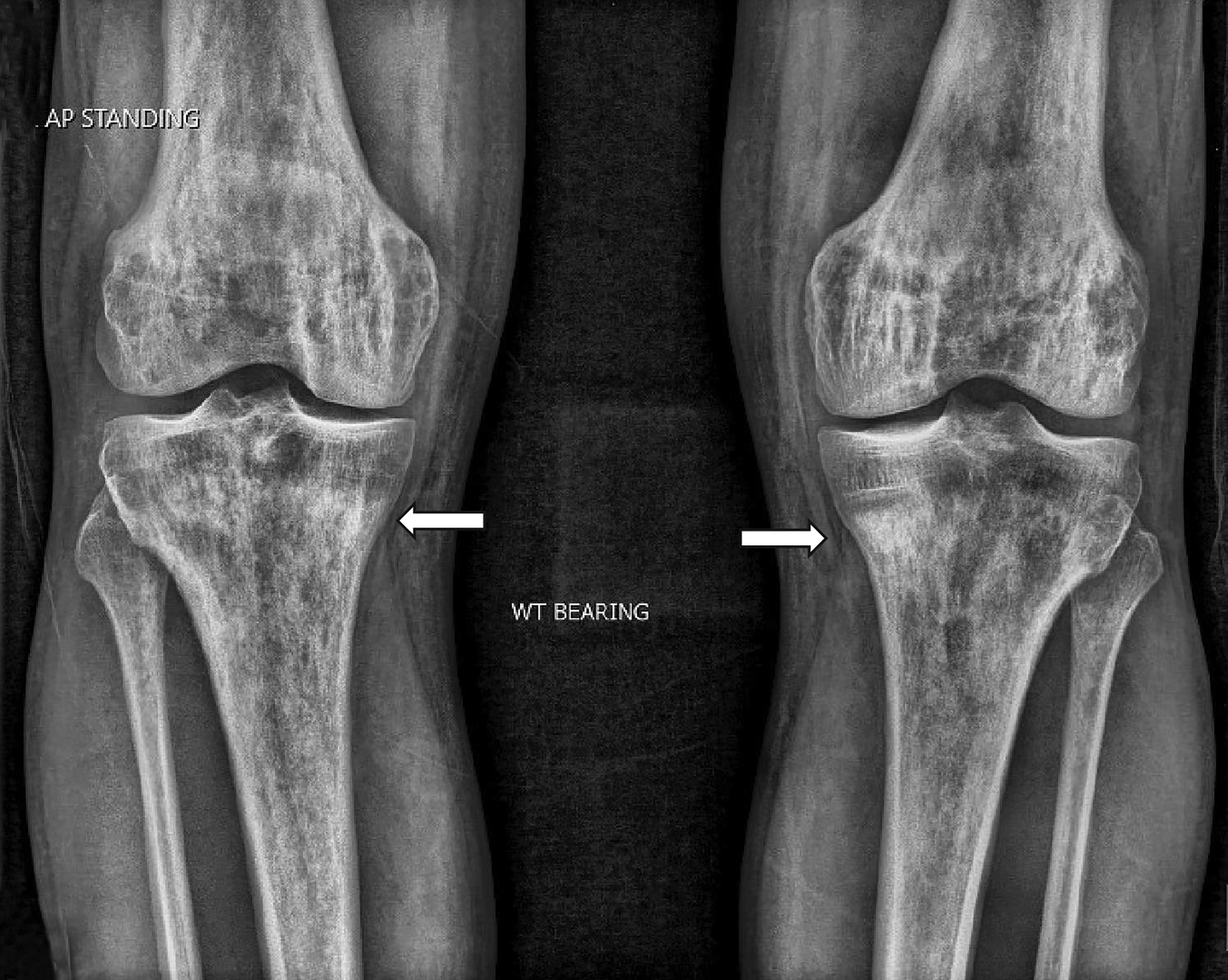
A biopsy of these masses revealed dense clusters of lipid-laden and foamy histiocytes with non-Langerhans features, Touton giant cells with multiple nuclei, and reactive inflammatory fibrosis. Genetic analysis disclosed BRAF-V600E mutation on chromosome 7, which controls cell growth and division in this particular entity. Further confirmation was provided by the characteristic osteosclerosis seen on the patient’s knee radiographs (Figure 3).
Diagnosis
Erdheim-Chester disease.
The differential diagnosis includes Langerhans cell histiocytosis, Rosai-Dorfman disease, lymphoma, orbital pseudotumors, and neurosarcoidosis.
Discussion
Erdheim-Chester disease is a rare neoplastic condition caused by genetic mutation of histiocytes that are normally found in the bone marrow, blood, liver, and spleen as an important part of the immune system. Their malignant transformation leads to infiltration of various organs by lipid-laden and foamy histiocytes and giant macrophages that incite an inflammatory reaction and fibrosis. 1,2
Since the disease’s initial description as lipoid granulomatosis in 1930, about a thousand isolated cases and small series of patients with Erdheim-Chester disease have been reported. It develops predominantly in men (3:1 male/female ratio) during the 5th to 7th decade. 1-4
About 68% of patients present with renal and retroperitoneal involvement. The characteristic finding is the bilateral infiltration of perinephric fat that is called the “hairy kidney sign.” The concomitant pathological process in the peri-pelvic area and around the proximal ureters can cause hydronephrosis, encase the abdominal aorta as the “Coated aorta sign,” and simulate the appearance of retroperitoneal fibrosis (Figure 1). However, that condition can be differentiated from retroperitoneal xanthogranulomatosis in Erdheim-Chester disease, which has perinephric infiltration and other coexisting skeletal and orbital lesions. Occasional involvement of other abdominal organs such as the mesentery, adrenal glands, liver, and pancreas may also occur. 3-6
About 50% of the reported cases had presented with symptoms of brain or eye involvement. This is often manifested as bilateral exophthalmos owing to histiocytic mass formation in the retrobulbar spaces (Figure 2). These lesions surround the optic nerves but do not invade ocular muscles or lacrimal glands. Their distinct MRI features as hypointense on T1 and T2 sequences and hyperintense on contrast-enhanced T1 with fat suppression will help in differentiating these entities from inflammatory orbital pseudotumors. 7-9
In addition to retrobulbar masses, some patients develop brain lesions, and their various patterns on CT and MRI have been reported. It is also noted that brain stem involvement results in ataxia, while pituitary gland infiltration leads to diabetes insipidus. 7,8
The most common presenting complaint—in 96% of reported cases—has been bone and joint pain. 3,4,10 This is caused by characteristic osteosclerotic changes best seen in the diaphyseal-metaphyseal area of the long bones, particularly in the femoral condyle and proximal tibia. About 8-10% of the cases also show some scattered lytic lesions caused by xanthogranulomas in the bone marrow. 3,10 These findings were demonstrated on the subsequently obtained knee radiographs of this patient (Figure 3).
Treatment of Erdheim-Chester disease consists of targeted immunotherapy with BRAF mutation inhibitors, interferon-alpha, and corticosteroids. 1-3 Until 2 decades ago, most patients had a relatively poor prognosis, owing to renal failure or cardiopulmonary complications, but the new treatments have increased overall 5-year survival from 68% to 83%. 1,5 Our patient died from congestive heart failure and pulmonary edema 4 years after presenting to our institution.
Conclusion
Diagnosis of Erdheim-Chester disease depends upon the histology and genetic analysis of biopsied tissue. However, radiological imaging studies play a crucial role in demonstrating various sites of involvement and their subsequent monitoring. Hence, radiologists should be familiar with the spectrum of presented features of this entity.
References
- Goyal G, Heaney M, Collin M. Erdheim-Chester disease: consensus recommendations for evaluation, diagnosis, and treatment in the molecular era. Blood. 2020;135(22):1929-1945. doi:10.1182/blood.2019003507.
- Mazor R, Manevich-Mazor M, Shoenfeld Y. Erdheim-Chester disease: a comprehensive review of the literature. Orphanet J Rare Dis. 2013;8:137. doi:10.1186/1750-1172-8-137.
- Veyssier-Belot C, Cacoub P, Caparros-Lefebvre D. Erdheim-Chester disease. Clinical and radiologic characteristics of 59 cases. Medicine. 1996;75(3):157-169. doi:10.1097/00005792-199605000-00005.
- Estrada-Veras J, O’Brien K, Boyd L. The clinical spectrum of Erdheim-Chester disease: an observational cohort study. Blood Adv. 2017;1(6):357-366. doi:10.1182/bloodadvances.2016001784.
- Aswani Y, Patel A, Zhan X. Imaging in Erdheim-Chester disease. Radiographics. 2024;44(9):1-19. doi:10.1148/rg240011.
- Nikpanah M, Kim L, Mirmomen S. Abdominal involvement in Erdheim-Chester Disease (ECD): MRI and CT imaging findings and their association with BRAFmutation v600e. Eur Radiol. 2018;28(9):3751-3759. doi:10.1007/s00330-018-5326-1.
- Drier A, Haroche J, Savatovsky J. Cerebral, facial, and orbital involvement in Erdheim-Chester disease: CT and MR imaging findings. Radiology. 2010;255(2):586-594. doi:10.1148/radiol.10090320.
- Sedrak P, Ketonen L, Hou P. Erdheim-Chester disease of the central nervous system: new manifestations of a rare disease. AJNR Am J Neuroradiol. 2011;32(11):2126-2131. doi:10.3174/ajnr.A2707.
- Fang Y, Shen B, Dai Q. Orbital inflammatory pseudotumor: new advances in diagnosis, pathogenesis, and treatment. Eur J Med Res. 2023;28(1):395. doi:10.1186/s40001-023-01330.0.
- Dion E, Graef C, Miquel A. Bone involvement in Erdheim-Chester disease: imaging findings including periostitis and partial epiphyseal involvement. Radiology. 2006;238(2):632-639. doi:10.1148/radiol.2382041525.
Disclosures
The authors have no conflicts of interest to disclose. None of the authors received outside funding for the production of this original manuscript and no part of this article has been previously published elsewhere.
Citation
. Erdheim-Chester Disease. Applied Radiology. 2026;55(2). doi:10.37549/AR-D-25-0128.