Exencephaly
Applied Radiology — Vol. 39 , Issue 12 , pp. 30 -31
DOI: 10.37549/AR1788
Published: December 1, 2010
Categories
CASE SUMMARY
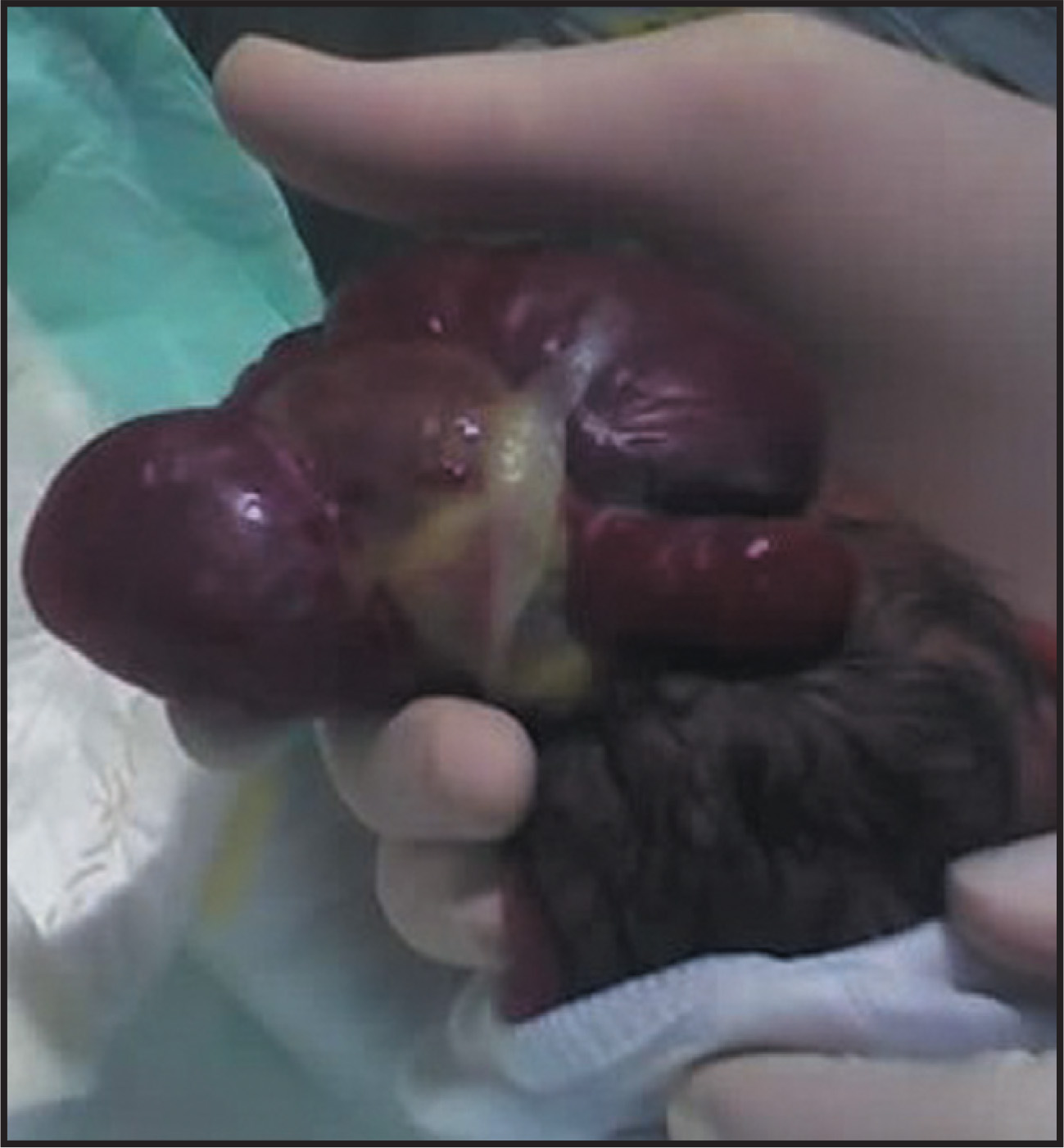
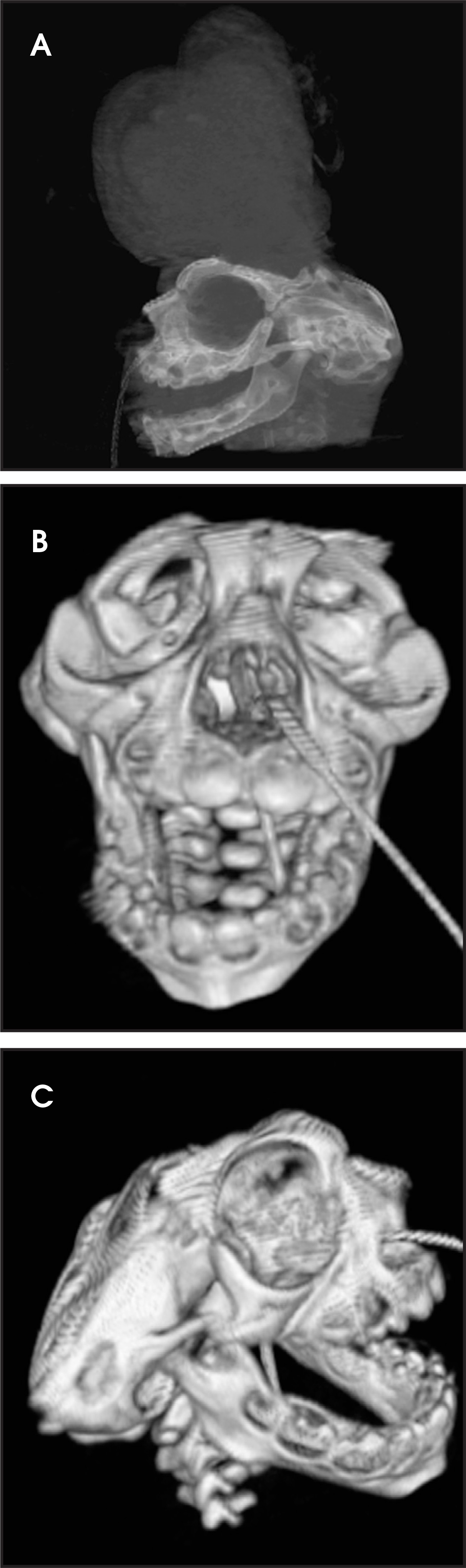
A 22-year-old G2P1 precipitously delivered a 37-week male infant at home. The infant was transferred to our tertiary care facility. The prenatal record was not available, but the patient’s mother gave a history of an abnormal prenatal ultrasound showing exencephaly. At diagnosis, the family was offered termination but declined. Admission examination demonstrated microcephaly with absence of the cranium above the orbital ridge with a large portion of neural tissue covered by dura protruding from the top of his head (Figure 1). Although computed tomography (CT) is not typically utilized to image this condition, it was requested immediately upon admission to better delineate the deformity, to determine prognosis, to guide clinical intervention (or withhold intervention) and to eventually assist in genetic counseling discussions (Figure 2).
After a meeting among the family, neurosurgeons, plastic surgeons and our ethics committee, the parents decided to provide palliative support. The patient expired after five days secondary to cardiac arrhythmia. Chromosomal analysis was normal.
DIAGNOSIS
Exencephaly (differential diagnosis includes acrania, acalvaria, anencephaly, large encephalocele or amniotic band syndrome.)1,3
IMAGING FINDINGS
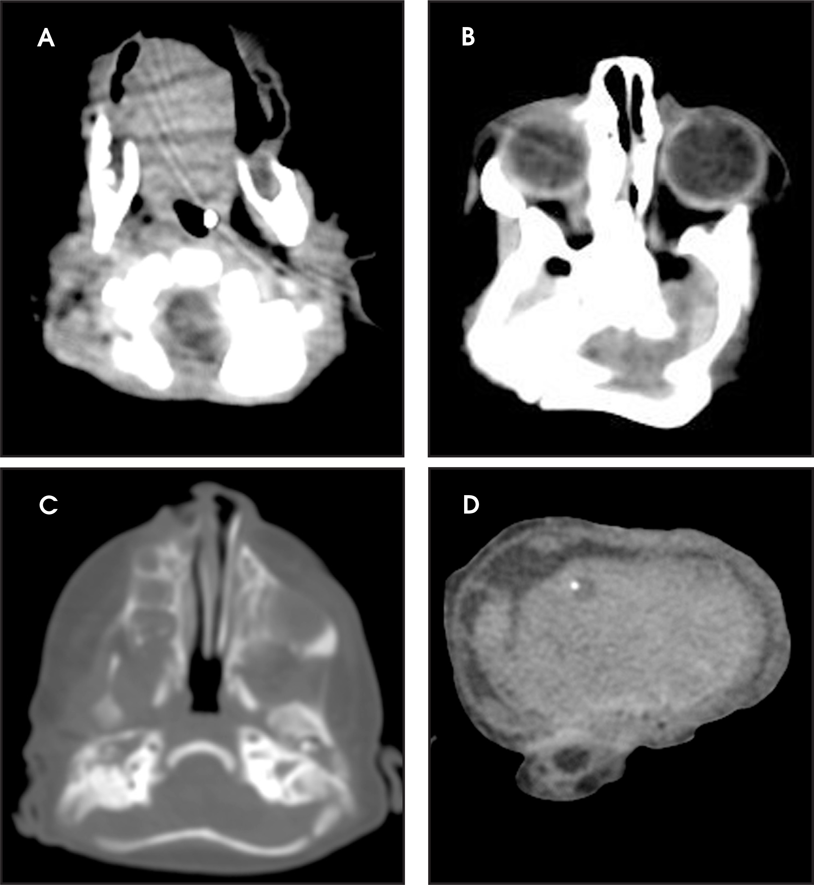
Disorganized brain tissue arising from the base of the cranium. The skull bones are malformed and the posterior cranial fossa is small. The flat bones of the skull are absent.
DISCUSSION
Exencephaly is a congenital malformation along a spectrum that includes acrania and anencephaly (acrania-exencephaly-anencephaly sequence).2,3 In acrania, the flat bones of the skull are at least partially absent, with complete but abnormal development of the cerebral hemispheres. Exencephaly demonstrates a large amount of disorganized brain tissue extending from a malformed skull base (Figure 3).3 Because this brain tissue is covered by only a vascular layer of epithelium, it can be slowly degraded by the amniotic fluid and degenerate into anencephaly.5 The incidence of exencephaly is much lower than that of anencephaly, but the etiology is the same, so the recurrence risk is the same as for other neural tube defects.6 Although the mineralization of the skull is not well established until after 10 weeks, there have been cases of prenatal diagnosis as early as 9 weeks, 3 days proven by postmortem endovaginal ultrasound.2,4 First trimester ultrasound findings of exencephaly include decreased size of the cranial pole compared with the chest, dorsally bulging cranial pole, irregularity of the surface of the cranium2 and echogenic amniotic fluid.3 The etiology of the sequence is not exactly clear, but failed migration of the mesenchyme during the 4th week of development or failed differentiation of the mesenchyme have been suggested.3 Although a significant number of fetuses with open neural tube defects are chromosomally abnormal, this child was chromosomally normal.
CONCLUSION
The acrania-exencephaly-anencephaly sequence is commonly diagnosed by prenatal ultrasound between 14 and 18 weeks, but some findings may suggest the diagnosis much earlier in gestation. Regardless of gestational age at diagnosis, the prognosis is poor.
References
- Cincore V, Ninios A, Pavlik J. Prenatal diagnosis of acrania associated with amniotic band syndrome. Obstet Gynecol. 2003;102:1176-1178.
- Becker R, Mende B, Stiemer B. Sonographic markers of exencephaly at 9 + 3 weeks of gestation. Ultrasound Obstet Gynecol. 2000;16:582-584.
- Bianca S, Ingegnosi C, Auditore S. Prenatal and postnatal findings of acrania. Arch Gynecol Obstet. 2005;271:256-258.
- Machado R, Brizot M, Carvalho M. Sonographic markers of exencephaly below 10 weeks’ gestation. Prenat Diagn. 2005;25:31-33.
- Chen C, Chang T, Lin Y. Prenatal sonographic diagnosis of acrania associated with amniotic bands. J Clin Ultrasound. 2004;32:256-260.
- Nawale A, Merchant S, Koteyar S. Exencephaly: A rare case diagnosed on antenatal ultrasound. Bombay Hospital Journal [serial online]. 2000;42.
Citation
. Exencephaly. Applied Radiology. 2010;39(12):30-31. doi:10.37549/AR1788.