Fetal MRI
Applied Radiology — Vol. 33 , Issue 2 , pp. 9 -25
DOI: 10.37549/AR1219
Published: February 1, 2004
Categories
Magnetic resonance imaging (MRI) has been used effectively to evaluate fetal anomalies only since the development of so-called fast imaging techniques in the mid-1990s.1-3 This article reviews the literature on the use of fetal MRI and provides a basic discussion of this technique, as well as a pictorial overview of the spectrum of fetal disorders that are amenable to MRI examination.
The roles of ultrasound and MRI in fetal diagnosis
In centers in which fetal MRI is performed, ultrasound (US) and MRI are considered complementary technologies, and MRI is utilized as an adjunct to US in the evaluation of fetal anomalies. Ultrasound remains the predominant modality for evaluating disorders related to the fetus and pregnancy overall. Ultrasound continues to have several obvious advantages over MRI. It is safe and relatively inexpensive and is a widely available technology that allows for real-time imaging.4 Compared with MR imaging, US provides superior spatial resolution, permits assessment of fetal well-being, and provides quantitative assessment of fetal and placental blood flow with Doppler US.
However, US does have important limitations. First, it is uniquely operator- and interpreter-dependent.4,5 In addition, relative to MRI, US provides a small field-of-view, and the resolution of US images is restricted by penetration through soft tissues and bone. Thus, the sensitivity of US in evaluating the fetus is reduced in obese patients and in women whose pregnancies are complicated by low amniotic fluid volume. Examination of intracranial anatomy is limited by the attenuation of the US beam through the bony skull in later gestation.6,7 Finally, US quality is influenced by fetal position.7 A prone, breech fetus may be much more difficult to examine than a vertex supine fetus. If the head is low in the maternal pelvis, it may also be difficult to assess using US.4
There is a growing body of literature on the use of MRI and has documented its usefulness in confirming or expanding upon US findings.5,8-11 In contradistinction to US, MRI visualization of the fetus is not significantly limited by maternal obesity, fetal position, or oligohydramnios, and visualization of the brain is not restricted by the ossified skull.12 It provides superior soft-tissue contrast resolution and the ability to distinguish individual structures such as lung, liver, kidney, bowel, and gray and white matter.12 Multiplanar imaging is easier with MRI than it is with US, where it is often a challenge to obtain images in three planes. MRI provides a large field-of-view, facilitating examination of fetuses with large or complex anomalies, and visualization of the lesion within the context of the entire body of the fetus. Finally, it is difficult to quantify, yet important not to underestimate, certain nebulous benefits that MRI may provide. First, MRI may boost diagnostic confidence for fetal disorders diagnosed by US. Second, MR images may be more easily understood by patients, clinical practitioners with more experience with MR than US, and by those without routine exposure to imaging, such as nurses, genetic counselors, and social workers.4 The value of these benefits remains a topic of intense debate.13 Nonetheless, centers in which fetal therapy is practiced have found that MRI often helps to facilitate management decisions and guide therapy, particularly when fetal surgery is a consideration or when delivery is expected to present unique challenges.14,15
MRI has significant disadvantages compared with US. It tends to be more expensive, can be difficult for those suffering from claustrophobia, is not as readily available, and provides less spatial resolution. Furthermore, MRI is exquisitely sensitive to fetal motion. Fast imaging techniques have over-come this to some extent, and the average MRI sequence, which historically required 15 minutes, now takes approximately 20 seconds to acquire. Nonetheless, the MRI arsenal that can be applied to fetal imaging remains limited. Finally, as this is a relatively new application, the long-term safety of MRI during pregnancy is not yet established, though there have been no reports of adverse effects to date.16-26 Intravenous gadolinium is not used, as there is placental transfer and it may be teratogenic at high doses.27,28
Technical considerations
Safety
While the long-term safety of MRI has not been fully established, it is believed, in general, that the benefit it provides in fetal management far outweighs any theoretical risks. No adverse biological effects have been reported at the energies currently delivered with MRI.16-26,29-31 Whether or not to obtain written consent reflects institutional bias: at Children’s Hospital Boston, consent is no longer obtained for performance of routine fetal MRI, while at Massachusetts General Hospital, it is. Most imaging occurs at or after 18 weeks’ gestation, when fetal size and motion first allow reasonable visualization with current MRI techniques, avoiding exposure during the peak of organogenesis. Gadolinium is not administered.
Preparation
US should always be performed prior to MRI, in order to check fetal position, as fetal position may determine coil selection and position. It is also prudent to confirm the presence of fetal cardiac activity with US prior to MRI, for psychological and medico-legal reasons.4 Chemical immobilization of the fetus is not necessary in routine situations.
Every effort should be made to ensure that the pregnant patient is well-fed and hydrated. Patients are asked to empty their bladder prior to the MRI. They are placed in a position of comfort. If supine, a bolster behind the knees helps take pressure off the low back. Patients often benefit from having a companion in the room. The examination typically lasts 30 minutes.
Technique
A 1.5T magnet is used. Imaging at or after 18 weeks’ gestation is preferred. Beyond this period, the fetus is large enough to image many anomalies, and motion is usually not as problematic. The mainstay of fetal MR imaging has been fast single-shot T2-weighted imaging.32 In general, slice thicknesses of between 4 and 7 mm are used with 0 to 1 mm interslice spacing (repetition time [TR] 2150, echo time [TE] 80Ef, matrix 256 × 256). Body coils and larger phased-array coils have been used most commonly. Smaller surface coils are sometimes useful for directed imaging of specific parts of fetal anatomy if the area of interest is not too far from the surface. Typically, 10 to 15 slices are acquired in 20 to 25 seconds. As fetal motion is unpredictable, shorter sequences are optimal. An initial fast localizing sequence is performed, and sequences are then acquired in orthogonal planes relative to each previous sequence. It is most effective to be ready to set up each sequence as rapidly as possible. One does not have the luxury of time, and the most successful examination is usually performed by personnel who are thoroughly familiar with fetal anatomy and fetal anomalies.
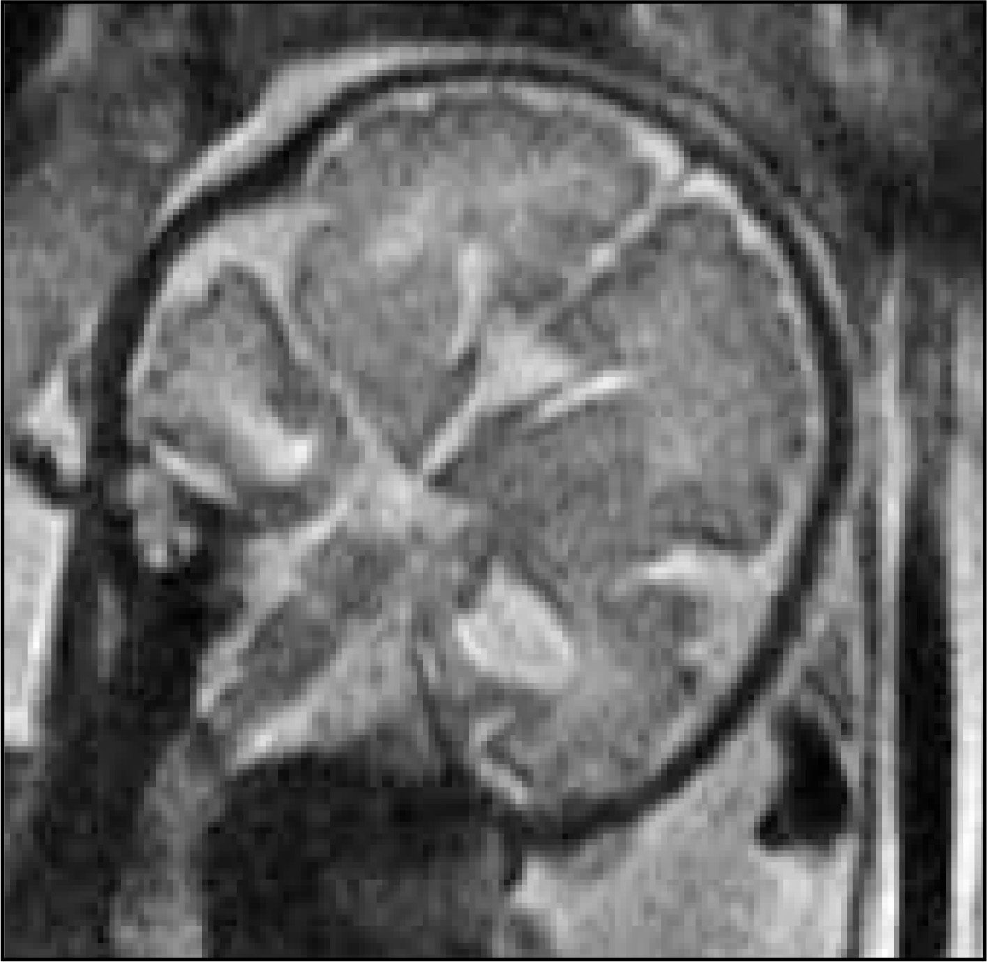
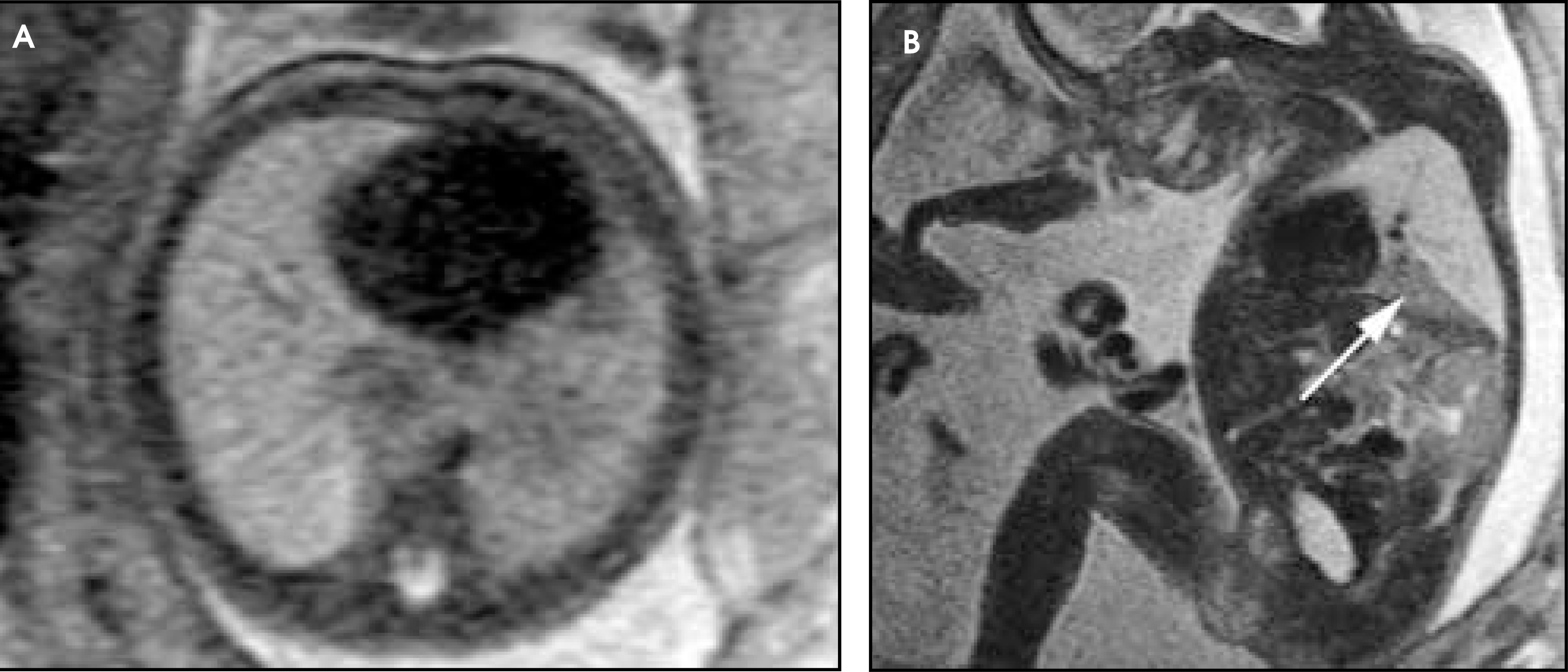
Fast T1-weighted imaging has also emerged into the fetal MRI arsenal. Children’s Hospital Boston uses an inversion recovery single-shot fast spin-echo (SSFSE) technique (TR 2530, TE 35.5, inversion time [TI] 2000, matrix 256 × 192) (Figure 1). Slice thickness selection is similar to the T2-weighted sequences. The T1-weighted technique has had particular utility in the brain, looking for hemorrhage, and in the abdomen, where the T1-weighted conspicuity of the liver has been reported to be helpful in assessing liver position in fetuses with congenital diaphragmatic hernia (CDH).11 Newer fast imaging techniques are developing rapidly and are expected to have important fetal applications. One must always be aware of the level of energy deposition, however, when considering such applications.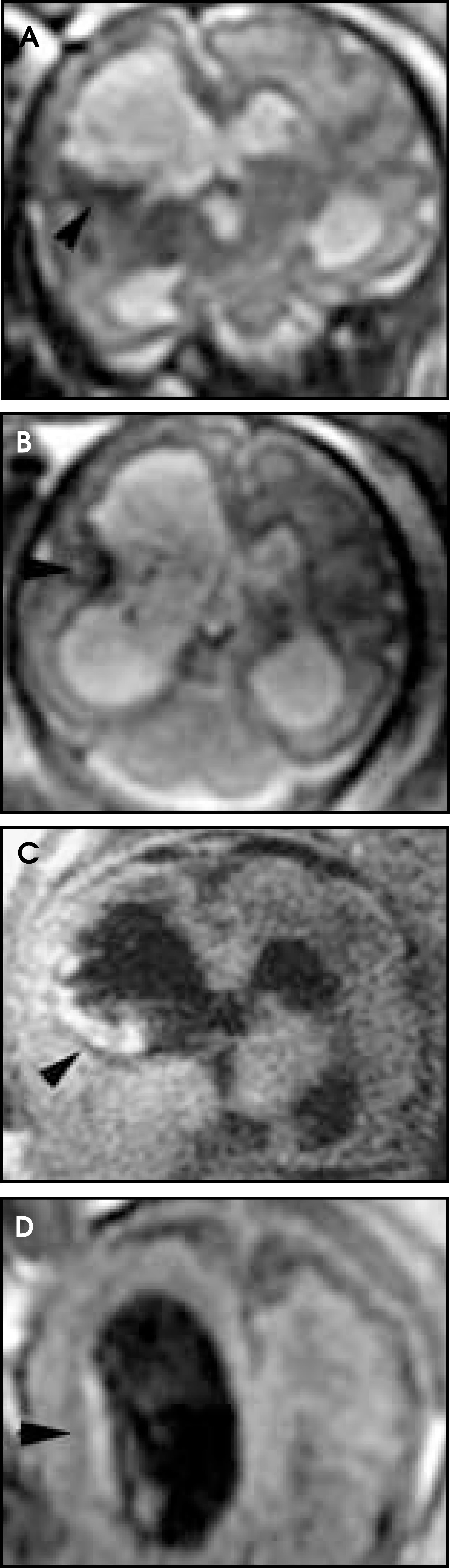
Prenatal MRI of the central nervous system, head and neck
The most common use of fetal MRI has been to evaluate central nervous system (CNS) abnormalities, where it has been found to influence or change management and counseling in up to 50% of cases.4,9,10 MRI has been particularly useful in looking at the posterior fossa, corpus callosum, and gray-white matter.8,12,33 Levine et al34 found that MRI changed the US diagnosis in 26 of 66 (40%) of CNS anomalies and changed patient counseling in 33 of 66 (55%). Simon et al9 found that 24 of 52 (46%) fetuses with CNS abnormalities were managed differently after the MRI studies were performed. This report echoes the conclusions of the others that MRI is an effective adjunct to US in the evaluation of CNS anomalies and is particularly useful in providing additional information and allowing more definitive diagnoses than can be provided by US alone.
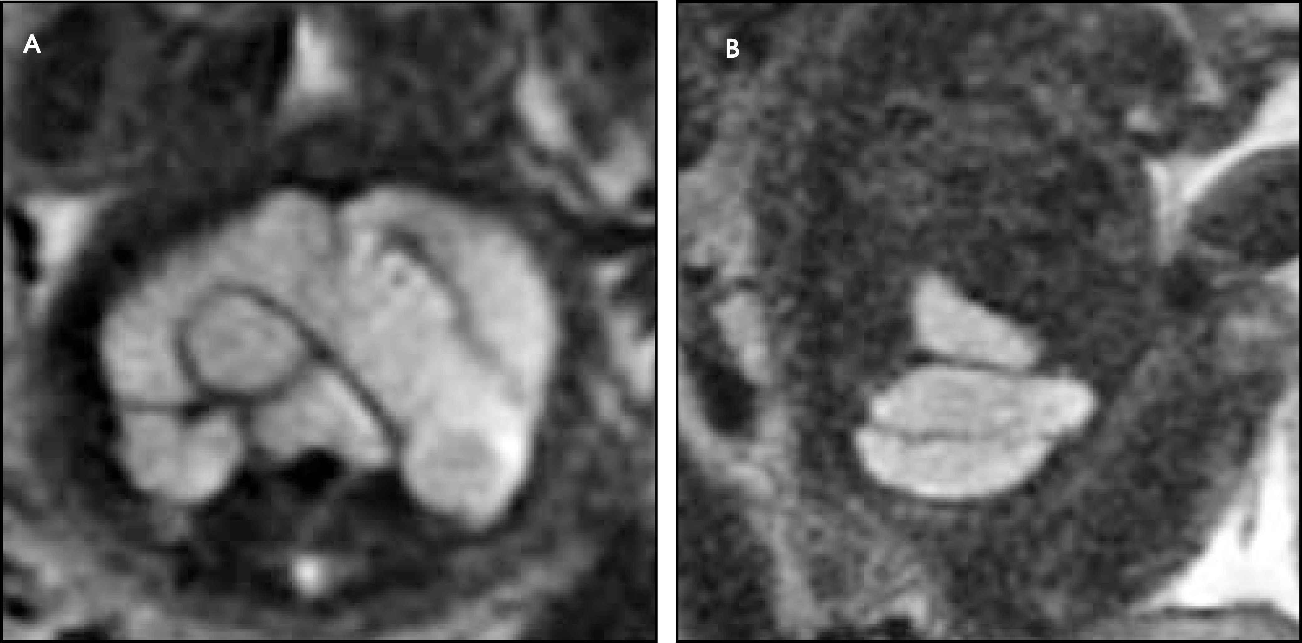
The mainstay of fetal CNS imaging has been fast single-shot T2-weighted image techniques. Normal anatomic structures can be seen routinely in second- and third-trimester fetuses (Figures 2 through 4). The single most common indication for fetal MRI has been evaluation of ventriculomegaly, where MRI is used to evaluate for associated abnormalities and underlying etiologies that are not as readily depicted with US.7 Agenesis of the corpus callosum, in particular, which is commonly associated with ventriculomegaly, can be a difficult second-trimester diagnosis with US but is often readily depicted with MRI (Figure 5).1,8,35 MRI has been found to be helpful in evaluating posterior fossa cystic lesions and in distinguishing between the Dandy-Walker malformation and variant, posterior fossa arachnoid cyst, and mega cisterna magna (Figures 6 and 7).12,33 Poutamo et al33 evaluated 19 fetuses with CNS abnormalities diagnosed by US and found that MRI provided additional information on anatomy of the posterior fossa in 9 of 19 (47%).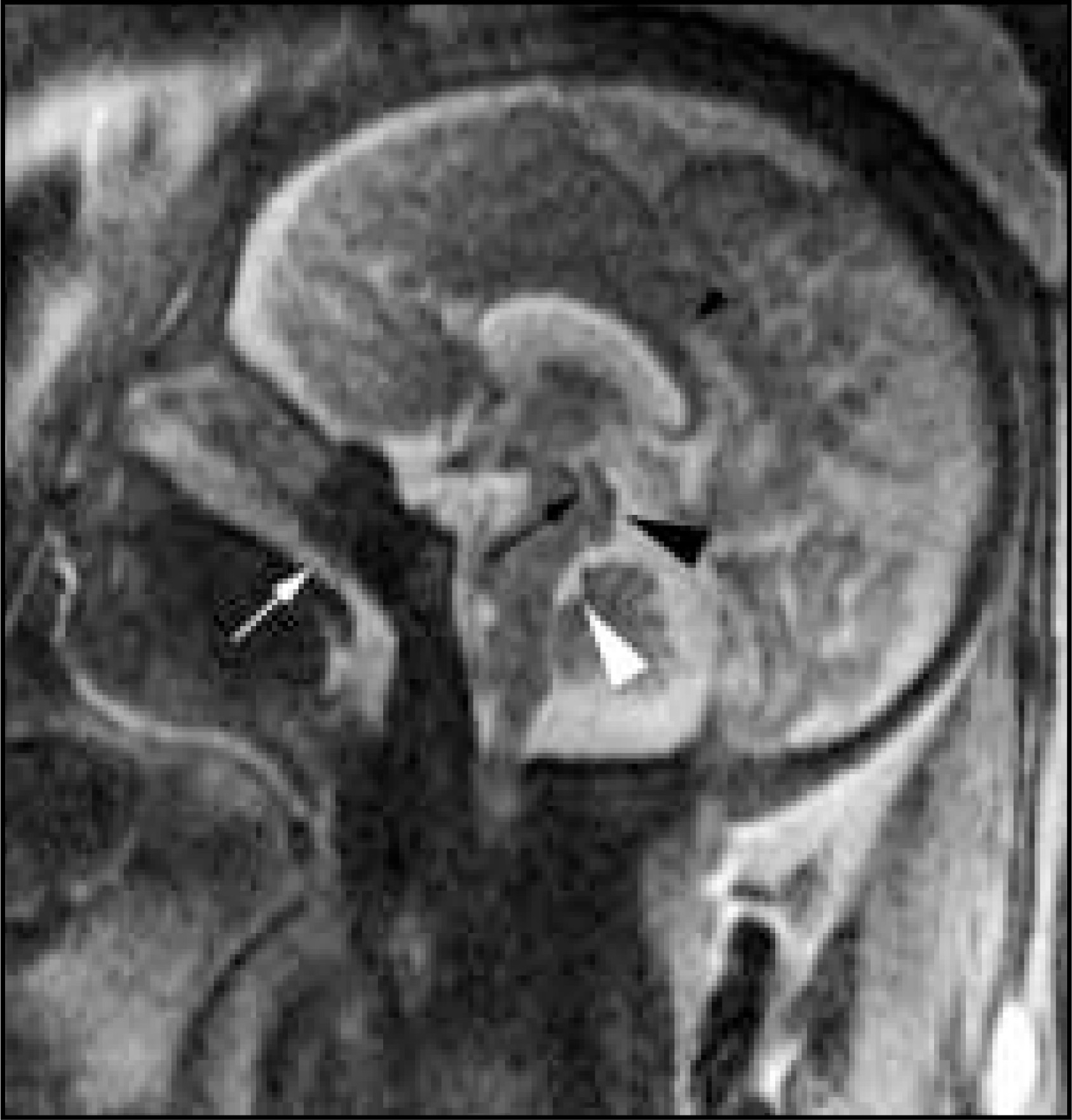
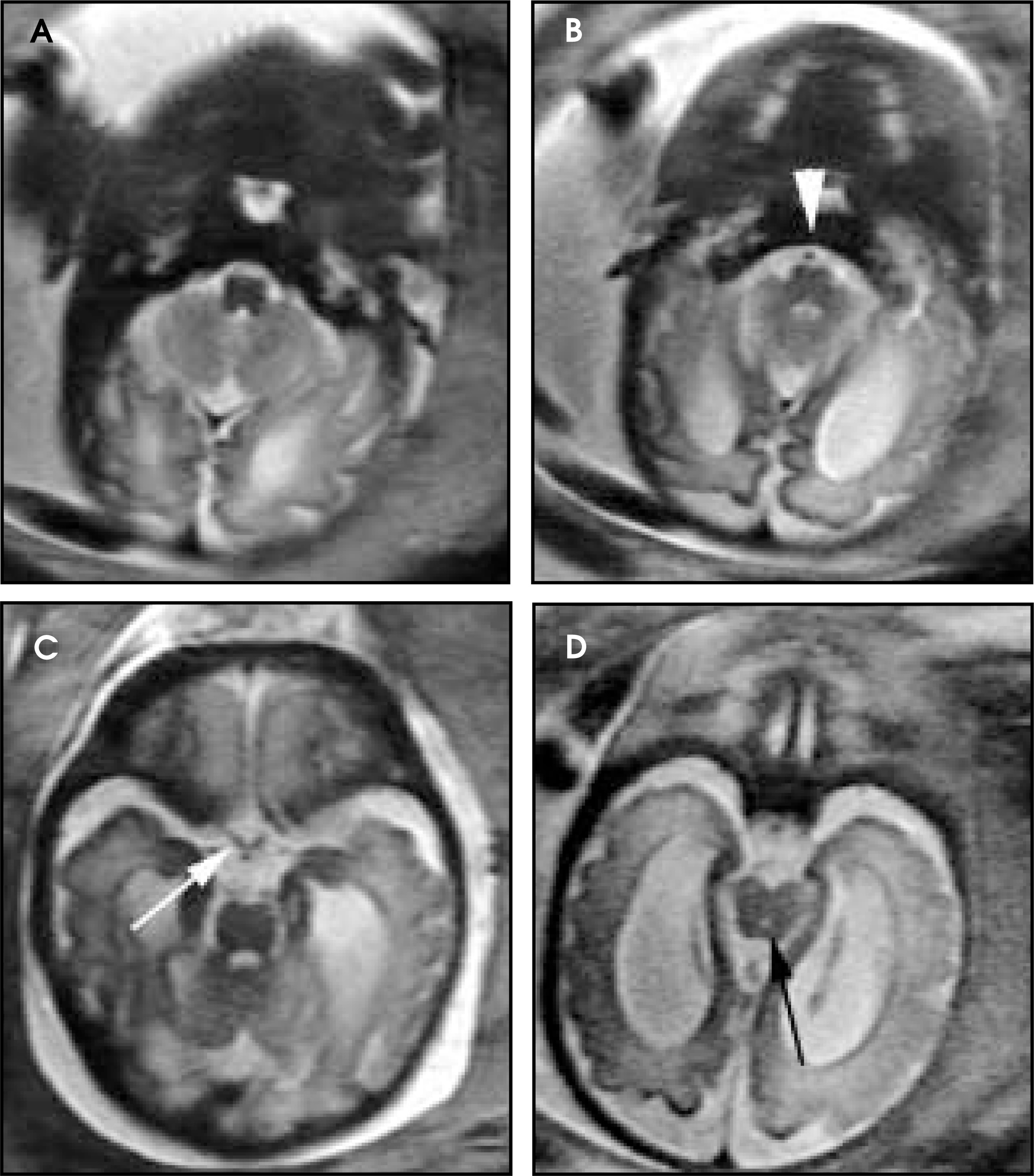
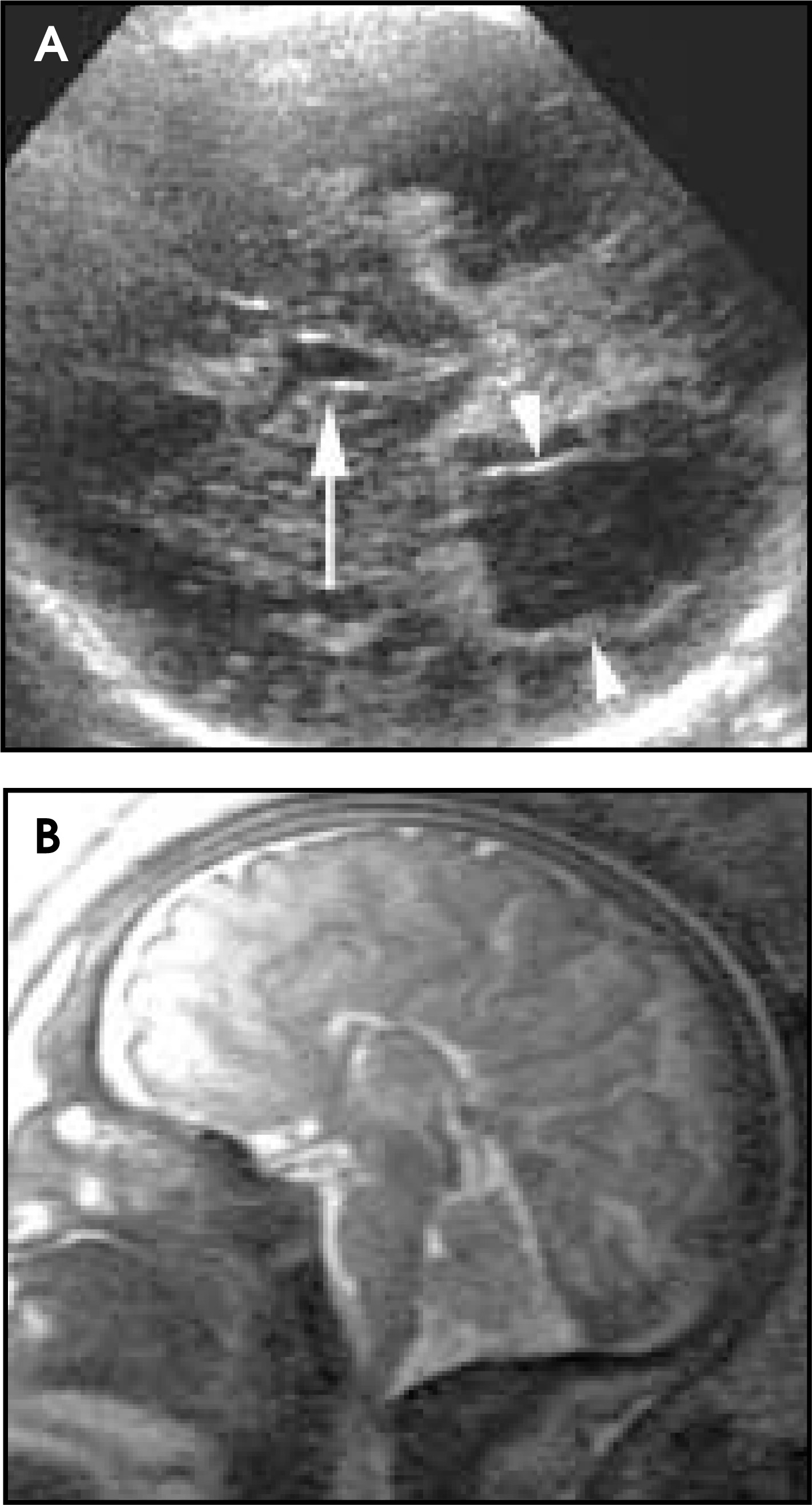
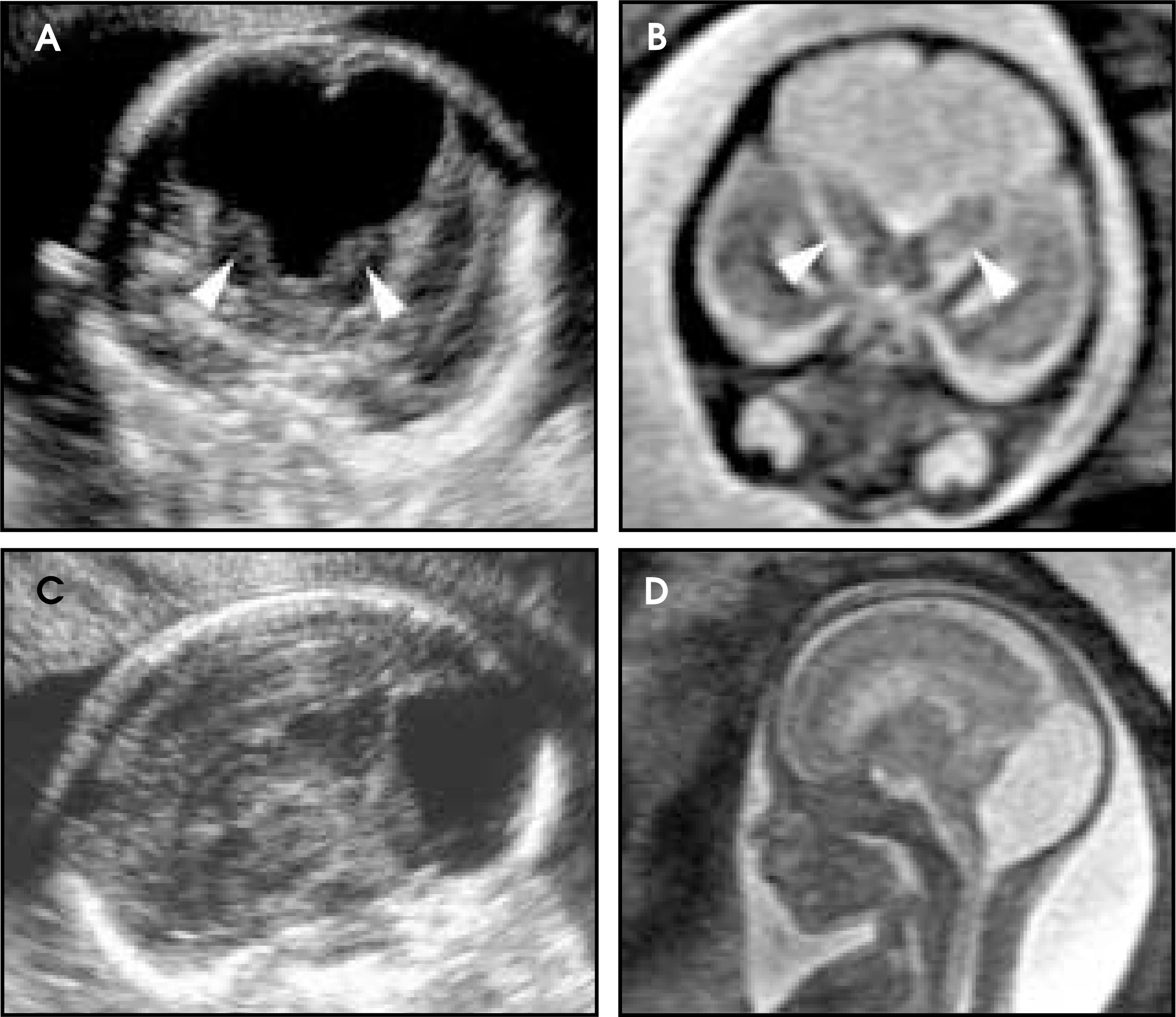
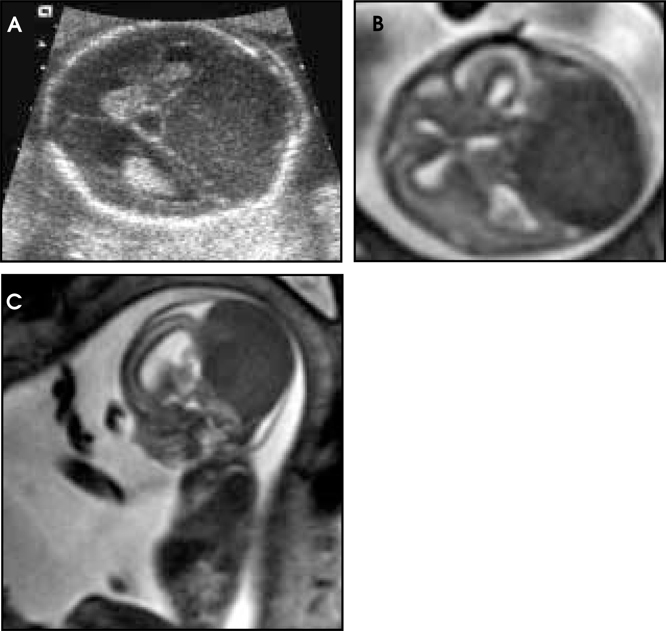
Several recent observations and technological developments hold promise in adding to the diagnostic and prognostic value in the application of MRI to the fetal CNS. Intrinsic ventricular morphology itself has been found to differ among various CNS abnormalities.36 The ability of MRI to provide information on cortical maturation may ultimately allow greater prognostic accuracy to be achieved when morphologic abnormalities, such as ventriculomegaly, agenesis of the corpus callosum, and Dandy-Walker malformation, are encountered, which historically have been associated with a wide spectrum of outcomes (Figure 8).37 Migrational abnormalities can now be identified in the third trimester and, in the future, may be detectable in the second trimester.7 Finally, MR spectroscopy has been applied to the fetal brain, and may be used clinically in the near future to provide important physiologic information in at-risk fetuses.38-40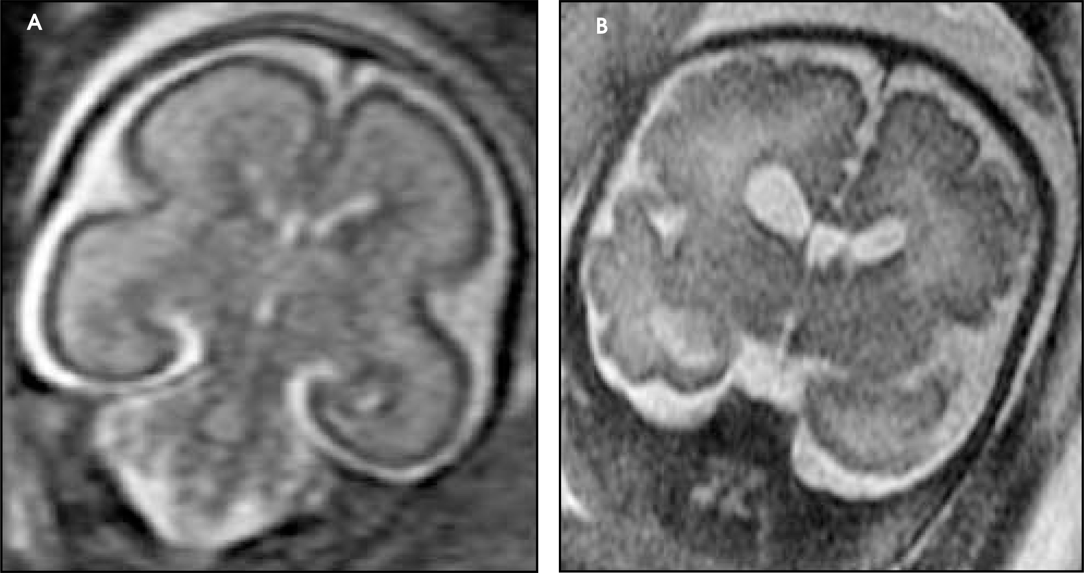
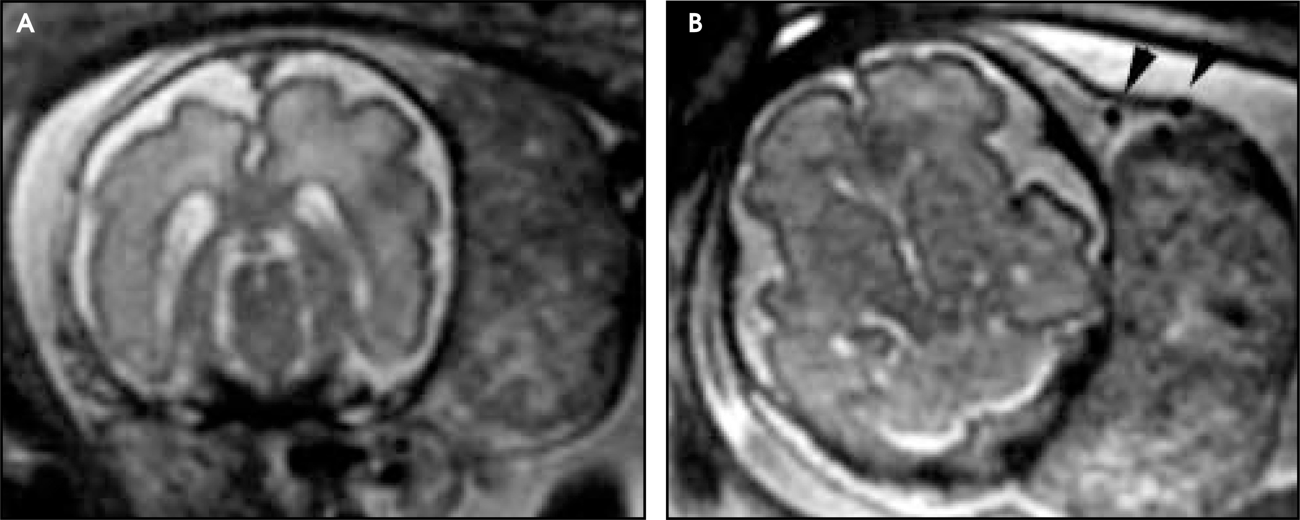
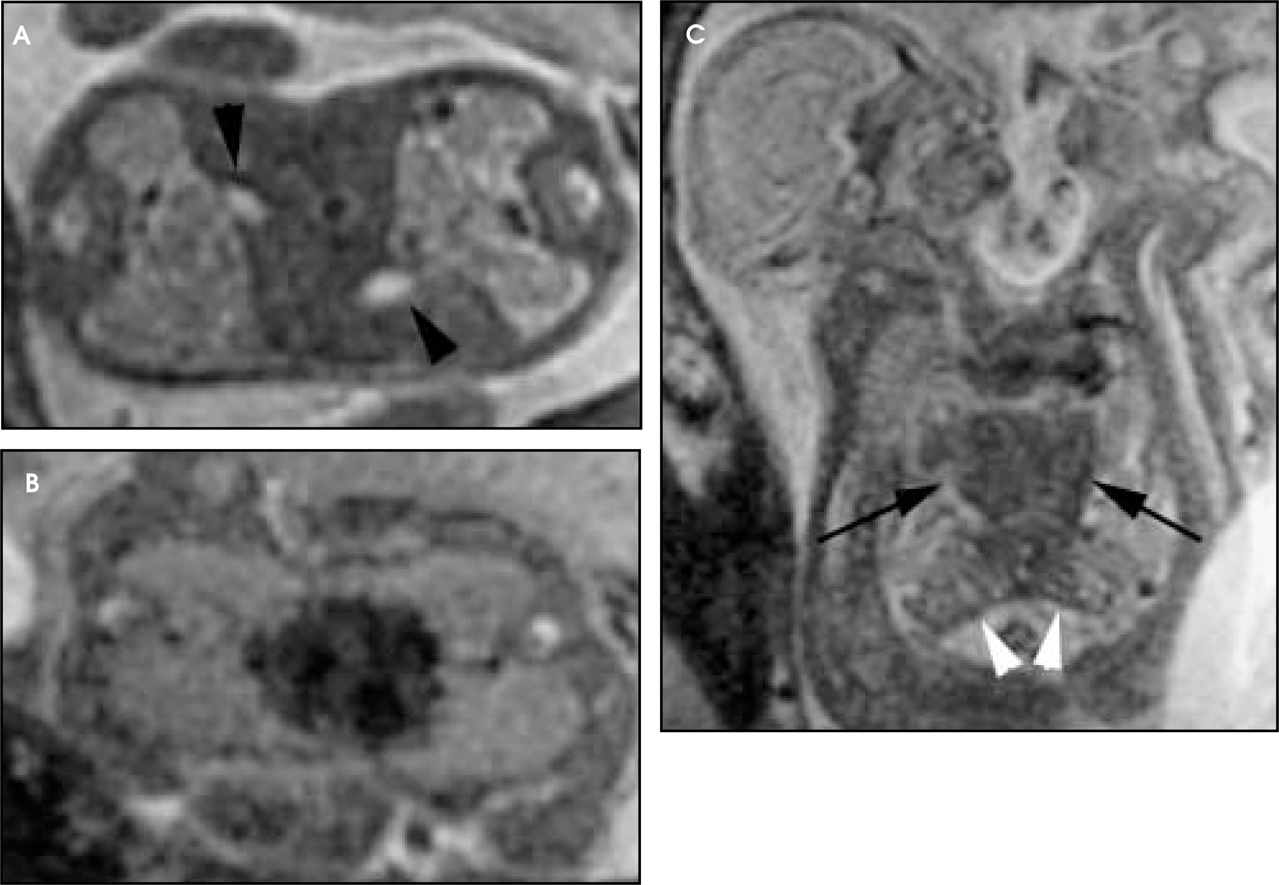
The fetal scalp, neck, and airway can be difficult to evaluate fully with US because of artifact related to adjacent bony structures as well as fetal position.33 MRI can be effective in evaluating abnormalities related to these structures and can provide information complementary to US. The fetal airway is fluid-filled and, therefore, discretely bright on T2-weighted images. Its location and course become quite important in the evaluation of cervical and thoracic masses in which the anatomy of the airway is a crucial determinant of method and location of delivery (Figures 9 and 10).7 In addition to aiding in delineating the airway, superior soft-tissue contrast resolution of MRI facilitates evaluation of scalp masses and cervical teratomas by sharply demonstrating the relationship of the masses to bone and pharyngeal soft tissues and by demonstrating extension into the mediastinum (Figure 11).33 In these situations, the aggregate information provided by both US and MRI allows for fuller appreciation of the anatomy than either modality alone would provide.7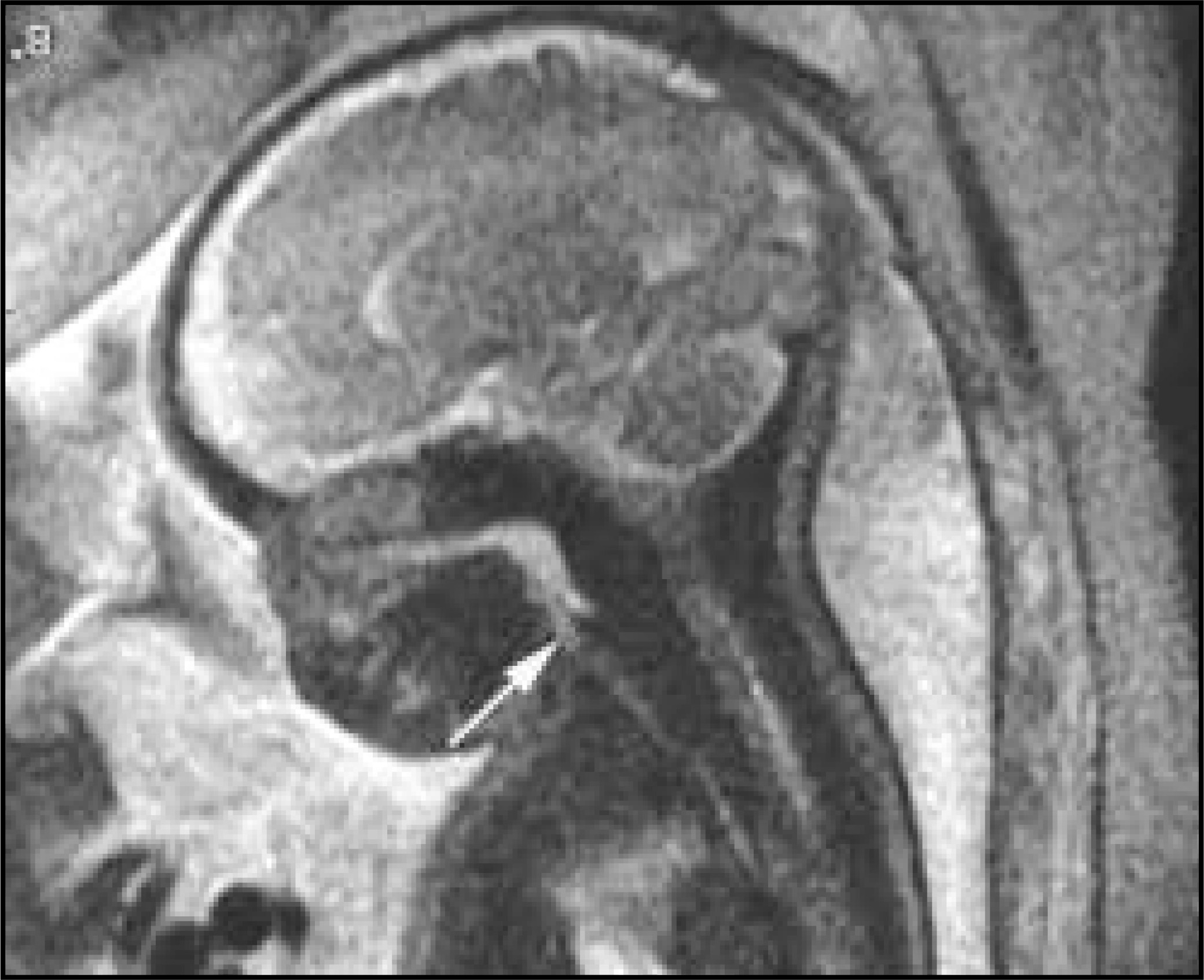
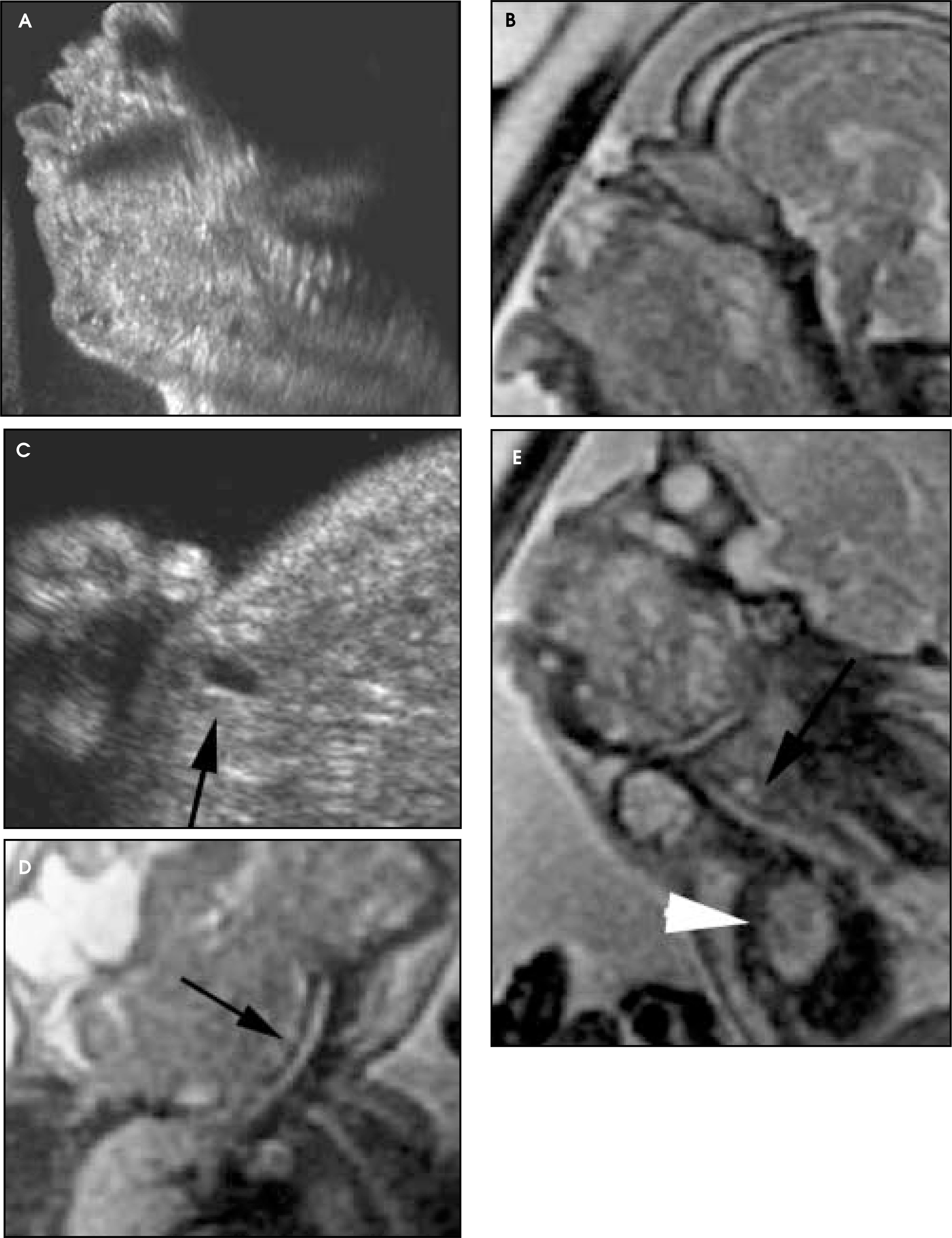
Ultrasound and MRI can be similarly complementary in evaluation of sacrococcygeal teratoma (Figure 12).7 Ultrasound, with its fine spatial resolution, often shows the details of the association of the lesion with the caudal aspect of the fetal spine and can also show its cystic and solid nature. Moreover, US has a distinct advantage in its ability to demonstrate lesion vascularity as well as to provide physiologic information vital for monitoring the development and progression of fetal hydrops. MRI better depicts lesions in the context of the whole body of the fetus and in multiple planes, which is important for surgical planning.41 In addition, MRI may provide additional information about the extension of these tumors into the pelvis, abdomen, or spinal canal, which may be underestimated by US because of obscuration by bony structures.41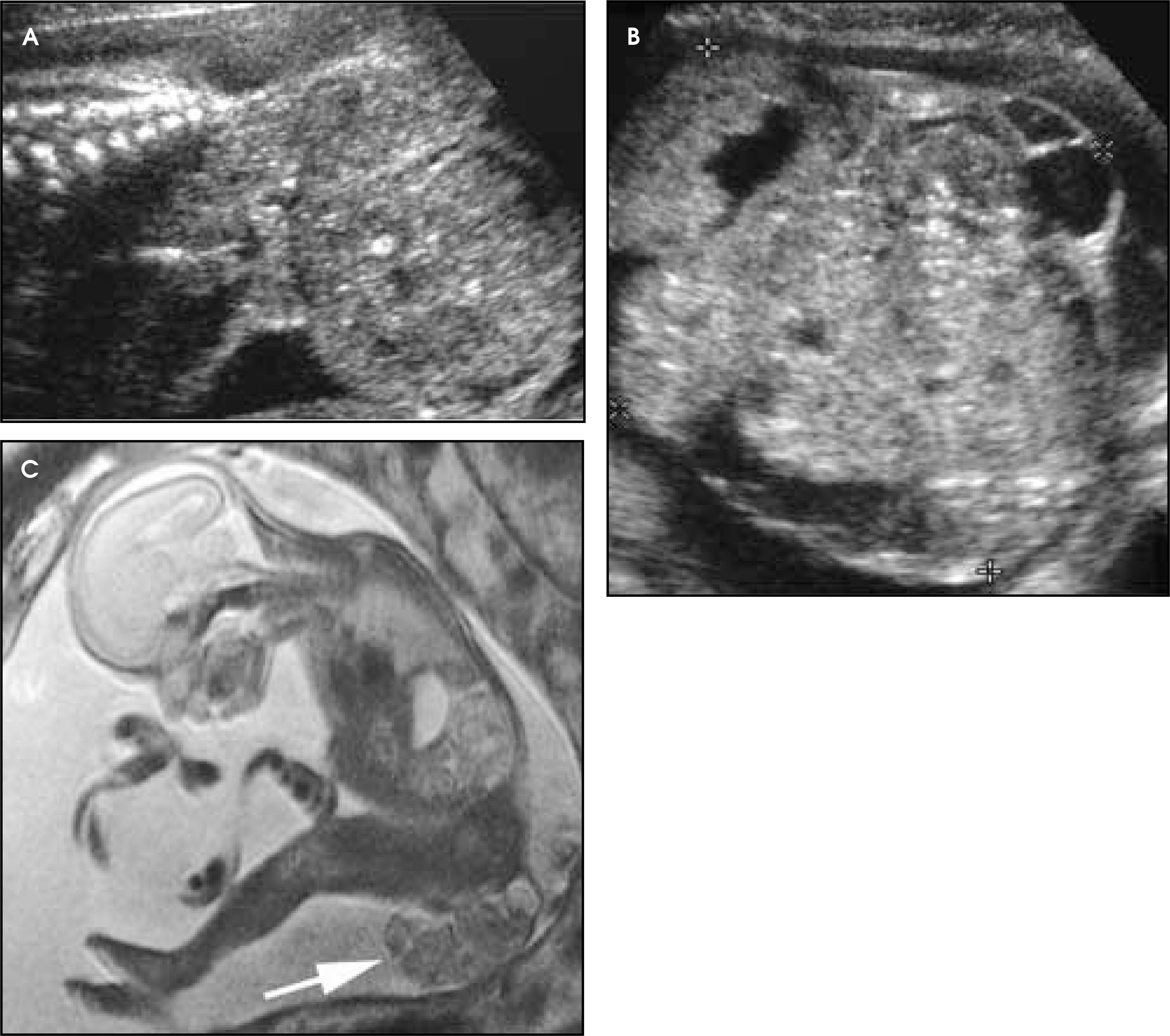
Whether MRI adds value in the routine management of neural tube defects (NTDs) has been argued. MRI may be more accurate than US in the setting of NTDs in the presence of maternal obesity. The MRI findings parallel characteristic US findings, including abnormal shape of the frontal bones, hydrocephalus, and Chiari II malformation (Figure 13).7 In general, the superior spatial resolution of US allows for more accurate delineation of the level of abnormality.42 MRI can detect subtle NTDs when US shows only the characteristic associated Chiari II malformation and fails to show the spinal lesion.1 In addition, some important dysraphic abnormalities, such as terminal myelocystocele, may not be associated with the Chiari II malformation.43 When in utero surgery is not a consideration, it is yet to be established whether MRI changes diagnosis or management in NTDs. MRI may be useful in the prenatal consultation for both the neurosurgeon and parents.1 Whether MRI will be used in the routine management of neural tube defects may depend on the outcome of currently ongoing trials assessing the outcomes of prenatal closure of neural tube defects.42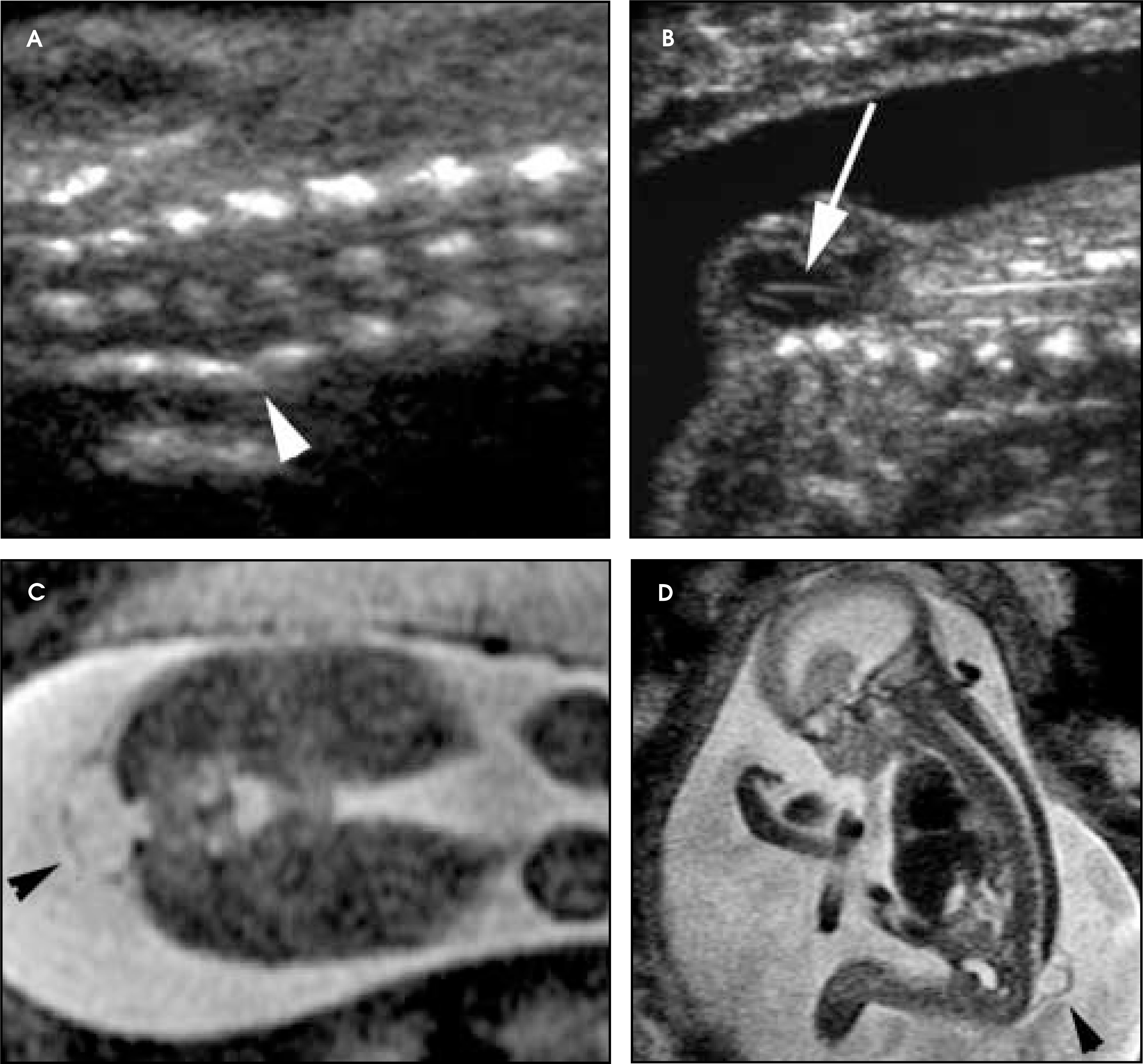
Prenatal MRI of the fetal body
After the CNS, the fetal thorax has received the most attention by those performing fetal MRI. Its role has been adjunctive to US, helping to define the anatomy of large masses, examine lesions with an atypical US appearance, and aid in the prognosis of CDH. Its emerging impact on fetal management and counseling for chest masses is approaching its use for CNS abnormalities.5 Hubbard et al11 reported 18 fetal chest masses diagnosed by US in which the subsequent prenatal MRI changed the diagnosis in 9 cases (50%). These cases included uncommon lesions, such as foregut cyst, bronchial atresia, tracheal atresia, and lung atresia, which were misdiagnosed as congenital cystic adenomatoid malformations (CCAM). This report revealed that the incorrect US diagnoses were related to image misinterpretation rather than to failure to detect the abnormality.
Normal fetal lungs are homogeneous in signal intensity on MRI, becoming progressively T2-bright with maturation (Figure 14). Because pulmonary maturation is so crucial to postnatal survival, much attention has been paid to establishing parameters that may allow MRI to evaluate lung development. These have included assessment of signal intensity, volumetric measurements, echoplanar imaging, and MR spectroscopy.44-48 As described earlier, MRI can depict the airway with excellent conspicuity, an asset that has proved invaluable in planning management for fetuses with cervical and thoracic masses. This has been of emerging importance as the ex utero intrapartum treatment (EXIT) delivery has been successfully used to deliver and manage these fetuses.15,49,50 In addition to the lungs, normal thoracic structures that can be evaluated with MRI include the thymus and thyroid gland.51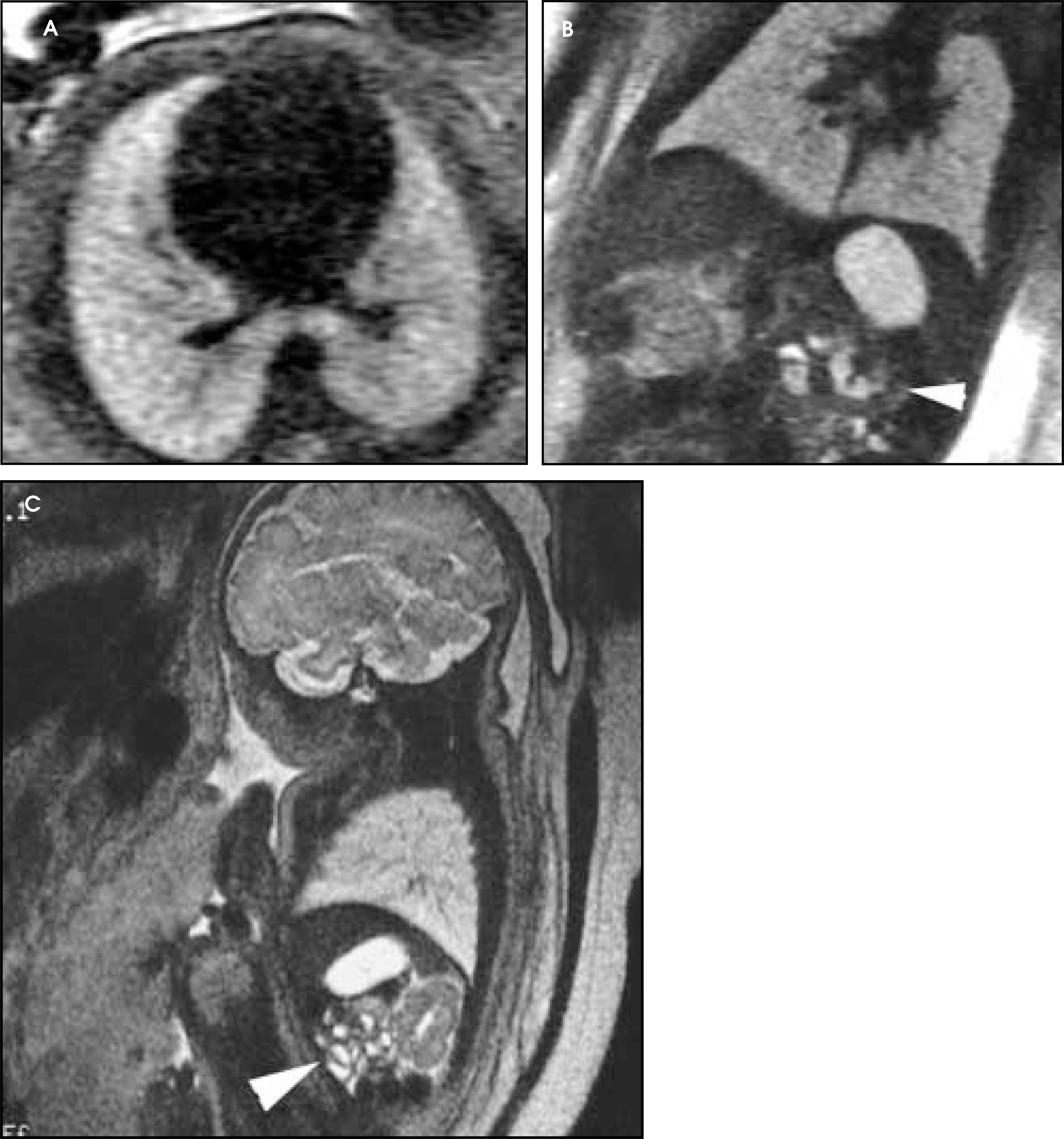
Congenital chest masses have characteristic, though overlapping, MRI appearances (Figures 15 and 16). The appearance of CCAM varies depending on whether the lesions are microcystic or macrocystic.7 The lesions can have multiple large cysts with discrete walls or can be solid lesions with scattered small cysts. Congenital cystic adenomatoid malformations tend to be higher in T2-weighted signal intensity than normal lungs. Bronchopulmonary sequestration can have an appearance similar to CCAM. Indeed, combined lesions are common. Pulmonary sequestration, found predominately in the left lower lobe, often appears as a bright echogenic mass on US and as a well-demarcated, wedge-shaped region of high signal intensity on T2-weighted MRI. Ultrasound may be able to demonstrate a systemic feeding vessel.7 Congenital lobar emphysema is the least frequently encountered of the congenital chest masses.52,53 The affected side may appear on MRI as a distended hemithorax that is higher in T2 signal intensity than the contralateral side.53 The multiple imaging planes allowed by MRI are useful to demonstrate overdistention of the upper lobe with collapse of the lower lobe.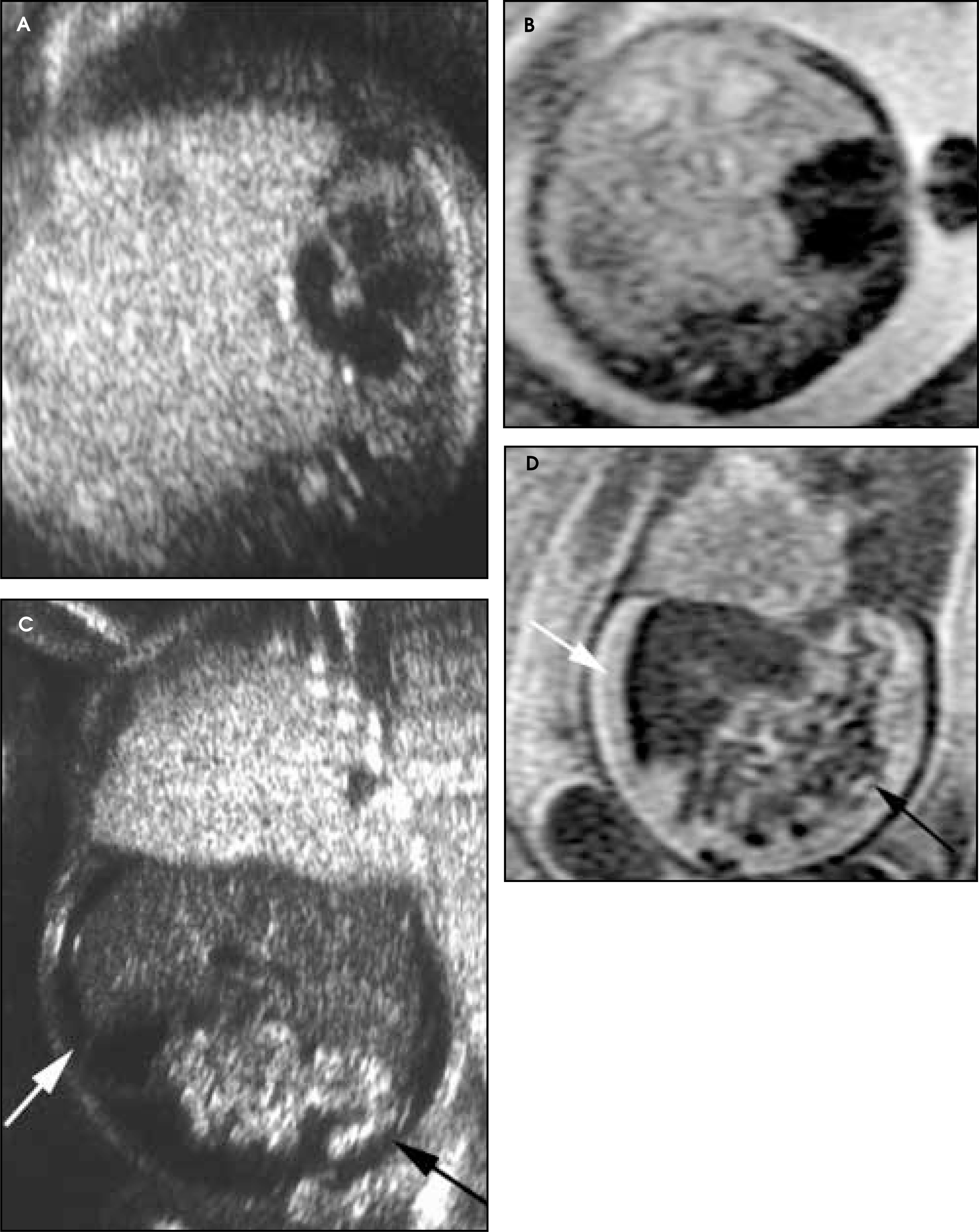
Assessment of fetal lung development with MRI has been particularly pertinent to the evolving management of CDH, in which prognosis is closely related to the severity of pulmonary hypoplasia. Ultrasound-based criteria that have been used to predict the outcome of CDH include the presence of associated structural or chromosomal abnormalities, the presence of liver above the diaphragm, and comparison of the area of residual lung with the head circumference (the lung-to-head circumference ratio, or LHR).15,54,55 The presence of liver in the chest in fetuses with left-sided CDH markedly worsens prognosis and reportedly shares importance with LHR in evaluation of these lesions.7,15,56 Due to the conspicuity of the liver on both T1- and T2-weighted images, MRI has been found to be more sensitive than US in confirming liver position (Figure 17).5,15 Management of CDH has evolved in some centers to the degree that decisions are based largely on the location of the liver. Both MRI and US are critical in this setting.5,15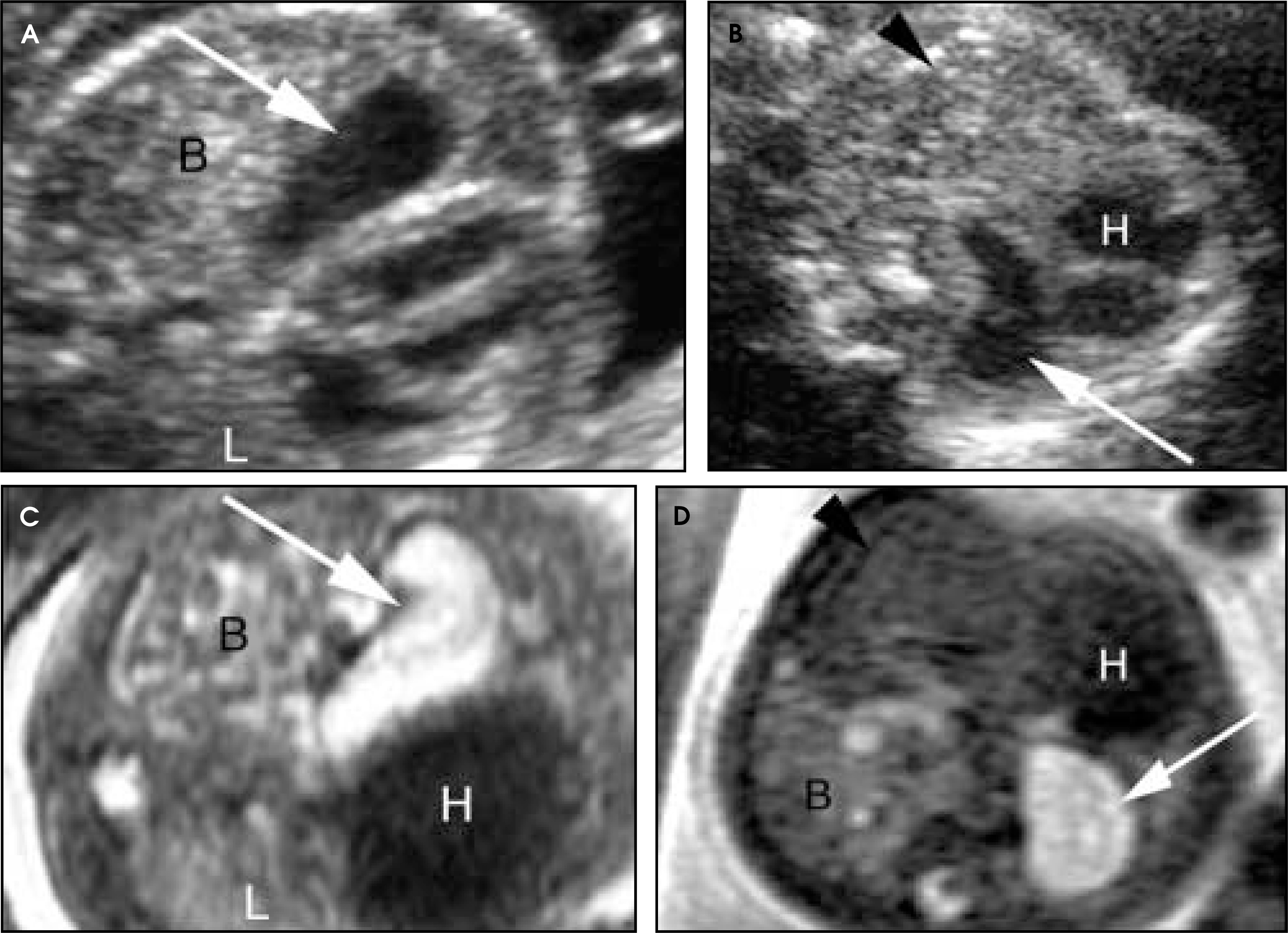
Several reports have described the use of MRI in evaluating a broad spectrum of genitourinary anomalies (Figure 18).51,57-60 MRI may be particularly useful in the assessment of pregnancies complicated by oligohydramnios, which can limit the diagnostic sensitivity of US.61 Poutamo et al,62 in a report that re-affirms the complementary relationship between US and MRI, performed MRI on 24 fetuses with oligohydramnios or genitourinary anomalies diagnosed by US and found that both MRI and US provided the correct diagnosis in 12 of 24 (50%). However, US, but not MRI, provided the correct diagnosis in 3 of 24 (12.5%), and MRI, but not US, provided the correct diagnosis in 8 of 24 (33.3%).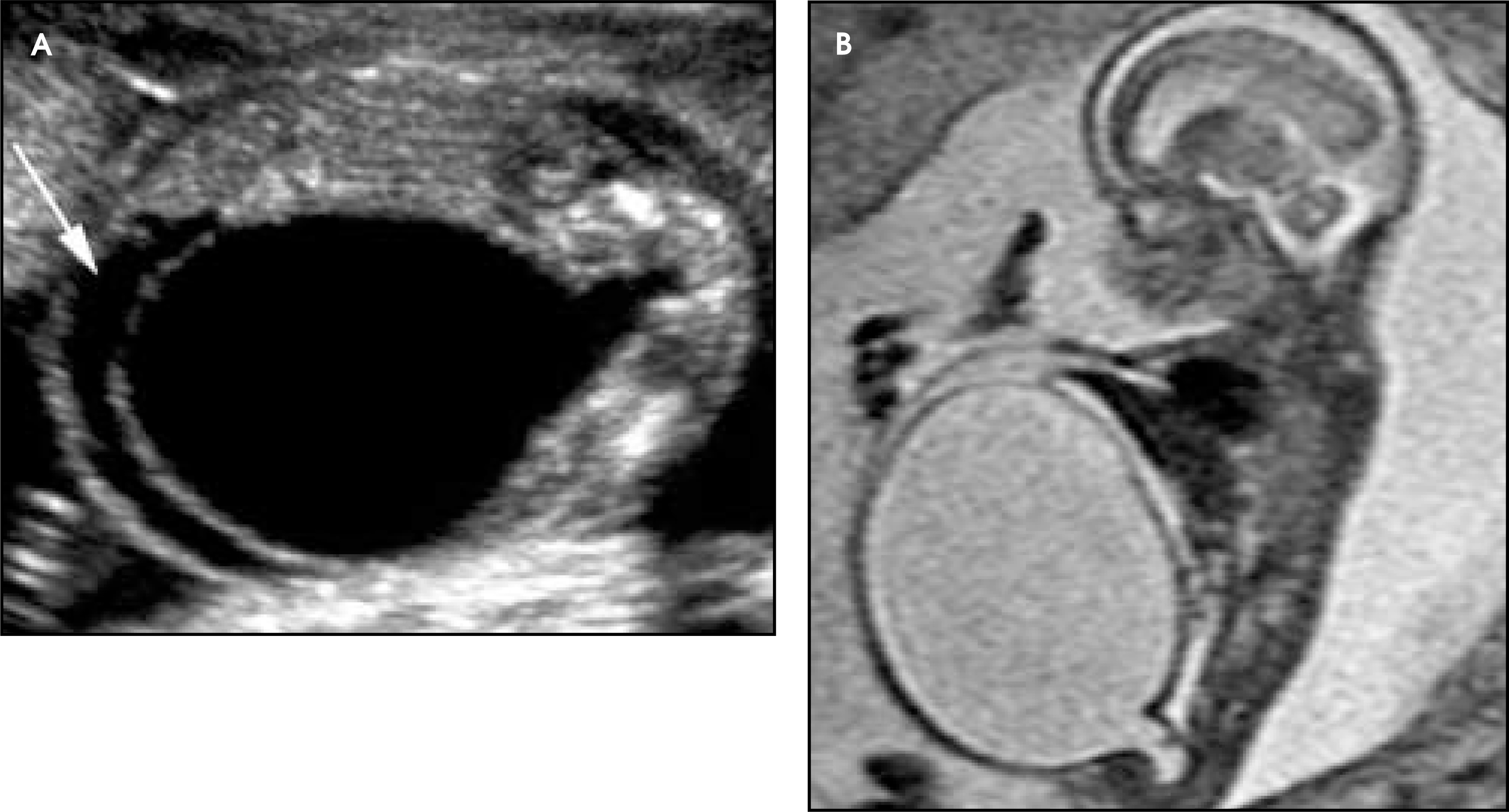
No research series has yet directly assessed the benefits that MRI may have over US in the evaluation of GI anomalies or remaining developing organ systems outside the thorax and urogenital system. Reports of MRI examinations of the fetal gastrointestinal tract have presented the appearance of normal bowel on both T1-weighted and T2-weighted images, and the MRI appearances of a number of abnormalities have been described, including omphalocele, gastroschisis, obstruction, intestinal and esophageal atresias, and hiatal hernia (Figures 19 and 20).63,64,51 Many reports and pictorial expositions have emerged in which MRI has been used to address a wide variety of other fetal anomalies, including skeletal dysplasias.65-67 The added utility of fetal MRI in these other settings has not yet been demonstrated. MRI has become a quite valuable adjunct to US in the antenatal evaluation of conjoined twins in whom postnatal separation is being anticipated. Several reports have indicated that MRI increases the precision and detail of the prenatal evaluation in these complex cases (Figure 21).68-71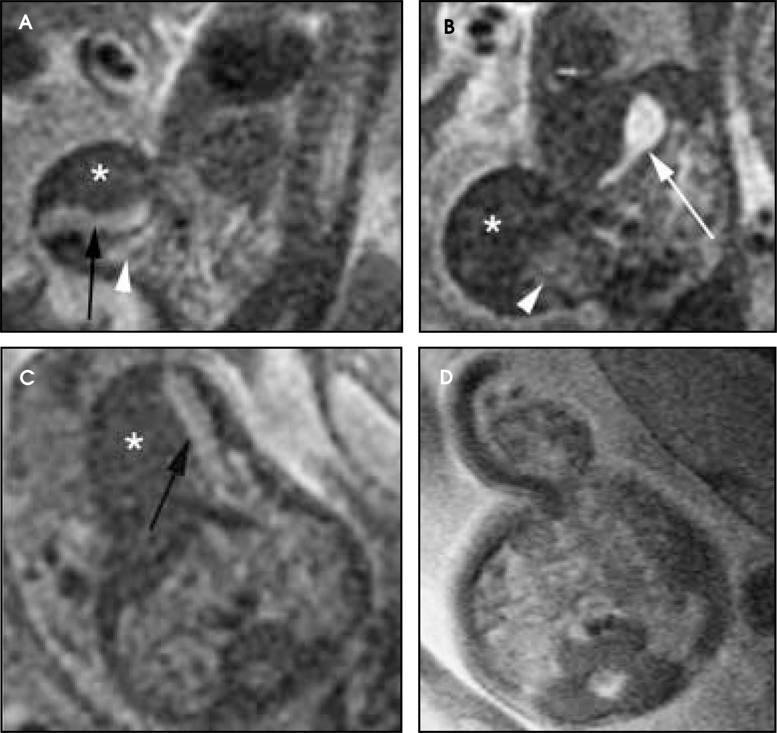
Conclusion
MRI has emerged as an important adjunct to US in the evaluation of fetal anomalies. It is unlikely that MRI will ever supplant US in the primary evaluation of pregnancy status and fetal well-being. Nonetheless, as fetal therapy evolves, parents demand greater understanding and involvement in decision-making and more pediatric subspecialists become involved in antenatal management, the demand for fetal MRI as a supplement to US will likely parallel the increased demand for MRI occurring postnatally in pediatric centers. Accordingly, more studies are needed to evaluate the impact of fetal MRI on the diagnosis of fetal anomalies, clinical decision-making, parental understanding, and neonatal outcomes.
References
- Levine D. Fetal magnetic resonance imaging. Top Magn Reson Imaging. 2001;12:1-2.
- Huisman T, Martin E, Kubik-Huch R, Marincek B. Fetal magnetic resonance imaging of the brain: Technical considerations and normal brain development. Eur Radiol. 2002;12:1941-1951.
- Hill M, Lande I, Larsen J. Prenatal diagnosis of fetal anomalies using ultrasound and MRI. Radiol Clin North Am. 1988;26:287-307.
- Levine D. Ultrasound versus magnetic resonance imaging in fetal evaluation. Top Magn Reson Imaging. 2001;12:25-38.
- Hubbard A. Magnetic resonance imaging of fetal thoracic abnormalities. Top Magn Reson Imaging. 2001;12:18-24.
- Levine D, Barnes P, Madsen J. Fetal central nervous system anomalies: MR imaging augments sonographic diagnosis. Radiology. 1997;204:635-642.
- Hubbard A, Simon E. Fetal imaging. Magn Reson Imaging Clin N Am. 2002;10:389-408.
- Levine D, Barnes P, Madsen J. Fetal CNS anomalies revealed on ultrafast MR imaging. AJR Am J Roentgenol. 1999;172:813-818.
- Simon E, Goldstein R, Coakley F. Fast MR imaging of fetal CNS anomalies in utero. AJNR Am J Neuroradiol. 2000;21:1688-1698.
- Coakley F, Hricak H, Filly R. Complex fetal disorders: Effect of MR imaging on management—Preliminary clinical experience. Radiology. 1999;213:691-696.
- Hubbard A, Adzick N, Crombleholme T. Congenital chest lesions: Diagnosis and characterization with prenatal MR imaging. Radiology. 1999;212:43-48.
- Stazzone M, Hubbard A, Bilaniuk L. Ultrafast MR imaging of the normal posterior fossa in fetuses. AJR Am J Roentgenol. 2000;175:835-839.
- Malinger G, Lev D, Lerman-Sagie T. Is fetal magnetic resonance imaging superior to neurosonography for detection of brain anomalies?. Ultrasound Obstet Gynecol. 2002;20:317-321.
- Coakley F. Role of magnetic resonance imaging in fetal surgery. Top Magn Reson Imaging. 2001;12:39-51.
- Coleman B, Adzick N, Crombleholme T. Fetal therapy: State of the art. J Ultrasound Med. 2002;21:1257-1288.
- Wolff S, Crooks L, Brown P. Tests for DNA and chromosomal damage induced by nuclear magnetic resonance imaging. Radiology. 1980;136:707-710.
- Reid A, Smith F, Hutchison J. Nuclear magnetic resonance imaging and its safety implications: Follow-up of 181 patients. Br J Radiol. 1982;55:784-786.
- Schwartz J, Crooks L. NMR imaging produces no observable mutations or cytotoxicity in mammalian cells. AJR Am J Roentgenol. 1982;139:583-585.
- Kanal E, Shellock F, Talagala L. Safety considerations in MR imaging. Radiology. 1990;176:593-606.
- Kanal E, Gillen J, Evans J. Survey of reproductive health among female MR workers. Radiology. 1993;187:395-399.
- Shellock F, Kanal E. Guidelines and recommendations for MR imaging safety and patient management. III. Questionnaire for screening patients before MR procedures. The SMRI Safety Committee. J Magn Reson Imaging. 1994;4:749-751.
- Baker P, Johnson I, Harvey P. A three-year follow-up of children imaged in utero with echo-planar magnetic resonance. Am J Obstet Gynecol. 1994;170:32-33.
- Athey T. FDA regulation of the safety of MR devices: Past, present, and future. Magn Reson Imaging Clin N Am. 1998;6:791-795.
- Myers C, Duncan K, Gowland P. Failure to detect intrauterine growth restriction following in utero exposure to MRI. Br J Radiol. 1998;71:549-551.
- Chew S, Ahmadi A, Goh P, Foong L. The effects of 1.5T magnetic resonance imaging on early murine in-vitro embryo development. J Magn Reson Imaging. 2001;13:417-420.
- Levine D, Zuo C, Faro C, Chen Q. Potential heating effect in the gravid uterus during MR HASTE imaging. J Magn Reson Imaging. 2001;13:856-861.
- Okuda Y, Sagami F, Tirone P. [Reproductive and developmental toxicity study of gadobenate dimeglumine formulation (E7155) (3)—Study of embryo-fetal toxicity in rabbits by intravenous administration]. J Toxicol Sci. 1999;24:79-87.
- Novak Z, Thurmond A, Ross P. Gadolinium-DTPA transplacental transfer and distribution in fetal tissue in rabbits. Invest Radiol. 1993;28:828-830.
- Sonigo P, Rypens F, Carteret M. MR imaging of fetal cerebral anomalies. Pediatr Radiol. 1998;28:212-222.
- O’Connor M. Intrauterine effects in animals ex-posed to radiofrequency and microwave fields. Teratology. 1999;59:287-291.
- Clements H, Duncan K, Fielding K. Infants exposed to MRI in utero have a normal paediatric assessment at 9 months of age. Br J Radiol. 2000;73:190-194.
- Chen Q, Levine D. Fast fetal magnetic resonance imaging techniques. Top Magn Reson Imaging. 2001;12:67-79.
- Poutamo J, Vanninen R, Partanen K. Magnetic resonance imaging supplements ultrasonographic imaging of the posterior fossa, pharynx and neck in malformed fetuses. Ultrasound Obstet Gynecol. 1999;13:327-334.
- Levine D, Barnes P, Madsen J. Central nervous system abnormalities assessed with prenatal magnetic resonance imaging. Obstet Gynecol. 1999;94:1011-1019.
- Bennett G, Bromley B, Benacerraf B. Agenesis of the corpus callosum: Prenatal detection usually is not possible before 22 weeks of gestation. Radiology. 1996;199:447-450.
- Levine D, Trop I, Mehta T, Barnes P. MR imaging appearance of fetal cerebral ventricular morphology. Radiology. 2002;223:652-660.
- Levine D, Barnes P. Cortical maturation in normal and abnormal fetuses as assessed with prenatal MR imaging. Radiology. 1999;210:751-758.
- Kok R, van den Berg P, van den Bergh A. Maturation of the human fetal brain as observed by 1H MR spectroscopy. Magn Reson Med. 2002;48:611-616.
- Kok R, van den Berg P, van den Bergh A. MR spectroscopy in the human fetus. Radiology. 2002;223:584.
- Kok R, van den Bergh A, Heerschap A. Metabolic information from the human fetal brain obtained with proton magnetic resonance spectroscopy. Am J Obstet Gynecol. 2001;185:1011-1015.
- Avni F, Guibaud L, Robert Y. MR imaging of fetal sacrococcygeal teratoma: Diagnosis and assessment. AJR Am J Roentgenol. 2002;178:179-183.
- Mangels K, Tulipan N, Tsao L. Fetal MRI in the evaluation of intrauterine myelomeningocele. Pediatr Neurosurg. 2000;32:124-131.
- Kolble N, Huisman T, Stallmach T. Prenatal diagnosis of a fetus with lumbar myelocystocele. Ultrasound Obstet Gynecol. 2001;18:536-539.
- Duncan K. Fetal and placental volumetric and functional analysis using echo-planar imaging. Top Magn Reson Imaging. 2001;12:52-66.
- Kuwashima S, Nishimura G, Iimura F. Low-intensity fetal lungs on MRI may suggest the diagnosis of pulmonary hypoplasia. Pediatr Radiol. 2001;31:669-672.
- Coakley F, Lopoo J, Lu Y. Normal and hypoplastic fetal lungs: Volumetric assessment with prenatal single-shot rapid acquisition with relaxation enhancement MR imaging. Radiology. 2000;216:107-111.
- Hubbard A, States L. Fetal magnetic resonance imaging. Top Magn Reson Imaging. 2001;12:93-103.
- Fenton B, Lin C, Ascher S, Macedonia C. Magnetic resonance spectroscopy to detect lecithin in amniotic fluid and fetal lung. Obstet Gynecol. 2000;95:457-460.
- Bouchard S, Johnson M, Flake A. The EXIT procedure: Experience and outcome in 31 cases. J Pediatr Surg. 2002;37:418-426.
- Mychaliska G, Bealer J, Graf J. Operating on placental support: The ex utero intrapartum treatment procedure. J Pediatr Surg. 1997;32:227-230.
- Shinmoto H, Kashima K, Yuasa Y. MR imaging of non-CNS fetal abnormalities: A pictorial essay. RadioGraphics. 2000;20:1227-1243.
- Quinton A, Smoleniec J. Congenital lobar emphysema—the disappearing chest mass: Antenatal ultrasound appearance. Ultrasound Obstet Gynecol. 2001;17:169-171.
- Olutoye O, Coleman B, Hubbard A, Adzick N. Prenatal diagnosis and management of congenital lobar emphysema. J Pediatr Surg. 2000;35:792-795.
- Paek B, Coakley F, Lu Y. Congenital diaphragmatic hernia: Prenatal evaluation with MR lung volumetry—preliminary experience. Radiology. 2001;220:63-67.
- Metkus A, Filly R, Stringer M. Sonographic predictors of survival in fetal diaphragmatic hernia. J Pediatr Surg. 1996;31:148-151.
- Walsh D, Hubbard A, Olutoye O. Assessment of fetal lung volumes and liver herniation with magnetic resonance imaging in congenital diaphragmatic hernia. Am J Obstet Gynecol. 2000;183:1067-1069.
- Fradin J, Regan F, Rodriquez R, Moore R. Hydronephrosis in pregnancy: Simultaneous depiction of fetal and maternal hydronephrosis by magnetic resonance urography. Urology. 1999;53:825-827.
- Takeuchi K, Moriyama T, Funakoshi T, Maruo T. Prenatal diagnosis of fetal urogenital abnormalities with oligohydramnios by magnetic resonance imaging using turbo spin echo technique. J Perinat Med. 1998;26:59-61.
- Hutcheson J, Canning D, Hubbard A. Magnetic resonance imaging of fetal urinoma. Urology. 2002;60:697.
- Miller O, Lashley D, McAleer I, Kaplan G. Diagnosis of urethral obstruction with prenatal magnetic resonance imaging. J Urol. 2002;168:1158-1159.
- Levine D, Goldstein R, Callen P. The effect of oligohydramnios on detection of fetal anomalies with sono-graphy. AJR Am J Roentgenol. 1997;168:1609-1611.
- Poutamo J, Vanninen R, Partanen K, Kirkinen P. Diagnosing fetal urinary tract abnormalities: Benefits of MRI compared to ultrasonography. Acta Obstet Gynecol Scand. 2000;79:65-71.
- Langer J, Hussain H, Khan A. Prenatal diagnosis of esophageal atresia using sonography and magnetic resonance imaging. J Pediatr Surg. 2001;36:804-807.
- Saguintaah M, Couture A, Veyrac C. MRI ofthe fetal gastrointestinal tract. Pediatr Radiol. 2002;32:395-404.
- Suzumura H, Kohno T, Nishimura G. Prenatal diagnosis of hypochondrogenesis using fetal MRI: A case report. Pediatr Radiol. 2002;32:373-375.
- Teng S, Guo W, Sheu M, Wang P. Initial experience using magnetic resonance imaging in prenatal diagnosis of osteogenesis imperfecta type II. A case report. Clin Imaging. 2003;27:55-58.
- Ueno K, Tanaka M, Miyakoshi K. Prenatal diagnosis of atelosteogenesis type I at 21 weeks' gestation. Prenat Diagn. 2002;22:1071-1075.
- Casele H, Meyer J. Ultrafast magnetic resonance imaging of cephalopagus conjoined twins. Obstet Gynecol. 2000;95:1015-1017.
- Kingston C, McHugh K, Kumaradevan J. Imaging in the preoperative assessment of conjoined twins. RadioGraphics. 2001;21:1187-1208.
- Mackenzie T, Crombleholme T, Johnson M. The natural history of prenatally diagnosed conjoined twins. J Pediatr Surg. 2002;37:303-309.
- Spielmann A, Freed K, Spritzer C. MRI of conjoined twins illustrating advances in fetal imaging. J Comput Assist Tomogr. 2001;25:88-90.
Citation
. Fetal MRI. Applied Radiology. 2004;33(2):9-25. doi:10.37549/AR1219.