Food signs in musculoskeletal radiology
Applied Radiology — Vol. 34 , Issue 10 , pp. 17 -24
DOI: 10.37549/AR1373
Published: October 1, 2005
Categories
Today’s radiologist has little time to engage in the pursuit of gustatory and gastronomical pleasures during the course of his or her busy workday. While midday visceral cravings may continue unabated, intellectual satiety is readily achieved by recognizing the bounty of food signs in musculoskeletal radiology. Discussions of the underlying pathology responsible for these specific radiographic signs provide food for thought and are seasoned with differential diagnoses, where appropriate, on which the reader may ruminate.
Breakfast
Pancake vertebra
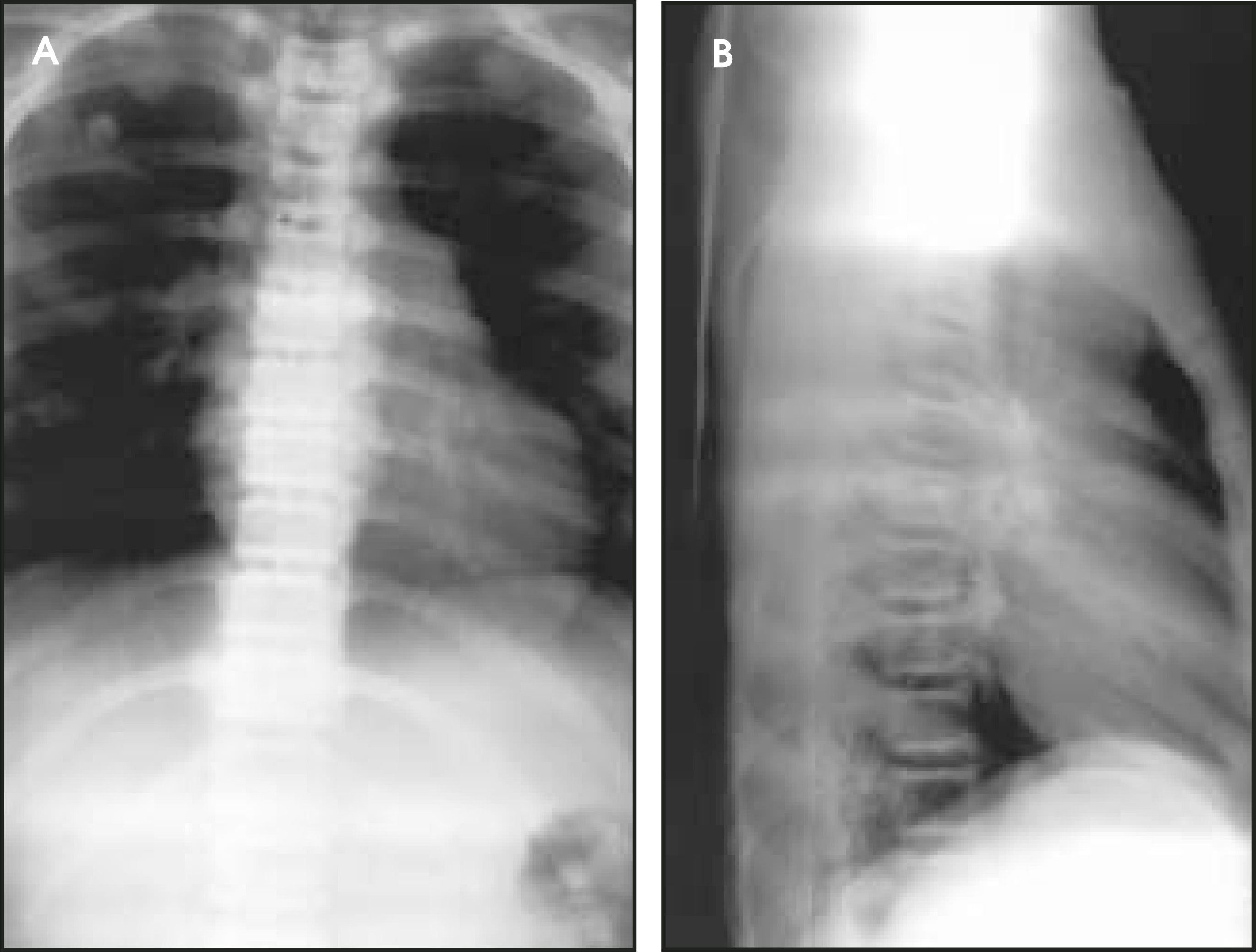
The term “pancake vertebra” (Figure 1) refers to pronounced, diffuse flattening of an entire vertebral body (vertebra plana).1 Such flattening may be seen with a variety of processes. In adults, osteoporosis, metastatic tumor, and multiple myeloma are among the more common causes. In children, eosinophilic granuloma should be considered. Other etiologies include leukemia, lymphoma, hemangioma, osteomyelitis, avascular necrosis, and traumatic fracture.2
Sausage digit
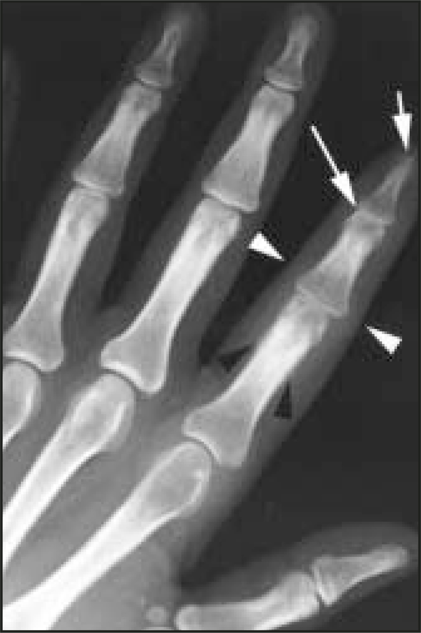
A “sausage digit” (Figure 2) refers to diffuse soft-tissue swelling of an entire finger (or toe) mimicking the shape of a sausage, as seen on physical examination or on radiographs. It is perhaps most characteristically associated with psoriatic arthritis, which may cause inflammation of both the distal and proximal interphalangeal joints as well as the tendon sheaths of a digit1,3; however, a sausage digit may be produced by a variety of different forms of arthritis or dactylitis. Infectious tenosynovitis should be considered in a patient without osseous or articular abnormalities. In the foot, reactive arthritis (one of the components of Reiter’s syndrome) or gout may produce a sausage digit.
Banana fracture
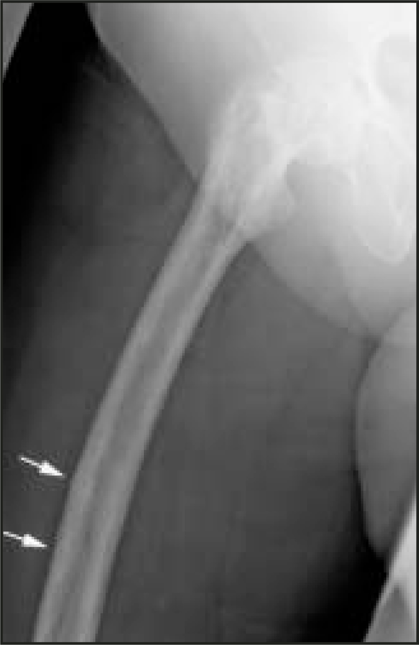
“Banana fractures” (Figure 3) occur in long bones affected by Paget’s disease.4 These cortical insufficiency fractures are so named because they occur along the tensile (convex) surface of the weakened and often bowed pagetoid bone, analogous to the site where the skin of a banana would break upon “snapping” it open. The lateral cortex of the proximal femur is a common site for banana fractures; conventional stress fractures of the proximal femur, in contrast, usually occur along the compressive (concave) surface.
Lunch
Sandwich vertebrae
“Sandwich vertebrae” (Figure 4) occur in patients with osteopetrosis, a hereditary bone disorder due to deficient osteoclast activity. In addition to diffusely increased bone density, on radiographs one may observe osteosclerosis of the superior and inferior vertebral body end-plates, resembling the bread of a sandwich.5 A similar pattern may be seen in patients with myelosclerosis or in patients with renal osteodystrophy (“rugger jersey spine”).
Piece-of-cheese lunate (and spilled teacup)
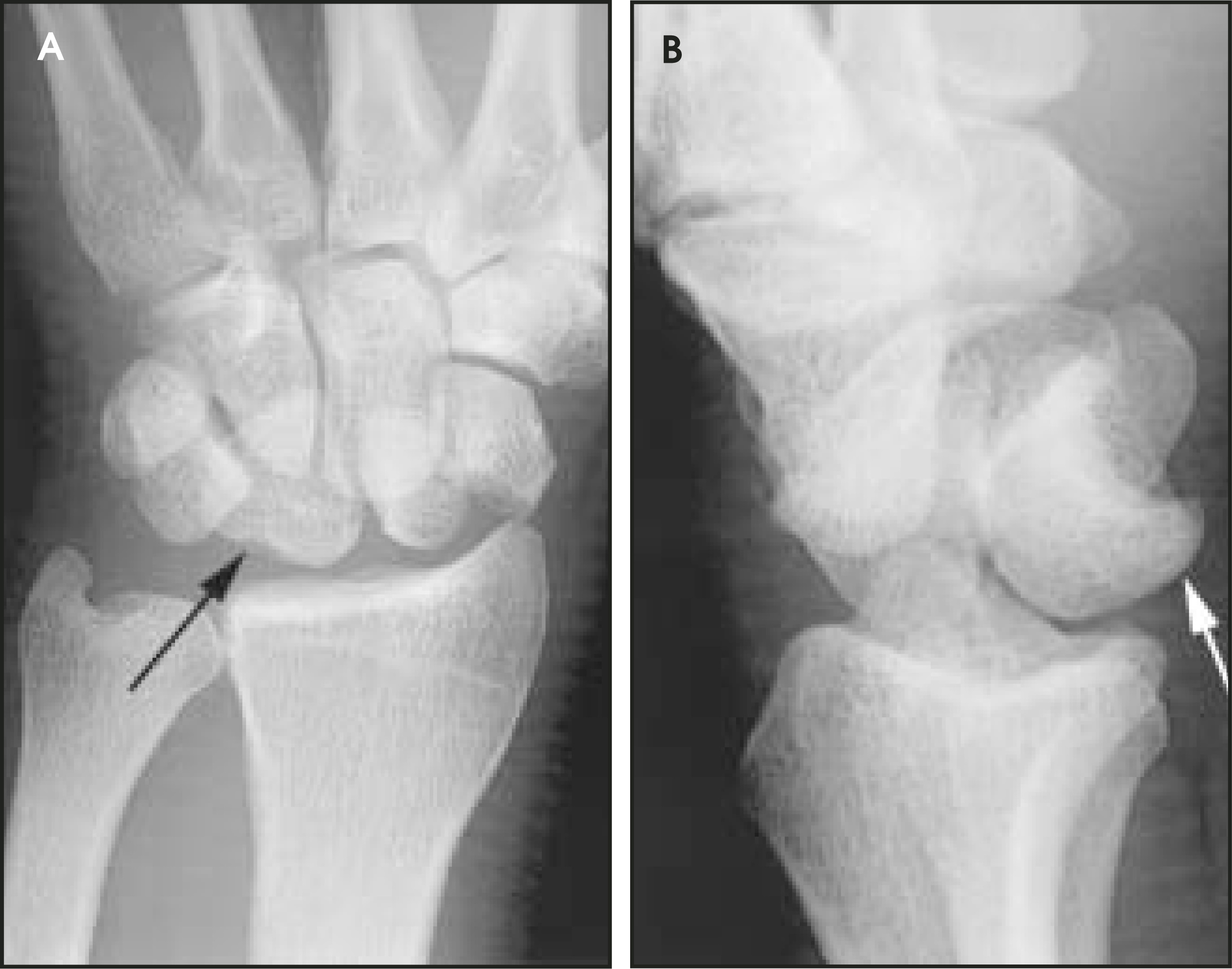
The lunate bone typically has a teacup-shaped appearance on lateral radiographs and a quadrilateral shape on posteroanterior (PA) views. The lunate often assumes a triangular shape on the PA projection (resembling a triangular slice of cheese)6 in patients who have sustained a perilunate or lunate dislocation (Figure 5A). These dislocations can be further evaluated on the lateral projection (Figure 5B). With perilunate dislocation, the radius and lunate will maintain a relatively normal relationship, while the capitate will appear dorsally dislocated. With lunate dislocation, the radius and capitate remain relatively collinear, while the lunate is displaced volarly. In our experience, a triangular-shaped lunate can also be seen on the PA view in patients with the dorsal intercalated segmental instability pattern.
Bite sign
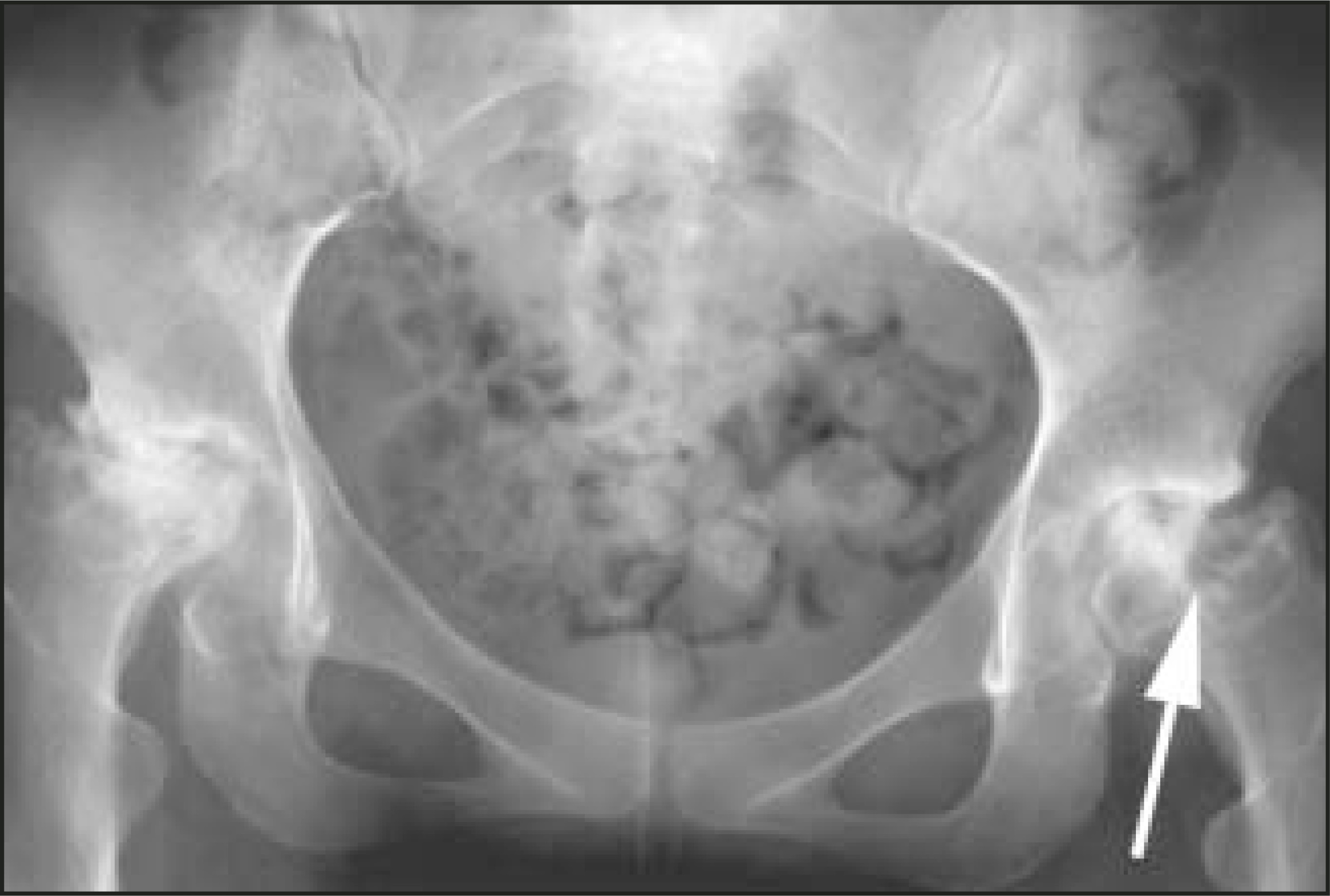
The “bite sign” (Figure 6) refers to neuropathic-like articular destruction seen with steroid arthropathy.7 In advanced cases of avascular necrosis (following subchondral collapse of the femoral head), the articular contour may become so deformed as to resemble a bite taken out of the bone. Although a variety of arthritides can result in large erosions that can mimic a “bite,” these processes will typically affect both sides of the joint and will not produce the femoral head sclerosis associated with avascular necrosis. Extra-articular bite-like erosions may also be seen in patients with tophaceous gout. A bite sign has also been described with chondromyxoid fibroma, a rare benign cartilaginous tumor of the bone. These tumors typically arise eccentrically within the metaphysis of a long bone, and may penetrate the cortex in a bite-like fashion.
Dinner
Hamburger bun and reverse hamburger bun signs
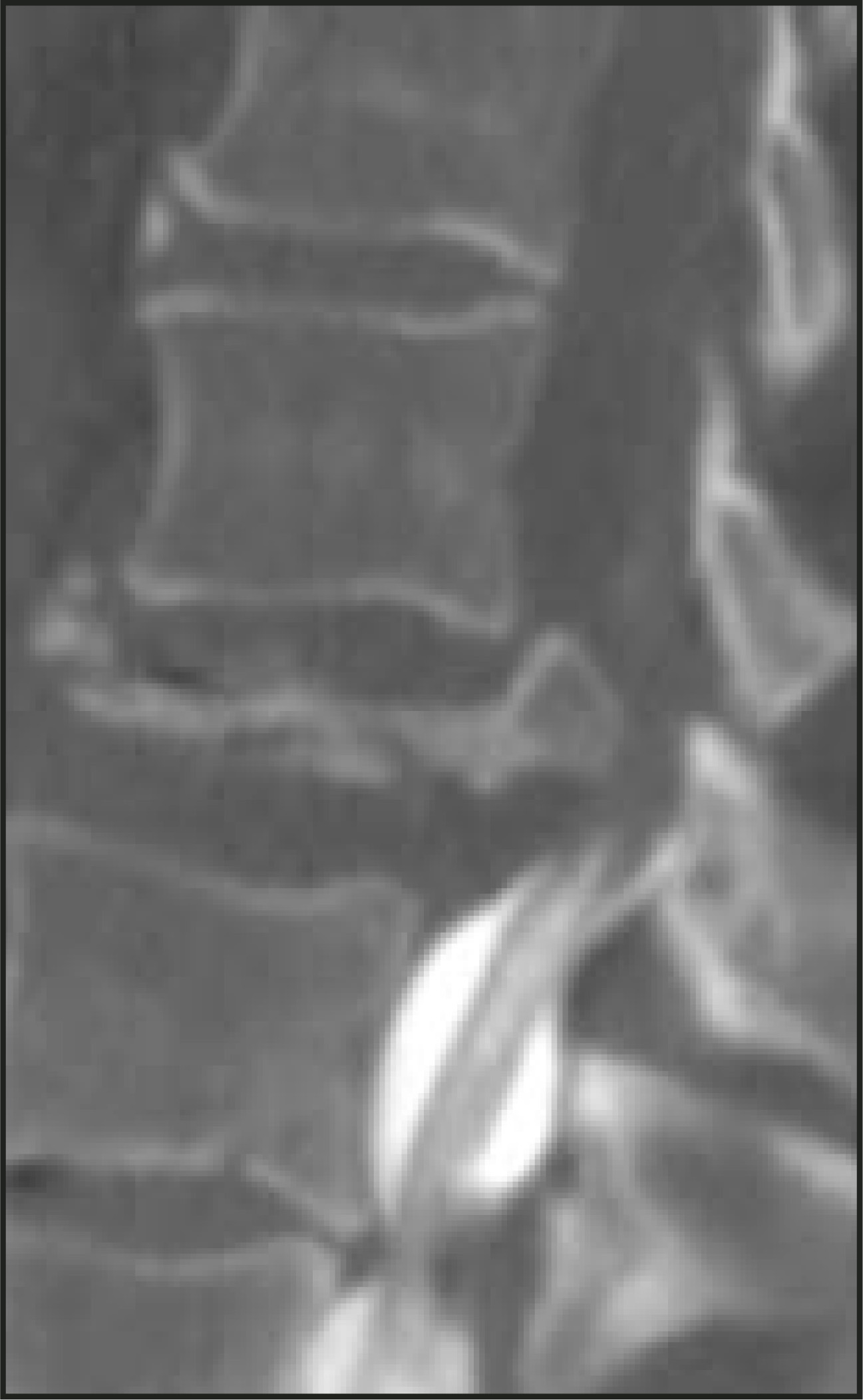
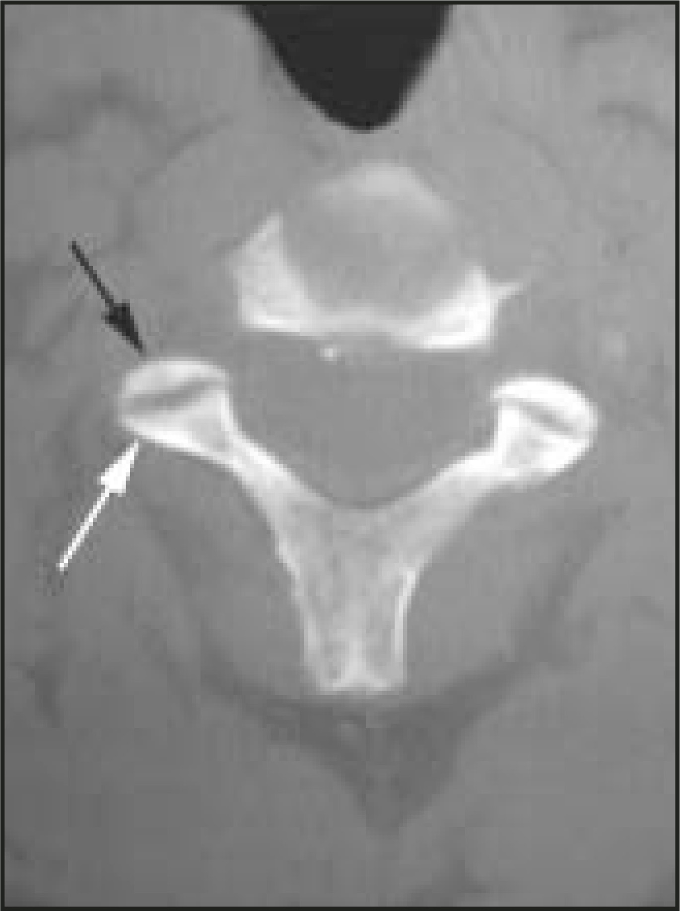
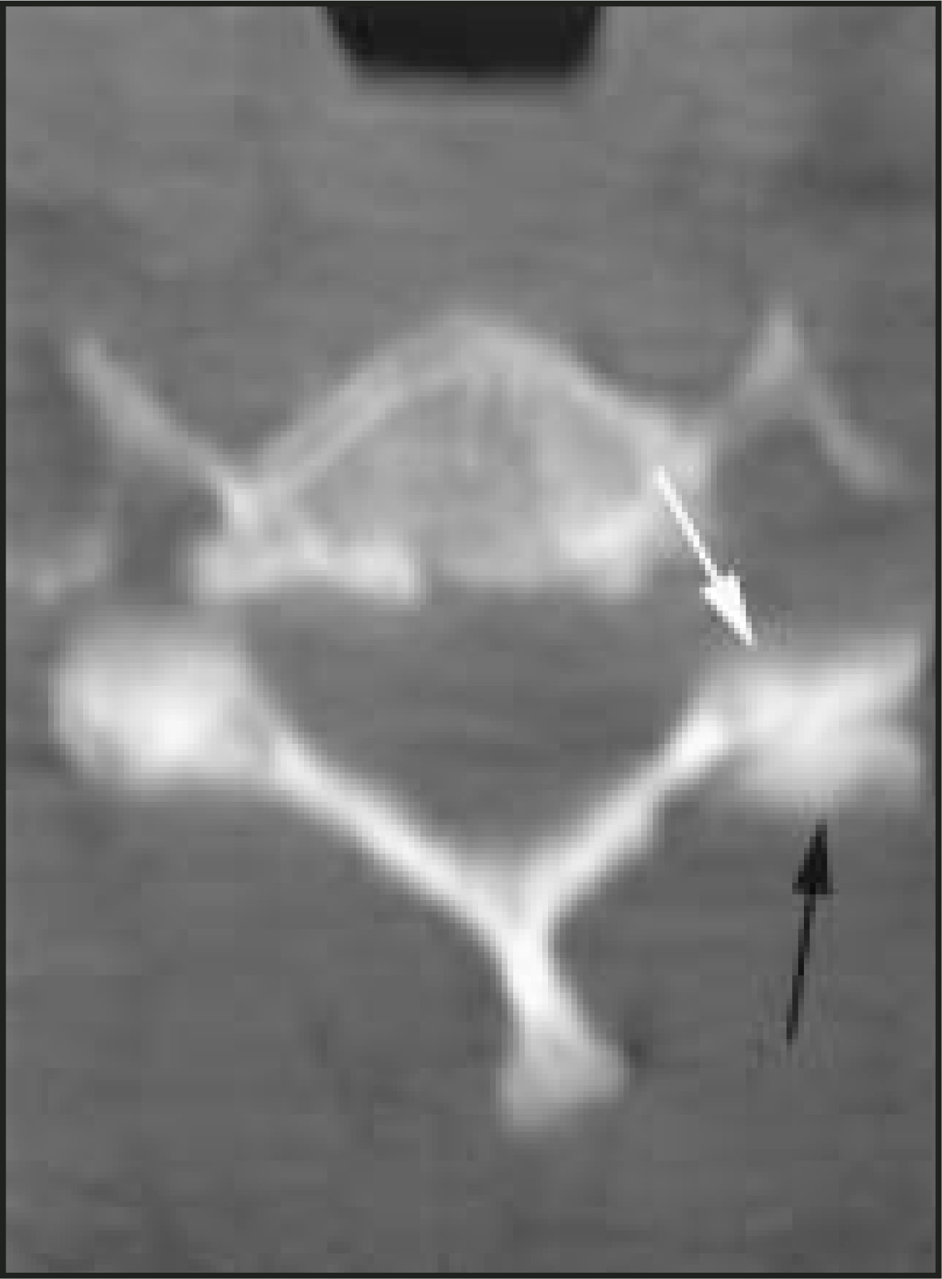
The “hamburger bun sign” (Figure 7) refers to the normal relationship between the superior and inferior articular facets of the cervical vertebrae, as seen on axial computed tomographic (CT) images.8 The superior facet forms the “top bun,” the inferior facet of the superjacent vertebra forms the “bottom bun,” and the intervening facet joint represents the “patty.” The absence of the hamburger bun sign is seen with dislocation of the facet joint; specifically, the superior facet may dislocate posterior to the inferior facet, producing an appearance that resembles the “bottom bun” resting on the “top bun” (Figure 8), also known as the “reverse hamburger bun” sign.
Onion-skin periostitis
“Onion-skin periostitis” (Figure 9) is seen with aggressive osseous processes, and is so-named because it manifests as multiple, thin periosteal layers similar to the concentric layers of an onion cut in cross-section. The pattern is believed to represent either an acceleration of the normal periosteal bone response or alternating periods of rapid and slow growth. It is found in a variety of aggressive musculoskeletal diseases, particularly malignant bone sarcomas (eg, osteosarcoma, Ewing’s sarcoma), lymphoma of bone, and acute osteomyelitis.9
Drumstick terminal phalanx
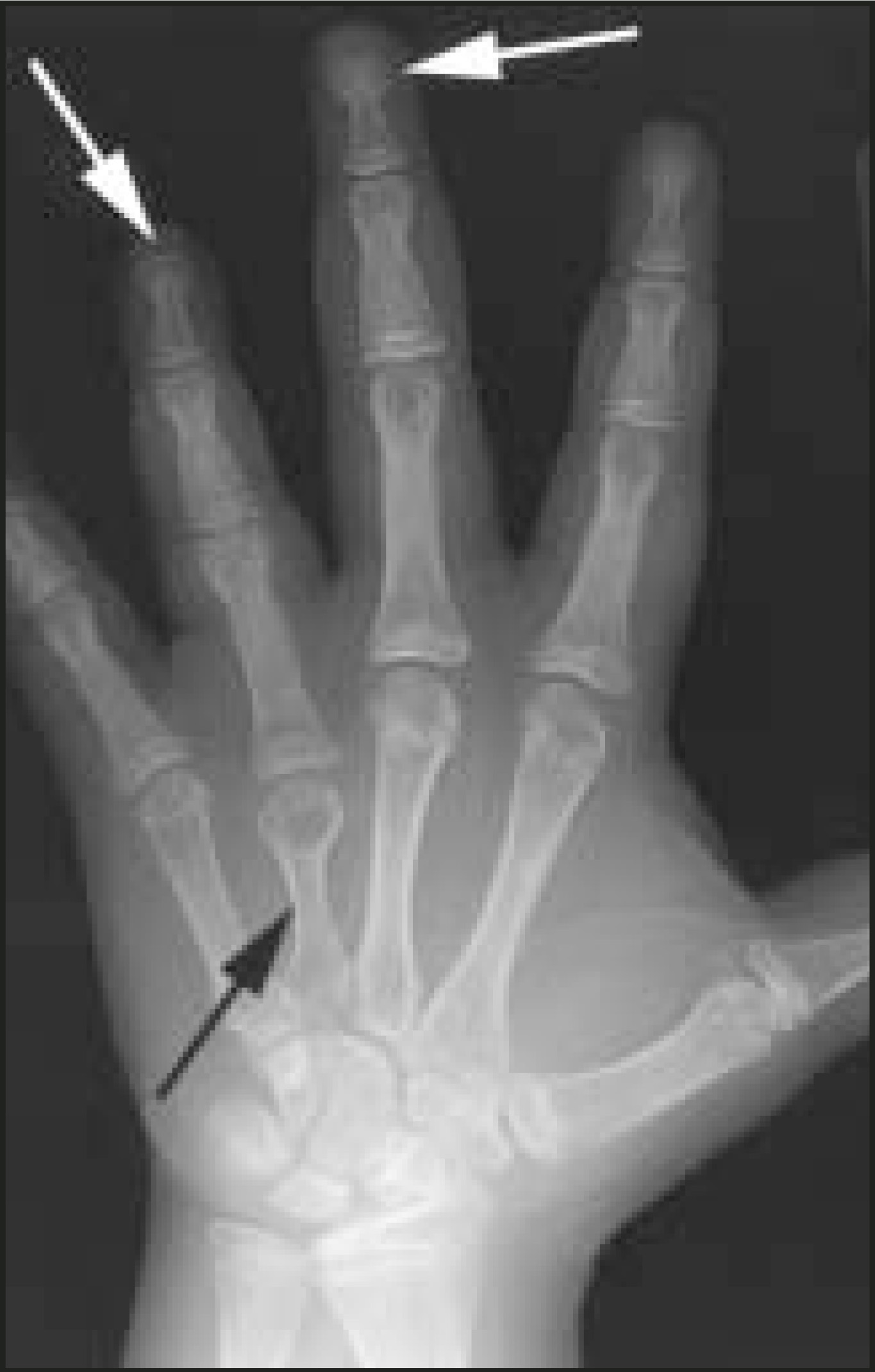
The term “drumstick terminal phalanx” (Figure 10) refers to rounding and broadening of the tufts of the distal phalanges of patients with Turner syndrome,10 figuratively similar to the widening of the meaty end of a chicken or turkey drumstick. Drumstick phalanges have also been described in patients with the rare genetic disorder Coffin-Lowry syndrome.
Salt-and-pepper calvarium
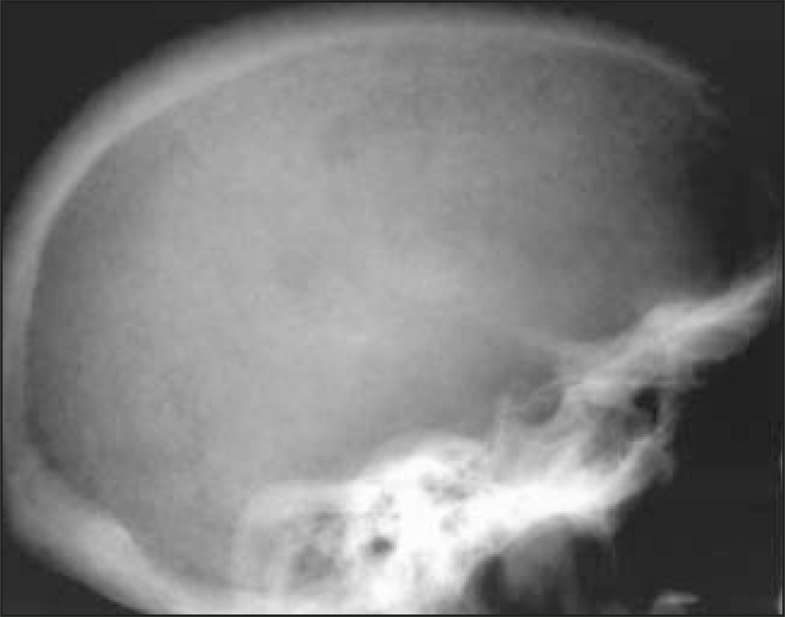
“Salt-and-pepper calvarium” describes the granular appearance of the skull in patients with hyperparathyroidism. This finding represents tiny spheres of trabecular bone resorption stimulated by the elevated levels of parathyroid hormone.11 On radiographs of the skull, the speckled radiolucencies look like scattered salt and pepper (Figure 11). Although most characteristically associated with hyperparathyroidism, the salt-and-pepper skull has also been described in patients with demineralization due to diffuse neoplasm and infection.2
Rice grains and rice bodies
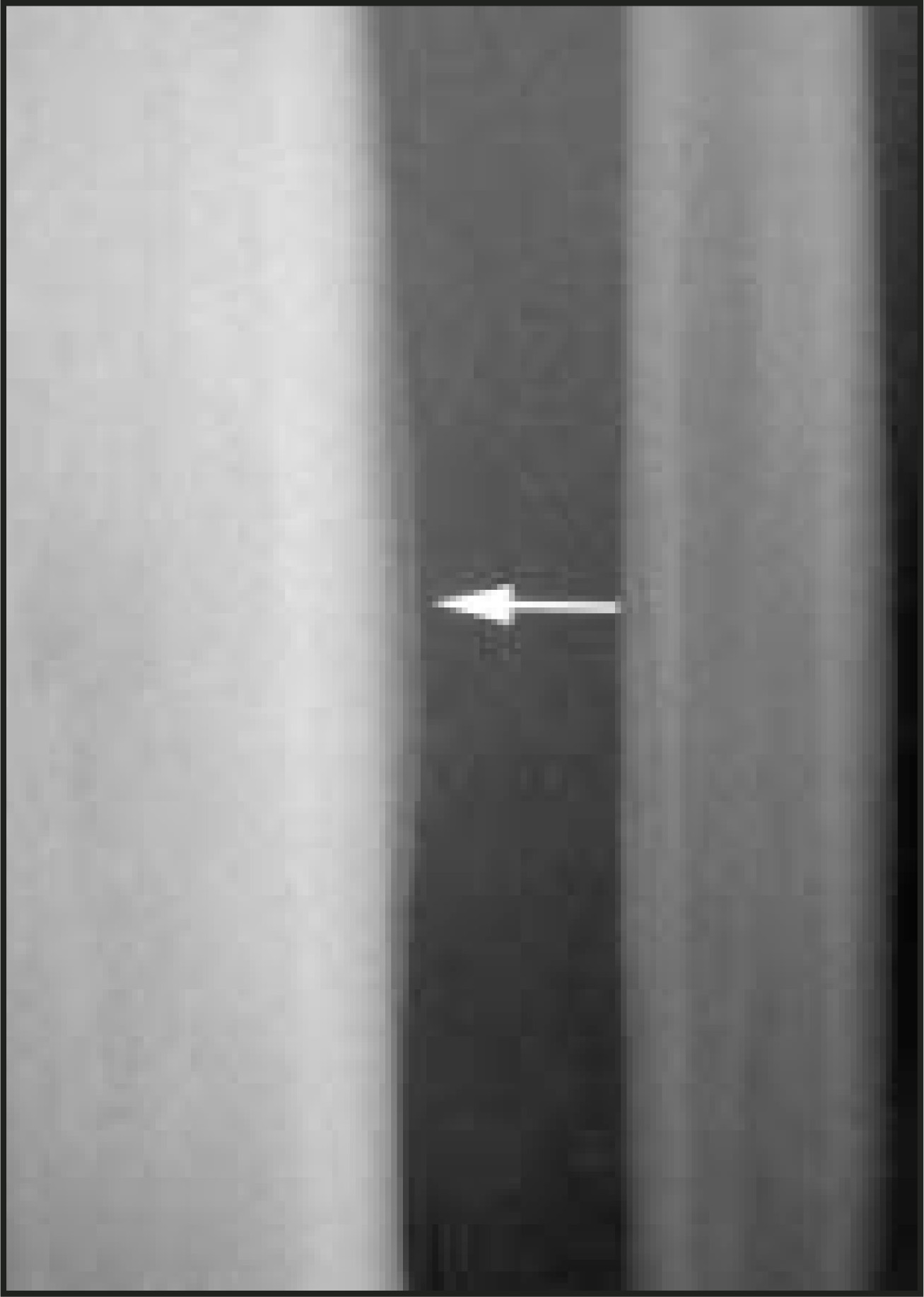
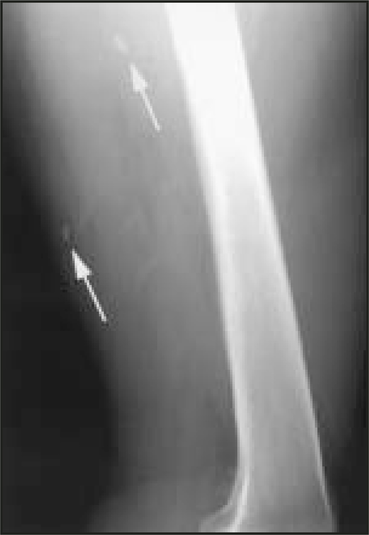
Ingestion of the parasite Taenia solium and the subsequent host response can lead to formation of granulomas (calcified dead larvae) within muscle, brain, and other tissues (cysticercosis). The radiographic appearance of these linear or ovoid calcified granulomas, which lie along the long axis of the muscle, has merited the term “rice grains” (Figure 12). The term “rice grains” has also been used to describe intra-articular loose bodies seen on radiographs of patients with synovial osteochondromatosis.
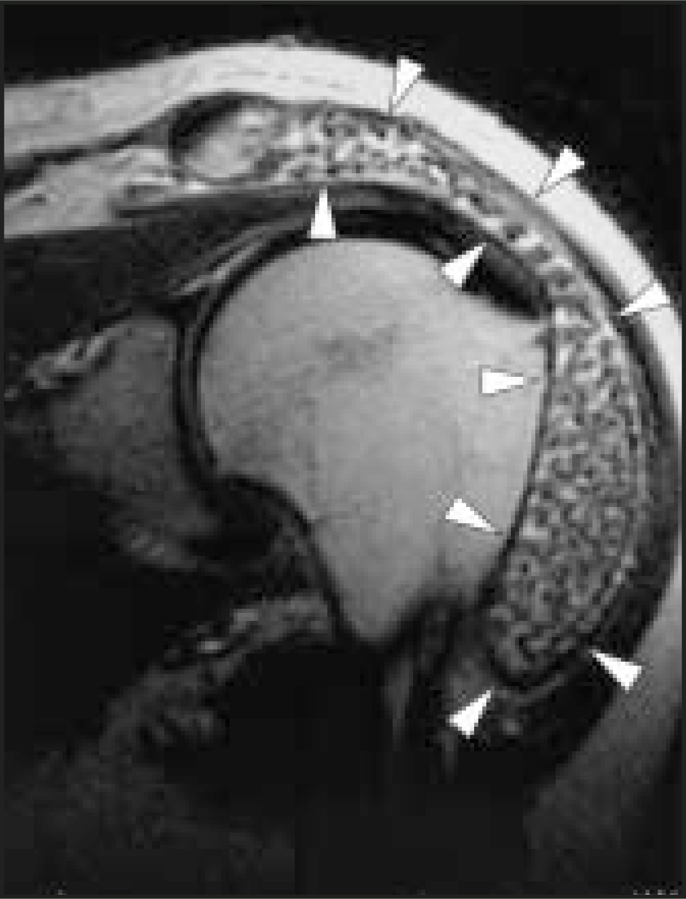
The term “rice bodies” has been used to describe the histologic appearance of intra-articular detached hypertrophied synovial villi in patients with rheumatoid arthritis, mycobacterial infection, and other arthritides.1,12 Rice bodies may also be seen within joints and bursae on magnetic resonance imaging (MRI) studies as multiple small nodules of low signal intensity on T2-weighted images (Figure 13).
Sweets and junk food
Slipping ice cream scoop
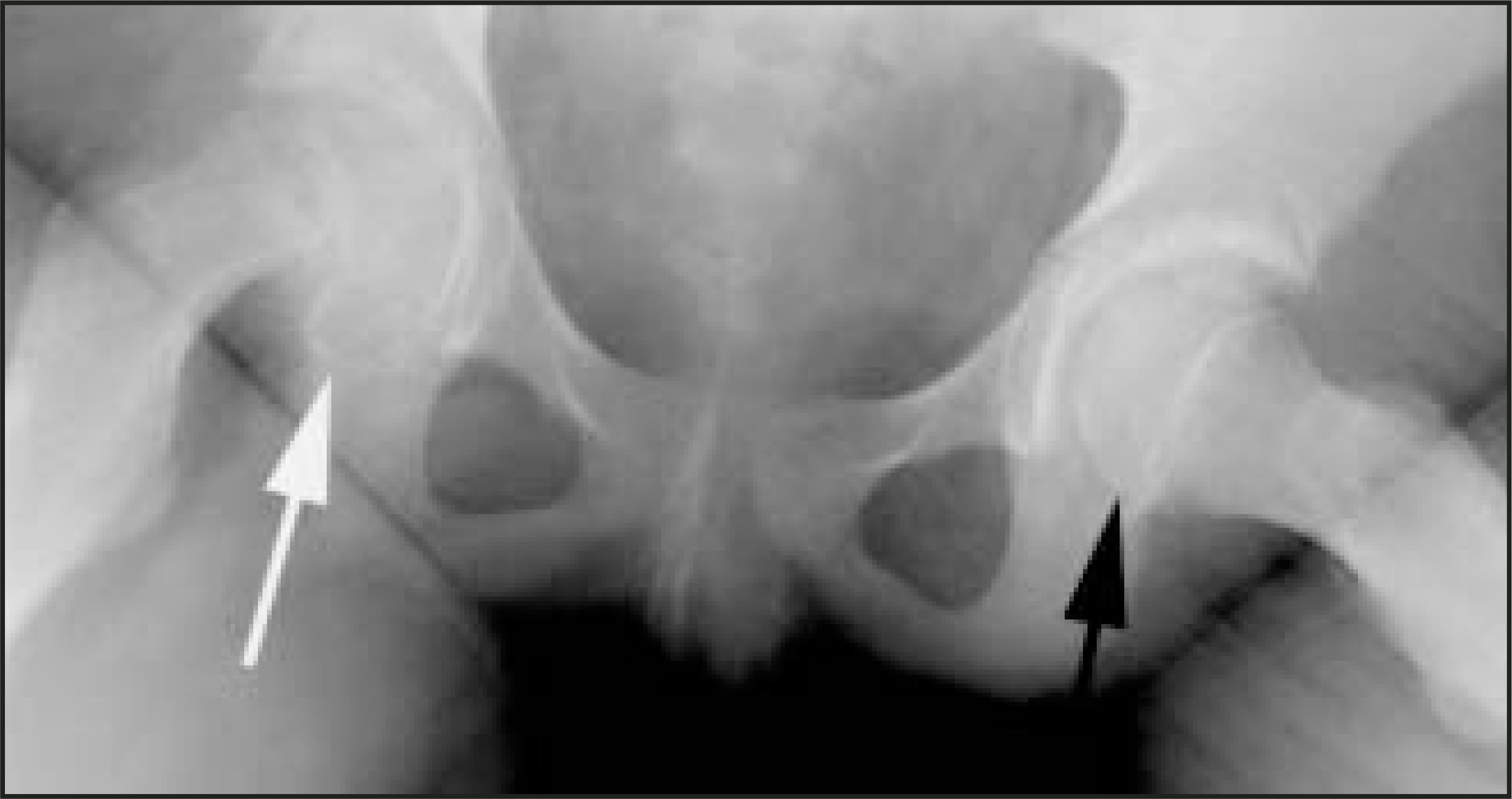
In children with slipped capital femoral epiphysis (SCFE), posteromedioinferior subluxation of the proximal femoral epiphysis with respect to the metaphysis has been described as resembling a scoop of ice cream slipping from its cone10,13 (Figure 14). In actuality, the femoral neck is subluxing anterosuperiorly, while the epiphysis remains situated within the acetabulum. This condition usually occurs during the adolescent growth spurt, when the physis is most susceptible to shearing stresses. Patients tend to be overweight, and boys are more commonly affected than are girls.
Licked candy stick
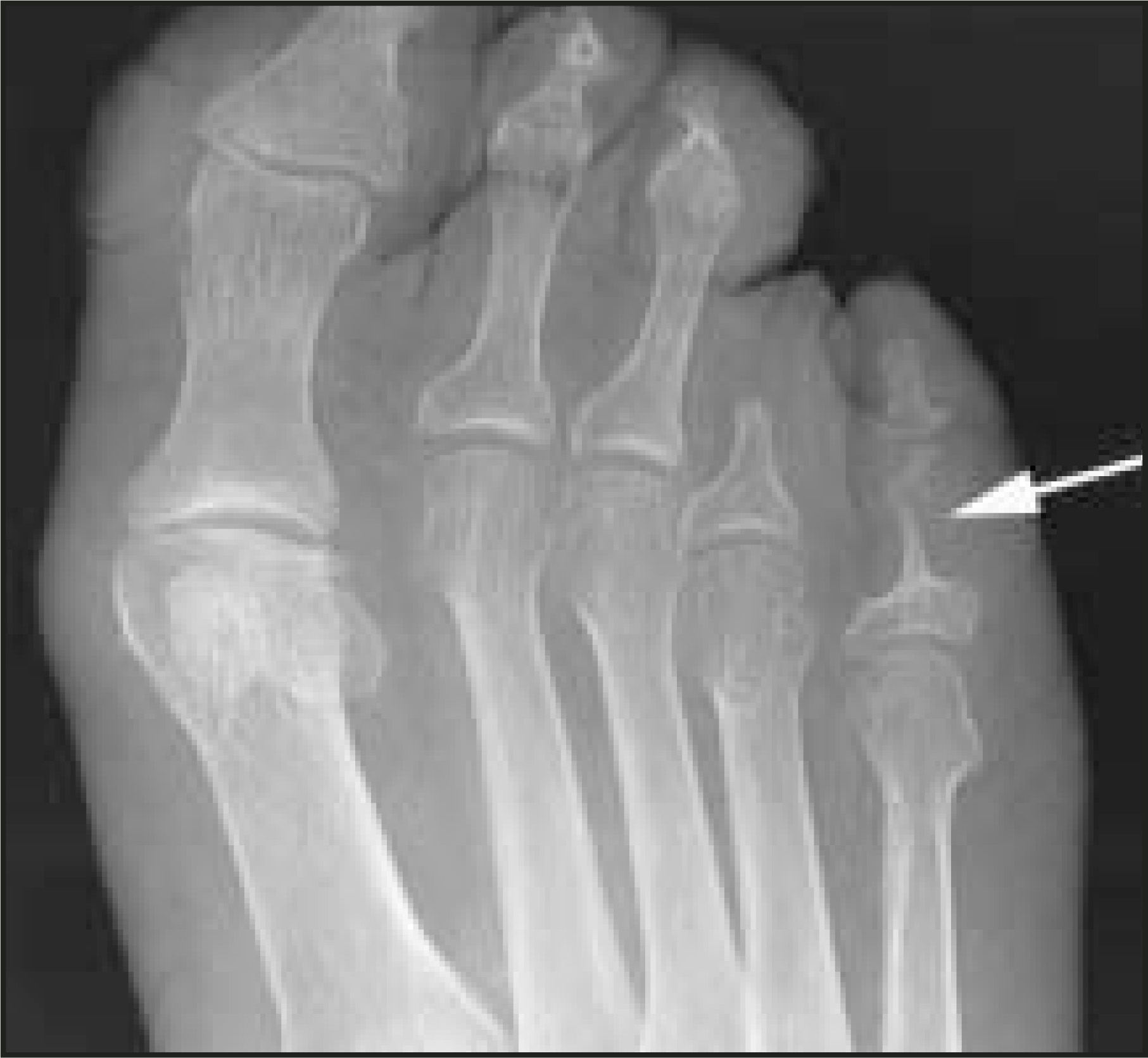
The “licked candy stick” describes the tapering of the distal ends of phalanges, metatarsals, metacarpals, or clavicles, that resemble the tip of a licked candy cane (Figure 15). This phenomenon is due to excessive bone resorption and is seen in patients with psoriatic arthritis, diabetes mellitus, rheumatoid arthritis, and leprosy.1,14
Popcorn calcification
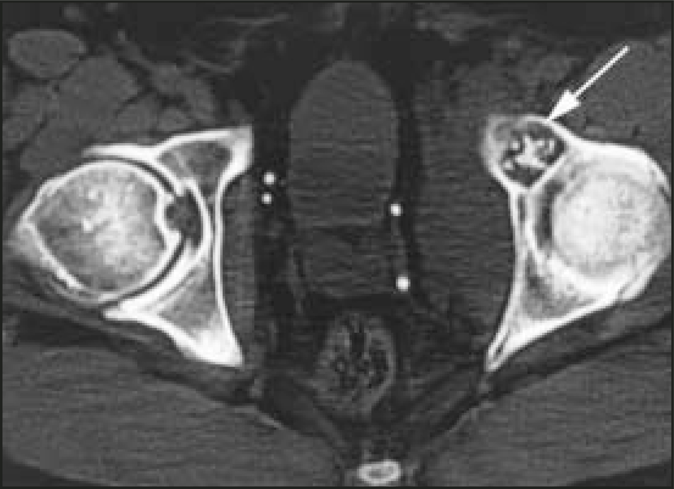
Cartilaginous tumors (such as osteochondromas, enchondromas, and chondrosarcomas) may produce irregularly shaped calcifications within their matrix that resemble popcorn on imaging studies (Figure 16). These “popcorn calcifications” can usually be distinguished from the “mashed potato,” or cloudlike, ossific matrix of bone-forming tumors such as osteosarcomas (Figure 17).15 The term “popcorn calcification” has also been used to describe the lucent areas with sclerotic margins seen in the metaphyses and epiphyses of patients with osteogenesis imperfecta.1
Parfait sign
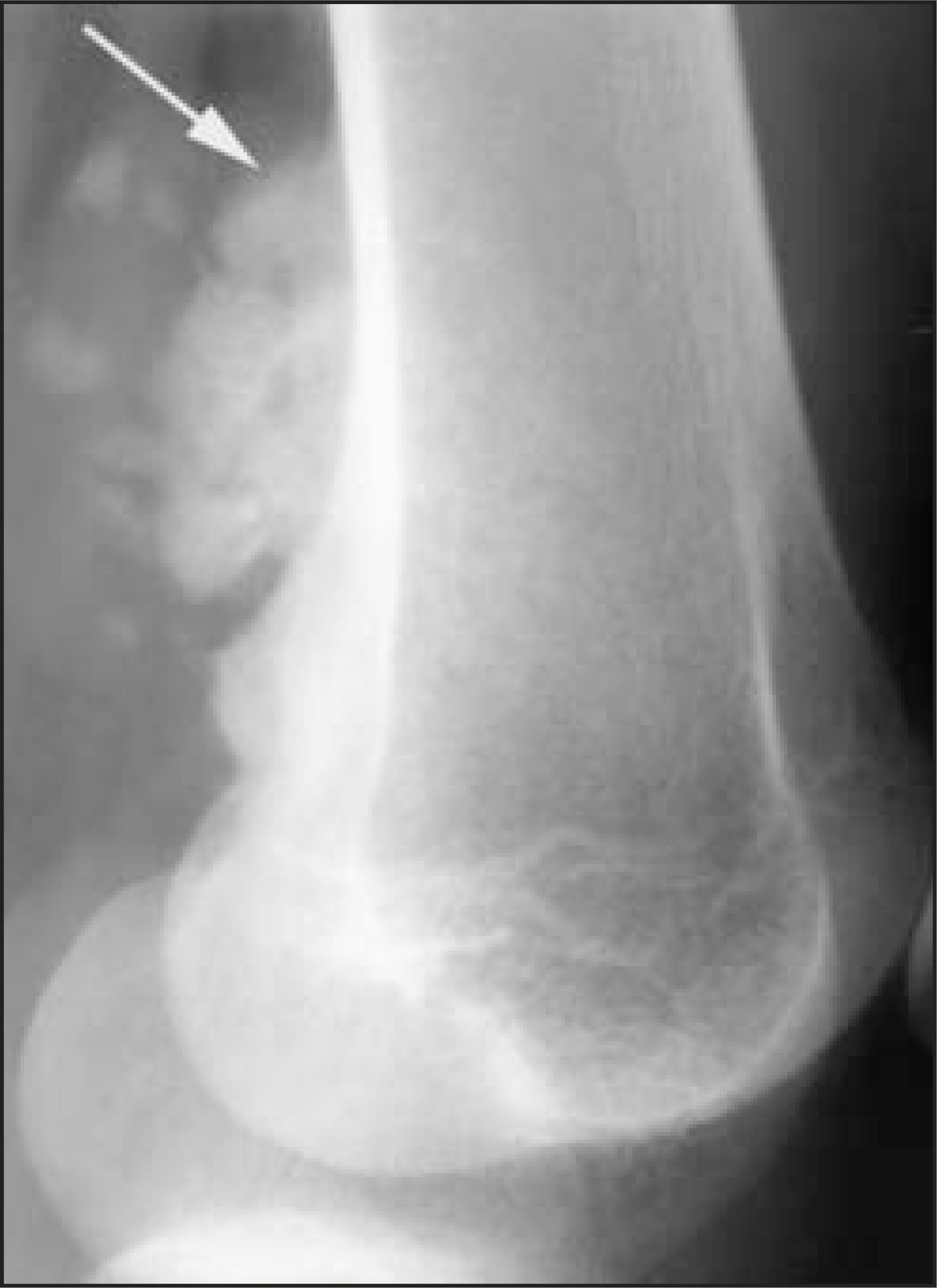
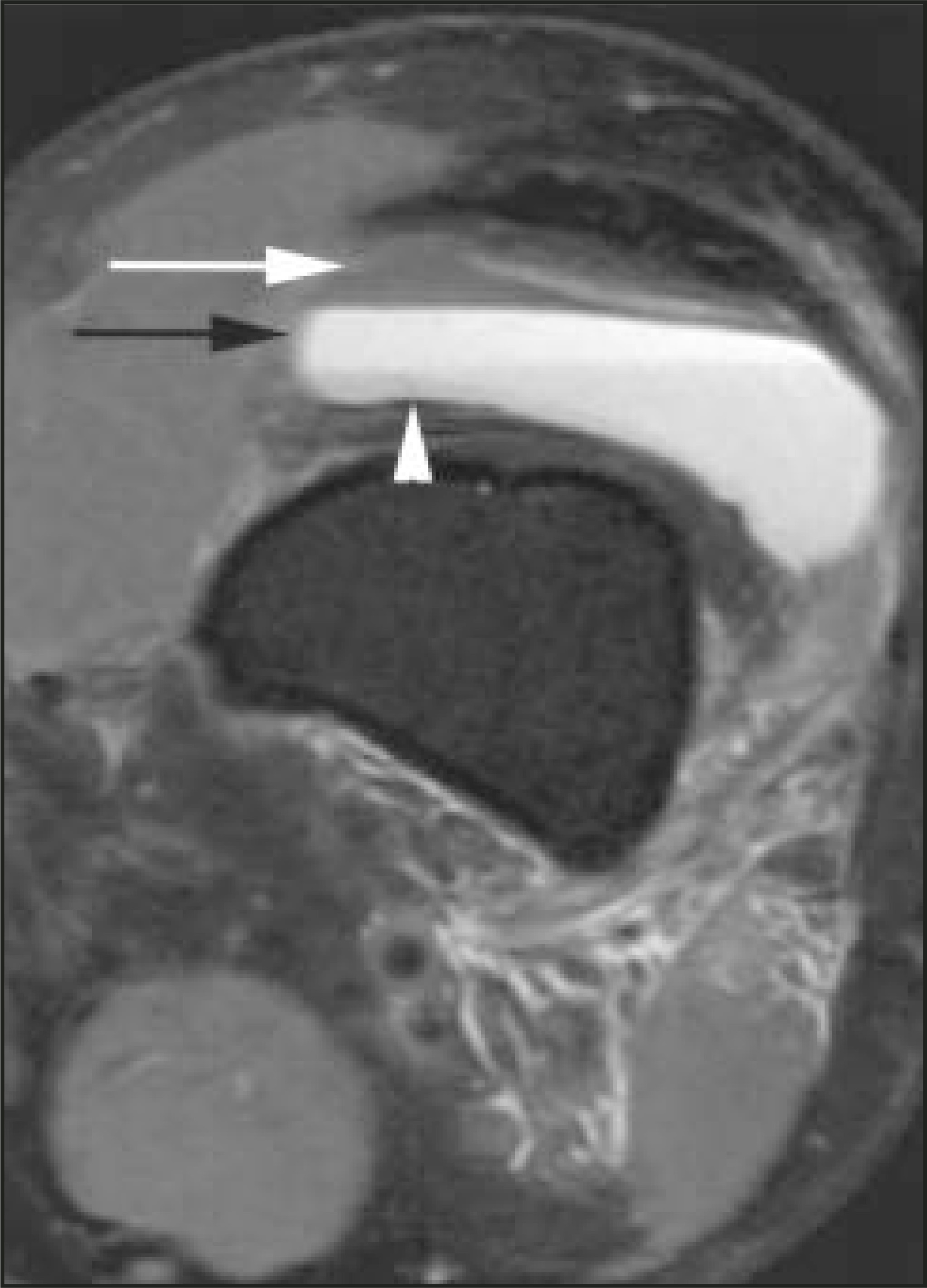
The term “parfait sign” has been used to describe the tri-level appearance of a lipohemarthrosis in the knee on MRI (Figure 18) and CT studies.16 The top (antidependent) layer represents floating fat that has escaped from the marrow through a cortical fracture and, hence, follows fat density and signal intensity on CT and MRI, respectively. The middle layer contains serum and the bottom (dependent) layer represents red blood cells; the signal intensities of these layers depend on the age of the blood products and the specific imaging sequences used. On CT, the dependent layer is often the most dense. On MRI studies, a thin band representing chemical shift artifact may also be noticed between the fat and serum layers.17
Conclusion
When reviewing prior to meal times, readers should be warned that the mind can absorb so long as the stomach can endure. To maximize absorption while dining at the buffet of these food signs in musculoskeletal radiology, readers are most welcome to snack while they learn. Recognition of these signs, served with an understanding of the relevant differential diagnoses and pathologies, will go a long way toward satisfying the radiologist’s ever-present hunger for learning and thirst for knowledge. Bon apetit!
References
- Resnick D. Diagnosis of Bone and Joint Disorders. 2002.
- Reeder M, Bradley W. Reeder and Felson’s Gamuts in Radiology: Comprehensive Lists of Roentgen Differential Diagnosis. 1993.
- Rothschild B, Pingitore C, Eaton M. Dactylitis: Implications for clinical practice. Semin Arthritis Rheum. 1998;28:41-47.
- Hamdy R. Paget’s Disease of Bone: Assessment and Management. 1981.
- Benichou O, Laredo J, de Vernejoul M. Type II autosomal dominant osteopetrosis (Albers-Schonberg disease): Clinical and radiological manifestations in 42 patients. Bone. 2000;26:87-93.
- Primiano G, Reef T. Disruption of the proximal carpal arch of the hand. J Bone Joint Surg Am. 1974;56:328-332.
- Miller W, Restifo R. Steroid arthropathy. Radiology. 1966;86:652-657.
- Daffner S, Daffner R. Computed tomography diagnosis of facet dislocations: The “hamburger bun” and “reverse hamburger bun” signs. J Emerg Med. 2002;23:387-394.
- Volberg F, Whalen J, Krook L, Winchester P. Lamellated periosteal reactions: A radiologic and histologic investigation. AJR Am J Roentgenol. 1977;128:85-87.
- Swischuk L, John S. Differential Diagnosis in Pediatric Radiology. 1995.
- Genant H, Heck L, Lanzl L. Primary hyperparathyroidism. A comprehensive study of clinical, biochemical and radiographic manifestations. Radiology. 1973;109:513-552.
- Chau C, Griffith J, Chan P. Rice-body formation in atypical mycobacterial tenosynovitis and bursitis: Findings on sonography and MR imaging. AJR Am J Roentgenol. 2003;180:1455-1459.
- Hubbard A. Imaging of pediatric hip disorders. Radiol Clin North Am. 2001;39:721-732.
- Imhoff A. Diabetic osteoarthropathy. Course and differential diagnosis [German]. Z Orthop Ihre Grenzgeb. 1988;126:169-178.
- Richardson M. Approaches to differential diagnosis in musculoskeletal imaging: Lucent lesions of bone. 2004.
- Hunter J. John Hunter’s MSK teaching file: Knee MRI. 2004.
- Kier R, McCarthy S. Lipohemarthrosis of the knee: MR imaging. J Comput Assist Tomogr. 1990;14:395-396.
Citation
. Food signs in musculoskeletal radiology. Applied Radiology. 2005;34(10):17-24. doi:10.37549/AR1373.