Frontoethmoidal Encephalocele
Journal of Pediatric Case Reports — Vol. 1 , Issue 7
Published: October 1, 2026
1 University of Cincinnati College of Medicine, Cincinnati, Ohio
2 Department of Radiology and Medical Imaging, Cincinnati Children’s Hospital Medical Center, Cincinnati, Ohio
3 Department of Radiology, Phoenix Children’s Hospital, Phoenix, Arizona
Abstract
Encephaloceles are rare congenital malformations characterized by a herniation of brain tissue and meninges through a skull defect. While clinical presentation and severity can vary widely depending on the size, location, and contents of the encephalocele, early diagnosis is critical for management and outcome planning. Imaging, particularly MRI, plays a central role in delineating the extent of the defect and identifying associated abnormalities. Definitive treatment involves surgical repair, which can significantly improve both functional and cosmetic outcomes. Long-term prognosis depends on the presence of associated anomalies, involvement of functional brain tissue, and development of complications such as hydrocephalus.
Keywords
neuroradiology, brain, congenital anomaly
Categories
Case Summary
A G1P0 woman presented for advanced fetal imaging after a second-trimester US identified a frontoethmoidal encephalocele. The neonate was delivered at 36 weeks and 3 days via cesarean section due to the encephalocele, preterm premature rupture of membranes, and the onset of preterm labor. Postnatally, an external ventricular drain and ventriculoperitoneal shunt were placed.
Imaging Findings
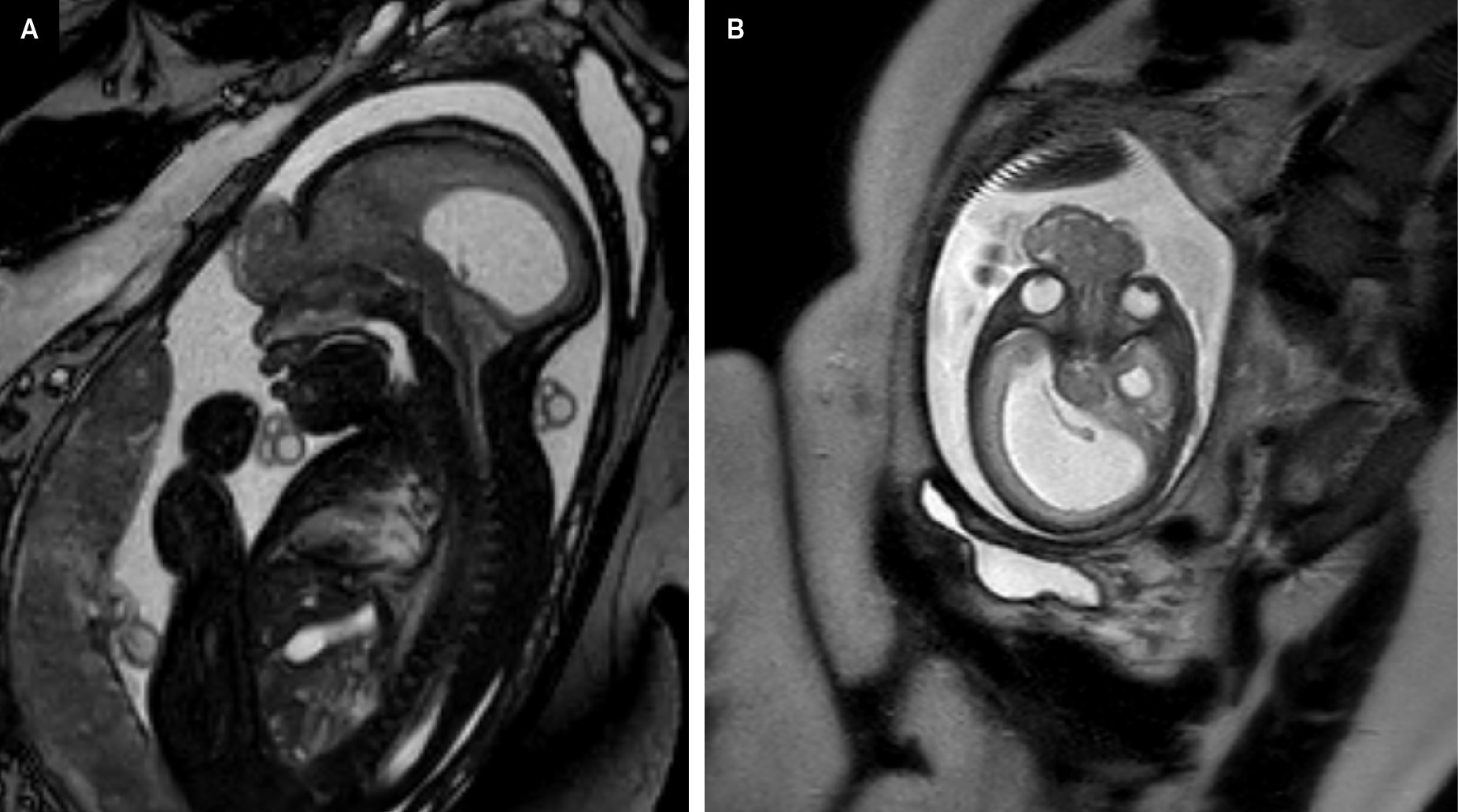
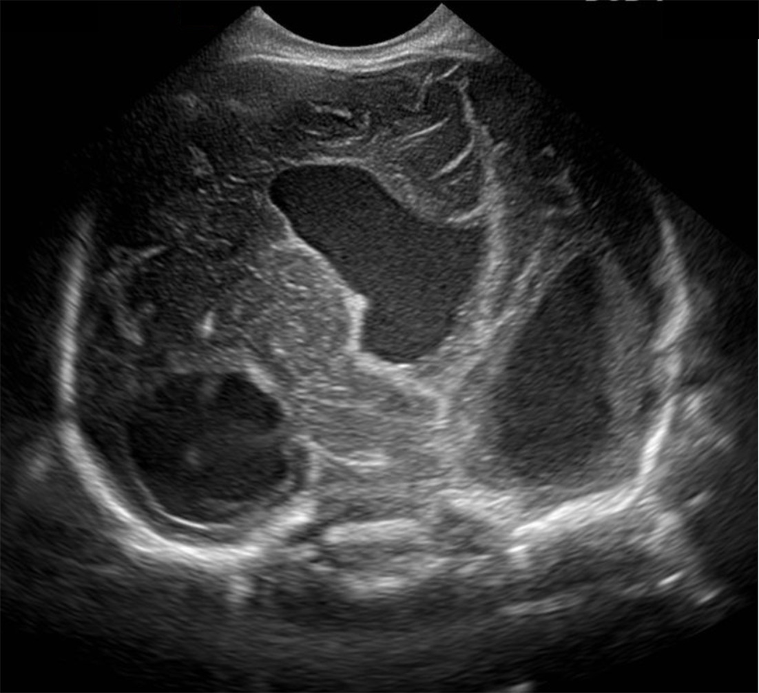
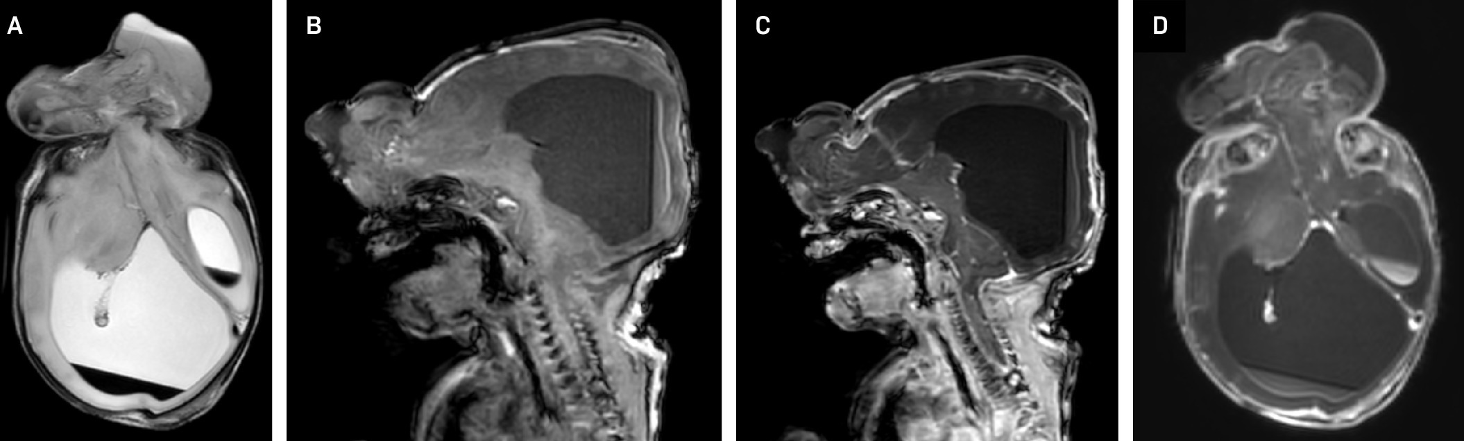
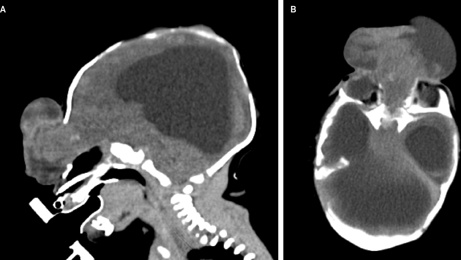
Fetal MRI (Figure 1) showed an anterior midline defect in the frontoethmoidal region at the level of the orbits extending from the nasal bone superiorly to the frontal area. A sac containing dysmorphic brain tissue with a small volume of cerebrospinal fluid extended from the bony defect of the skull. Postnatal head US (Figure 2) showed an enlarged, distorted ventricular system and dysmorphic cerebral tissue. Postnatal brain MRI (Figure 3) and head CT (Figure 4) confirmed the findings and identified intracranial hemorrhage.
Diagnosis
Frontoethmoidal encephalocele.
The differential diagnosis includes nasal glioma, dermoid cyst, nasal polyp, meningocele, mucocele, neurinoma, and vascular malformation.
Discussion
Encephaloceles are rare neural tube defects characterized by a sac-like protrusion of brain tissue and meninges covered by skin (or at least an epithelial layer) through a skull defect.1,2 Encephaloceles have a global prevalence between 0.8 and 4 per 10,000 live births, representing 10-20% of all craniospinal dysraphisms.3 Frontoethmoidal encephaloceles are more common in Southeast Asia, where the incidence is as high as 1 in 5000 live births. The etiology underlying this geographic predisposition is unknown, but likely multifactorial.1
Although encephaloceles can occur as isolated defects, they are associated with additional central nervous system or extracranial anomalies in 23-37.5% of patients.3 For example, herniation of the ventricular system into the encephalocele sac can lead to hydrocephalus. Encephaloceles have also been linked to genetic syndromes such as Meckel-Gruber syndrome, trisomy 13, and trisomy 18.3
Encephaloceles are classified by the location of herniation and may be anterior, occipital, basal, or involve the cranial vault.4 Occipital encephaloceles are the most common, accounting for approximately 75% of cases, followed by the frontoethmoidal (15%) and basal (10%) types.5 While typically considered neural tube defects, the exact pathophysiology remains unclear. Anterior encephaloceles are thought to result from abnormal neural crest cell development, whereas occipital encephaloceles are believed to arise from defective segmentation of the posterior cranial bones.3
Frontoethmoidal encephaloceles involve anterior herniation of brain tissue, typically from the frontal lobe, and meninges through a defect in the frontoethmoidal region. The nasofrontal subtype occurs at the junction between the frontal and nasal bones, usually near or through the foramen cecum.6 This generally results in a small anterior cranial fossa. Frontal encephaloceles almost always contain olfactory tissue.7 Clinically, they usually present at birth as a visible midline mass, which may enlarge with crying (a positive Furstenberg sign) and may be associated with cerebrospinal fluid rhinorrhea or recurrent meningitis.8
Multiple imaging modalities can aid in characterizing encephaloceles. MRI helps localize the defect, characterize the hernial sac contents, and identify other associated defects. Notably, defect size and hydrocephalus are key prognostic factors. Meanwhile, CT can better characterize bony anatomy. MR venogram or CTA are helpful in identifying venous abnormalities. Imaging of suspected encephaloceles is useful in ruling out other differential diagnoses. For instance, an encephalocele may appear similar to a nasal glioma, but present with intracranial extension and/or communication with the subarachnoid space on MRI.1,2,9
Prenatally, termination of a pregnancy may be chosen if the encephalocele is large or has a poor prognosis to minimize maternal risk. Larger encephaloceles with improved prognoses may require cesarean section to minimize birth trauma. For relatively small encephaloceles, vaginal delivery may be possible.10 Definitive postnatal treatment involves surgical intervention to remove the encephalocele and repair the associated dural and bony defects. In cases where hydrocephalus is present, placement of a ventriculoperitoneal shunt may be necessary.1 Surgical repair can lead to significant improvements in both functional and cosmetic outcomes. When the encephalocele is covered by intact skin, surgery may be safely delayed until early infancy to reduce the risks associated with anesthesia and excessive blood loss.11
Prognosis is variable depending on size, contents, infection, and associated defects. The mortality rate of encephaloceles is 29%. One study demonstrated that 83% of patients with encephaloceles developed mental and/or physical impairment. Approximately 20% of infants with congenital encephalocele have seizures.11
Conclusion
Encephaloceles are rare congenital malformations characterized by a herniation of brain tissue and meninges through a skull defect. While clinical presentation and severity can vary widely depending on the size, location, and contents of the encephalocele, early diagnosis is critical for management and outcome planning. Imaging, particularly MRI, plays a central role in delineating the extent of the defect and identifying associated abnormalities. Definitive treatment involves surgical repair, which can significantly improve both functional and cosmetic outcomes. Long-term prognosis depends on the presence of associated anomalies, involvement of functional brain tissue, and development of complications such as hydrocephalus.
References
- Hoving E. Nasal encephaloceles. child’s nervous system: chns: official journal of the international society for pediatric neurosurgery. . 2000;16(10-11):702-706. doi:10.1007/s003810000339.
- Dhirawani R, Gupta R, Pathak S, Lalwani G. Frontoethmoidal encephalocele: case report and review on management. Ann Maxillofac Surg. 2014;4(2):195-197. doi:10.4103/2231-0746.147140.
- Ghi T, Dall’asta A, Pilu G, et al. Obstetric Imaging: Fetal Diagnosis and Care. 2018:213-226. doi:10.1016/B978-0-323-44548-1.00041-3.
- Nugraha H, Sobana M, Gantini T. Nasofrontal encephalocele: a case report with literature and management review. Radiol Case Rep. 2024;19(5):1907-1912. doi:10.1016/j.radcr.2024.01.070.
- Lowe L, Booth T, Joglar J, Rollins N. Midface anomalies in children. Radiographics. 2000;20(4):907-922. doi:10.1148/radiographics.20.4.g00jl07907.
- Sarangi P. Frontoethmoidal meningoencephalocele - nasofrontal type. Case study. . 2025.
- Fenichel G, Fenichel G. Neonatal Neurology. 2007:167-186. doi:10.1016/B978-044306724-2.50010-2.
- Macagnan MS M, Gaillard F. Frontoethmoidal encephalocele. . 2016.
- Ilangovan GM T. Imaging of Encephalocele – “seeking a closure.”. . 2022.
- Liao S, Tsai P, Cheng Y. Prenatal diagnosis of fetal encephalocele using three-dimensional ultrasound. Journal of Medical Ultrasound. 2012;20(3):150-154. doi:10.1016/j.jmu.2012.07.005.
- Pontell M, Niklinska E, Bonfield C, Golinko M. Management of an open nasofrontal encephalocele during the first day of life. Childs Nerv Syst. 2022;38(1):207-210. doi:10.1007/s00381-021-05102-1.
Disclosures
The authors have no conflicts of interest to disclose. None of the authors received outside funding for the production of this original manuscript and no part of this article has been previously published elsewhere.
Citation
. Frontoethmoidal Encephalocele. Journal of Pediatric Case Reports. 2026;1(7). doi:10.37549/JPCR-26-0112.