Functional magnetic resonance imaging of the lung
Applied Radiology — Vol. 32 , Issue 2 , pp. 41 -48
DOI: 10.37549/AR1160
Published: February 1, 2003
Categories
Noninvasive imaging of human pulmonary function is an important step in the clinical evaluation of patients with various pulmonary diseases, including pulmonary embolism (PE), emphysema, interstitial lung disease, and asthma. Under normal circumstances, regional pulmonary ventilation (V) and perfusion (Q) are usually highly matched processes. Various disease states alter this normal relationship, causing V/Q mismatch. Noninvasive assessment of regional V/Q relationships is important in the diagnosis, evaluation, and treatment of patients with pulmonary disease.
There are several limitations of the clinical studies that are currently used to assess pulmonary function. Standard pulmonary function tests (PFTs) provide invaluable information about global lung function; however, they do not allow for determination of the spatial distribution of pulmonary function. Currently, nuclear medicine techniques based on the introduction of radioactive tracers are considered the gold standard for noninvasive functional imaging of the lung. However, these methods suffer from several significant limitations, including relatively poor spatial resolution, limitations on the acquisition of three-dimensional data, and the costs and risks associated with the radioactive tracers.
Magnetic resonance imaging (MRI) of the lung is challenging due to the effects of respiratory movement and the short transverse relaxation time (T2*) that results from the microscopic heterogeneity in the magnetic field. However, the recent development of high-speed gradient systems, coupled with the subsequent implementation of short echo-time acquisition has allowed for imaging of the lung parenchyma.1 As a result, several methods of functional MRI (fMRI) have recently been described to perform imaging of pulmonary perfusion and ventilation. The physical basis of these methods will be reviewed, including examples of potential clinical applications.
Perfusion MRI
Gadolinium-based methods
Extravascular-space gadolinium (Gd) chelates have been used for perfusion imaging in the central nervous system for more than a decade.2 In this approach, the tissue magnetization is observed dynamically using a rapid pulse sequence during the passage of a Gd compound following intravenous injection. The Gd causes shortening of both the longitudinal (T1) and transverse (T2*) relaxation time of the tissue; therefore the observed tissue signal intensity changes are a function of the weighting of the pulse sequence used for observation. For applications in the pulmonary circulation, T1-weighted rapid gradient-echo pulse sequences have been used, leading to signal enhancement during Gd passage.3-5 These methods offer the possibility of quantification of perfusion and have been validated in animal models.6
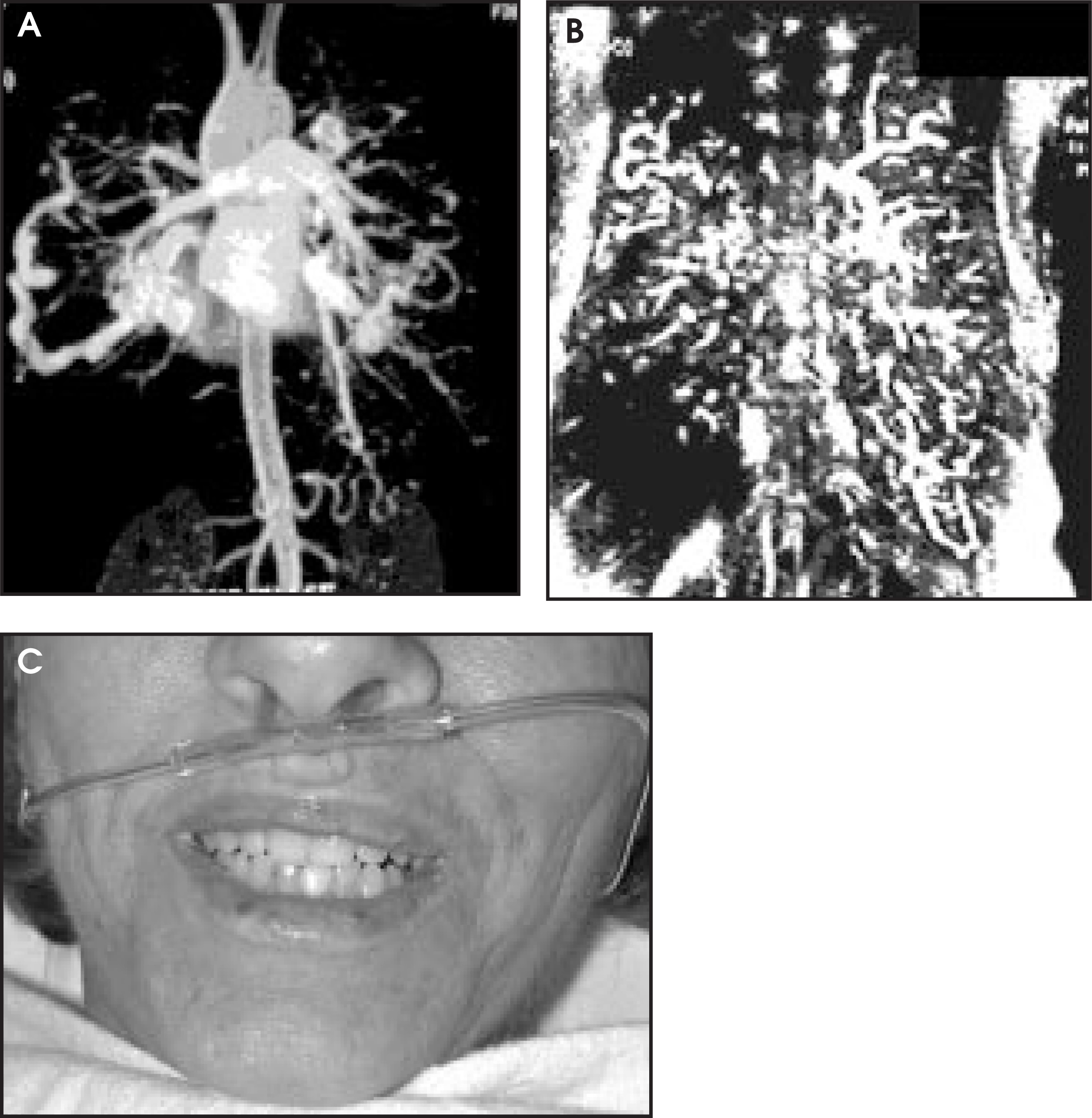
Advantages of the Gd-based perfusion methods include a relatively high contrast-to-noise ratio and spatial resolution, which offers high sensitivity for the detection of small perfusion defects (Figures 1 and 2). However, the injection of Gd inevitably results in some degree of background enhancement. This may degrade the quality of subsequent MR angiographic (MRA) or perfusion studies. Specifically, serial studies of perfusion to characterize dynamic processes are not feasible by this approach due to cumulative background enhancement. In addition, Gd-based techniques involve the risks and expenses of exogenous contrast administration. Nevertheless, Gd-enhanced MRI is a validated and robust method for pulmonary perfusion imaging.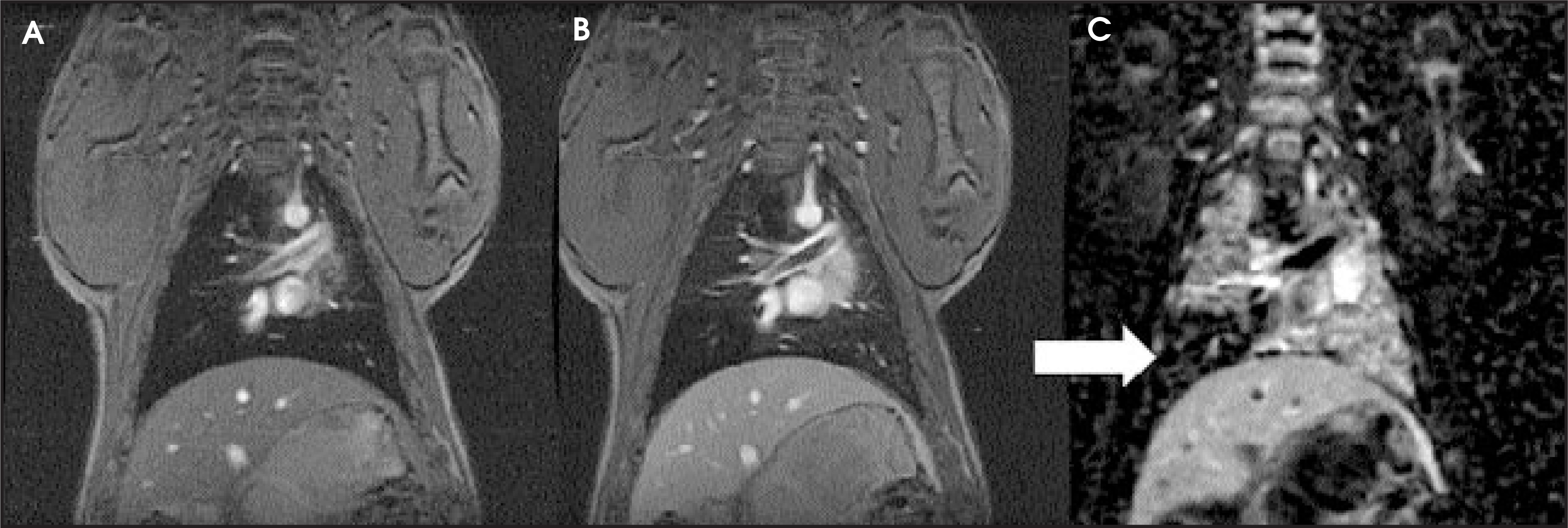
Spin-labeling methods
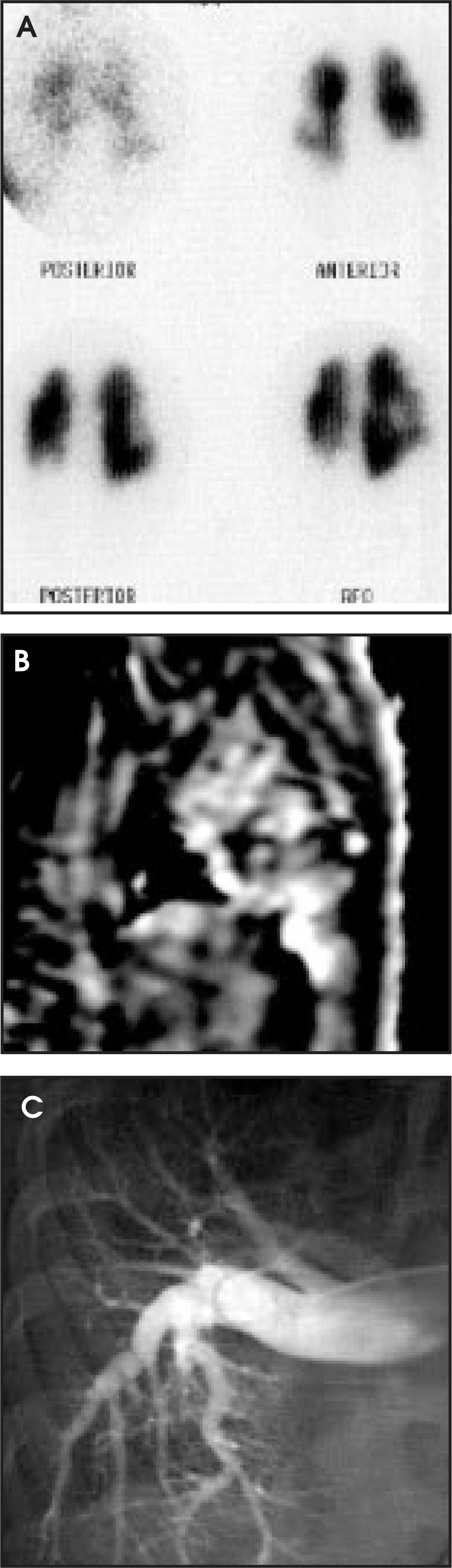
Magnetically labeled arterial water may be used as an endogenous contrast agent for perfusion MRI, which forms the basis for the so-called “arterial spin-labeling” (ASL) methods.7 Magnetic labeling may be achieved using a variety of methods, including spatial saturation,7 spatial inversion,8 or adiabatic inversion.9 Images acquired under conditions of arterial labeling are subtracted from images acquired under appropriate control conditions to generate a perfusion image, as shown in Figure 3. Solution of the modified Bloch equations may be performed to generate absolute values of perfusion.7,10 The ASL perfusion pulse sequences may be divided into continuous ASL (CASL)7 and pulsed ASL (PASL) methods.8,11 The CASL methods establish a steady state of arterial labeling, while the PASL methods do not. The CASL methods may be acquired in a steady state, thereby allowing for respiratory-triggered acquisition.12 The PASL methods do not seek to establish a steady state and, therefore, generally require breath-holding and cardiac gating. The PASL methods have also been applied successfully to the pulmonary circulation.13,14 In both approaches, blood is typically labeled either at the level of the mediastinum or the right/left pulmonary arteries. The ASL methods have been used with success to obtain high-resolution perfusion maps of the pulmonary circulation in many disease states. Due to their inherent sensitivity to changes in both perfusion and blood volume, these methods are expected to have high sensitivity to the detection of pulmonary embolism (Figures 4 and 5).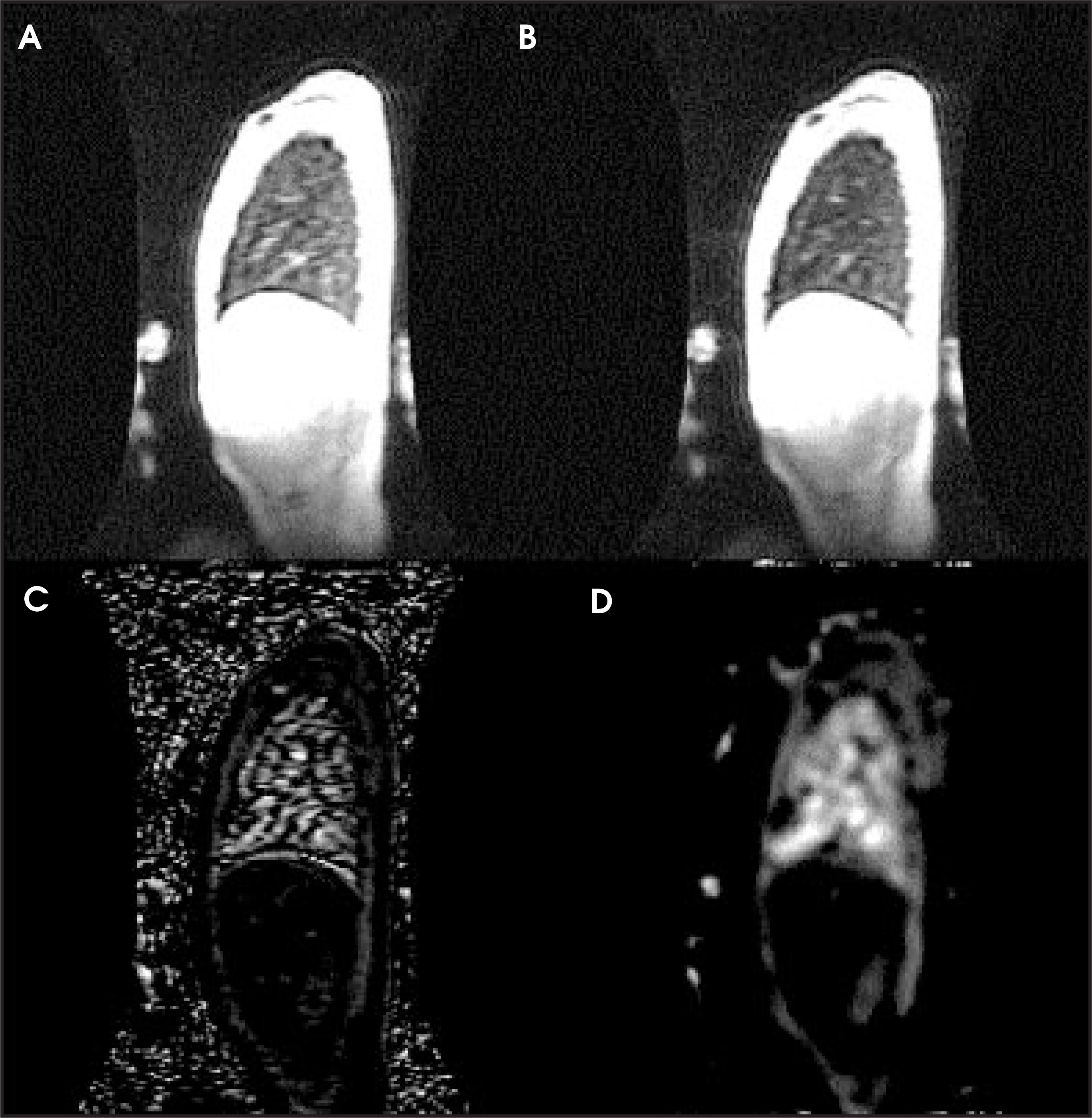
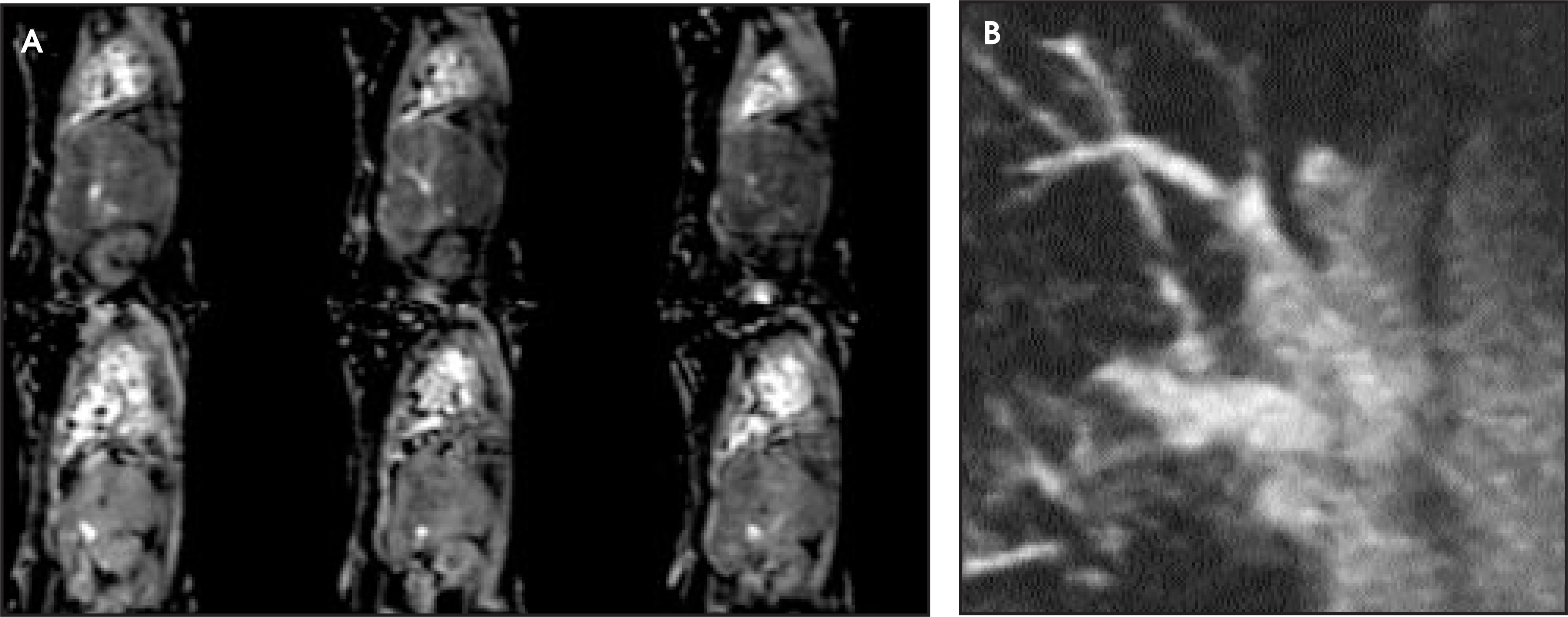
The ASL methods have several distinct advantages over Gd-based methods of perfusion imaging. As an entirely noninvasive method, ASL avoids the risks and expenses of an exogenous contrast agent. The ASL methods offer the possibility of quantification and may, therefore, be used to follow patients during therapy. Finally, ASL methods may be used repeatedly over time to study dynamic processes, as there is no degradation in the perfusion image as is seen with Gd-enhanced methods.15,16
Ventilation MRI
Laser-polarized noble gases
The laser-polarized noble gases helium (3He, a rare isotope of 4He) and xenon (129Xe) are inhaled contrast agents capable of creating high-resolution images of the gas distribution within the lung during a single breath hold.17-20Polarization is performed in a high-power laser apparatus under conditions of high temperature and pressure. The laser-polarized magnetization is five orders of magnitude larger than the thermal-equilibrium magnetization of protons in the lung, and this explains why the gases are visible in MRI despite their low density. However, the magnetization is in a nonequilibrium state and is therefore “nonrenewable.” Thus, the total available magnetization is fixed, and must be managed appropriately during the application of the MR pulse sequence. In addition to a magnet system capable of multinuclear “broadband” acquisition, a dedicated coil and transmit/receive switch tuned to the helium frequency (48.66 MHz at 1.5 T) must be available for 3He MRI.
As a result of the large, nonequilibrium polarization, the signal-to-noise ratio of ventilation MRI using hyperpolarized 3He is very high. The resulting ventilation images show with superb clarity the anatomic structure of the lungs, outlining the major and minor airways, pulmonary fissures, and vascular structures with exceptional detail (Figure 6). High spatial resolution has allowed for new capabilities in ventilation MRI, such as the noninvasive localization of air leaks that cause pneumothorax.21 In addition, 3He MRI has been used to measure pulmonary volumes with high accuracy, showing excellent agreement with pulmonary function tests.22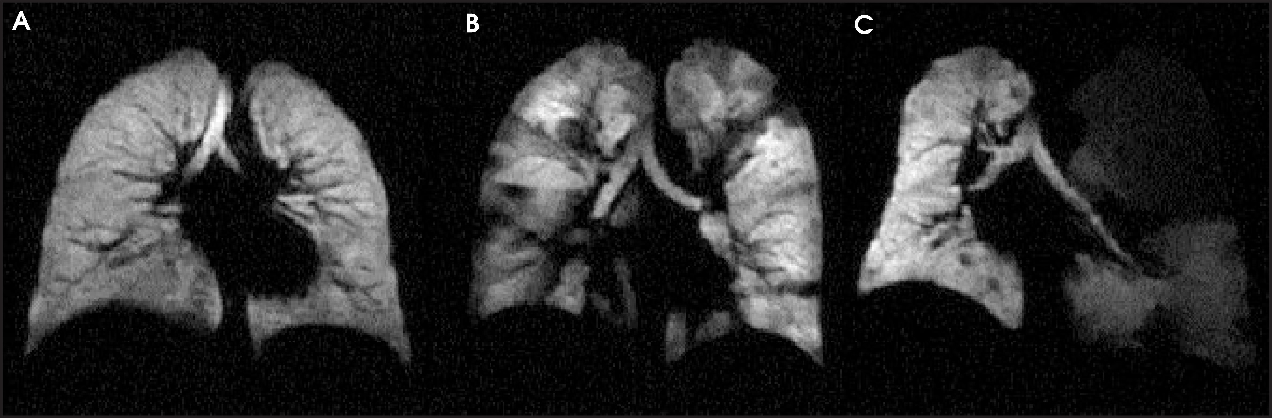
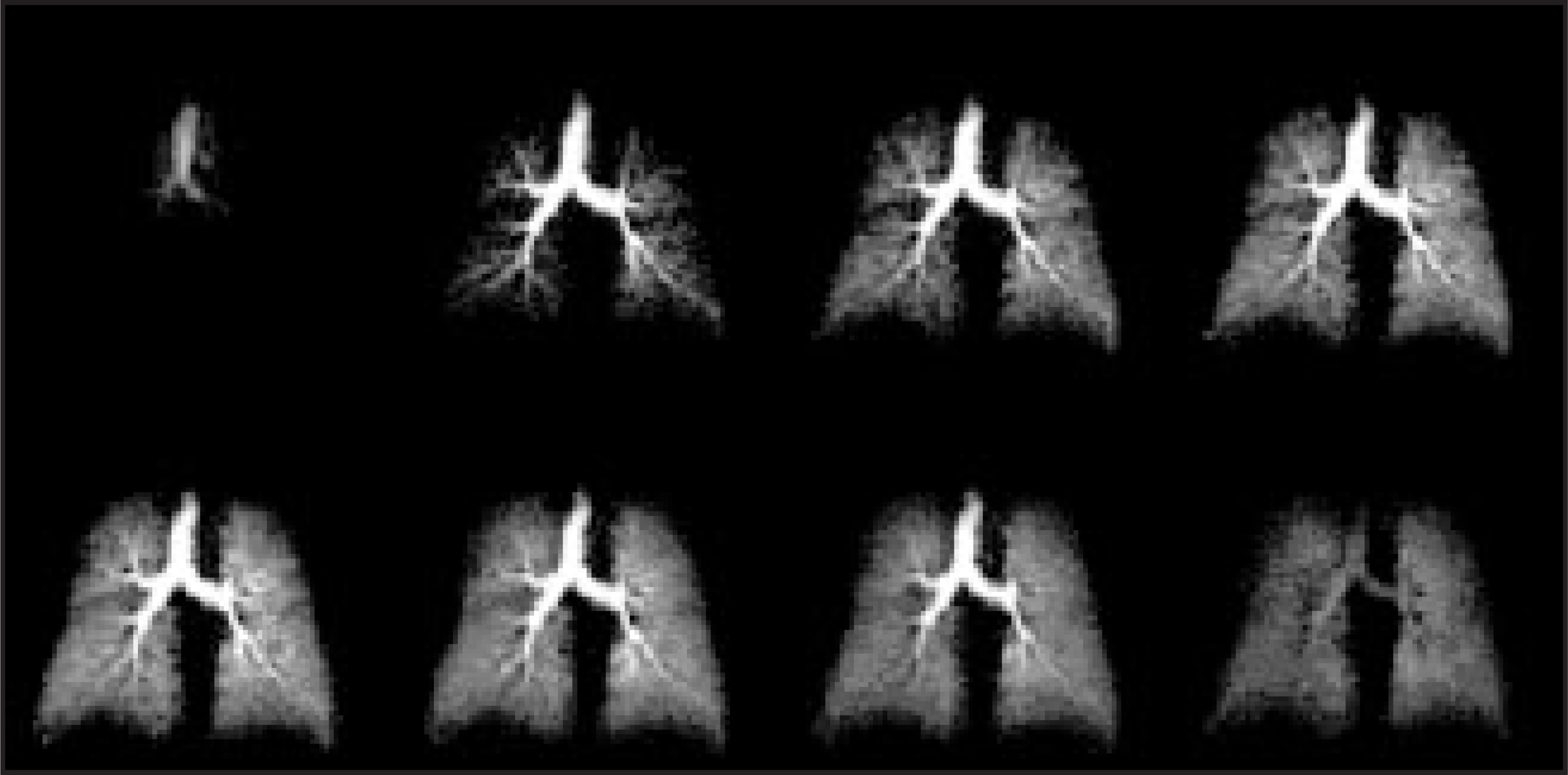
In addition to imaging the static distribution of gas within the lung, the unique properties of these gases provide additional contrast mechanisms for structural and functional imaging of the lung.23 Since 3He does not cross the alveolar membrane readily and has a high diffusion coefficient, it can be used to probe airspace morphology. Specifically, diffusion-weighted 3He MRI has been used to detect early changes in chronic obstructive pulmonary disease (COPD) (Figure 7).24,25 The T1 relaxation rate of 3He, which increases significantly in the presence of oxygen, has been exploited to measure regional intrapulmonary oxygen concentration.26-29 Images of the T1 relaxation rate of the gas may be used to map intrapulmonary oxygen concentration (Figure 8). The gas flow dynamics of 3He in the lung during inspiration and expiration can be measured with high temporal and spatial resolution using echoplanar imaging or interleaved spiral pulse sequences (Figure 9).30,31 The rapid exchange of 129Xe across alveolar membrane enables the integrity of the pulmonary diffusion barrier to be probed.32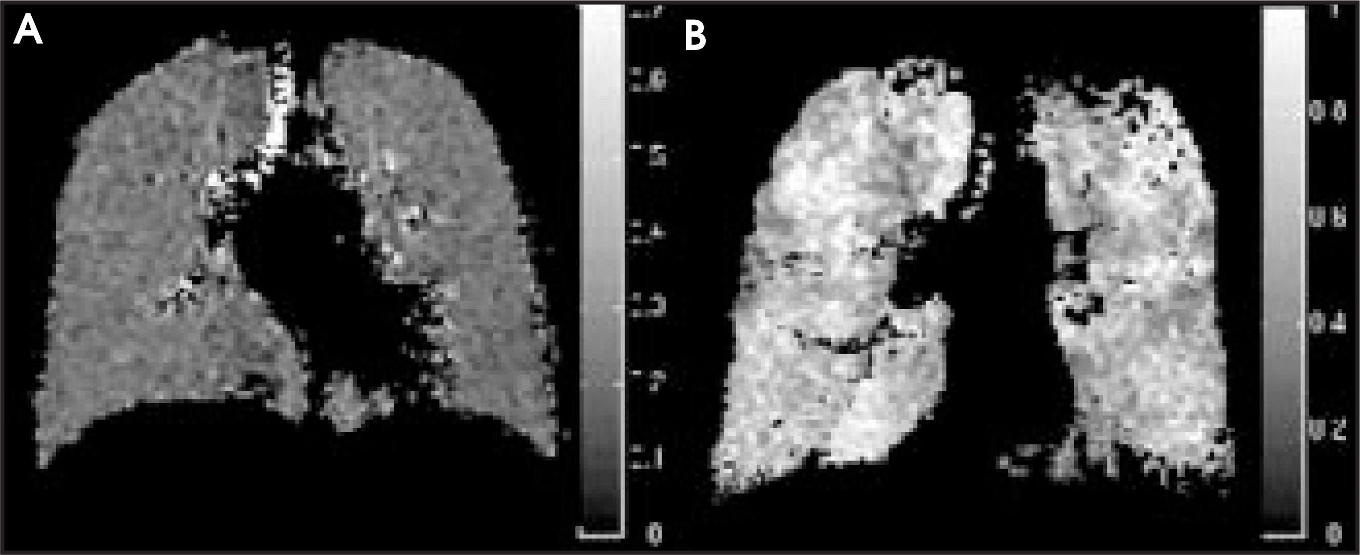
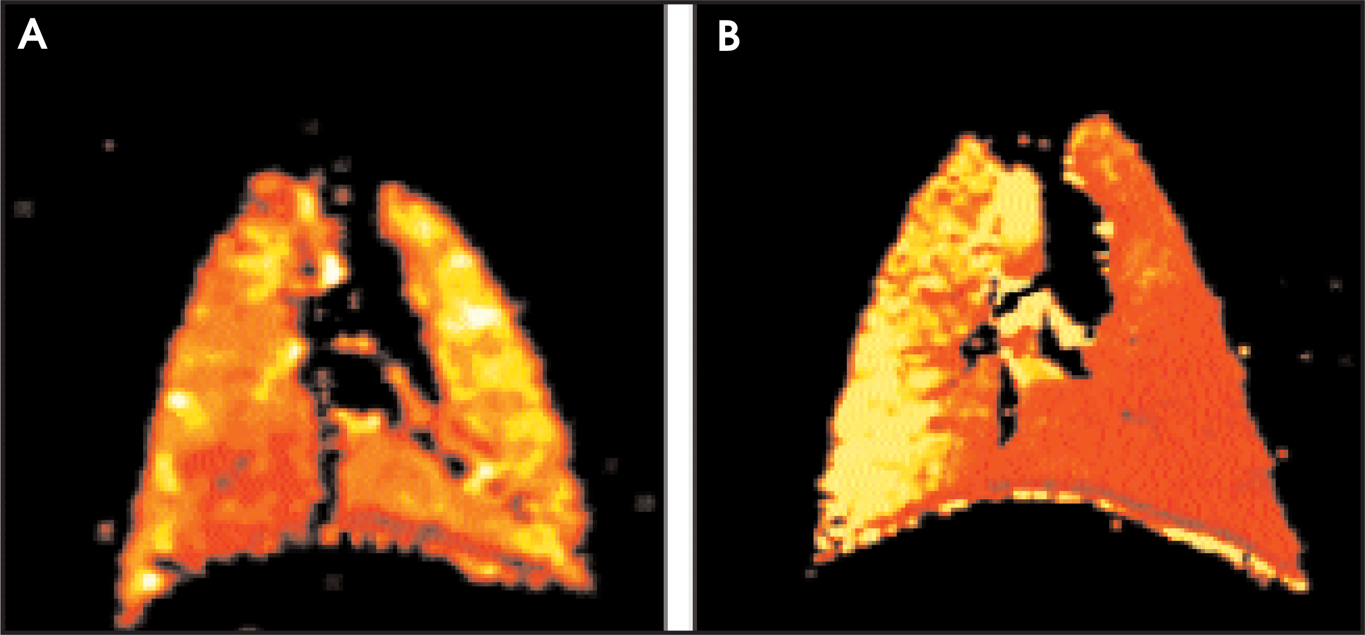
Preliminary studies of laser-polarized 3He imaging have shown potential for detecting ventilation defects in a variety of lung pathologies including COPD,33,34 asthma,35 cystic fibrosis,36 and obliterative bronchiolitis (OB), a form of chronic lung allograft rejection.37 Ventilation defects appear as areas of the lung that do not completely fill with 3He during a single inspiratory breath-hold. These ventilation defects represent regions of the lung distal to restrictions in the airway due to airway hyperactivity, mucous plugging, or altered alveolar oxygen content. 3He diffusion MRI has shown promise for detecting emphysematous changes in the lung (Figure 7), and has been shown to correlate with pulmonary function tests.25 As the lung parenchyma is destroyed, the changes in airspace morphology cause a decrease in the restriction of the gas’s motion, resulting in an increase in the apparent diffusion coefficient of emphysematous regions.
Oxygen-based methods
An alternative to MR ventilation imaging with laser-polarized noble gas uses inhaled oxygen (O2) as a paramagnetic agent.38 Though oxygen is weakly paramagnetic, its overall effect on the lung is considerable given the large surface area of the lung and the large difference in partial pressures between room air (21% oxygen) and 100% oxygen. MR signal modulation between inhaling room air and 100% oxygen results from the shortening of the longitudinal relaxation time (T1) of the lung caused by the increased concentration of dissolved oxygen in pulmonary tissues and blood after breathing 100% oxygen. An average oxygen-induced shortening to T1 of approximately 150 msec has been reported, leading to signal changes of 20% to 30%.39 T1-weighted MR sequences such as inversion recovery (IR) or multiple inversion recovery (MIR) have been applied to detect the signal difference in the lung as the subject alternatively inhales room air and 100% oxygen.39
To minimize misregistration artifacts, images with matched respiratory phases should be selected and subsequently averaged. The difference between the average images acquired during room air and 100% oxygen yields a qualitative oxygen-enhanced ventilation map. Additionally, a qualitative oxygen-enhanced ventilation map can be obtained using statistical analysis, an approach widely used in fMRI analysis to detect brain activation.40,41 This approach exploits the box-car pattern of the signal modulation of the lung between periods of inhalation of room air and 100% oxygen.
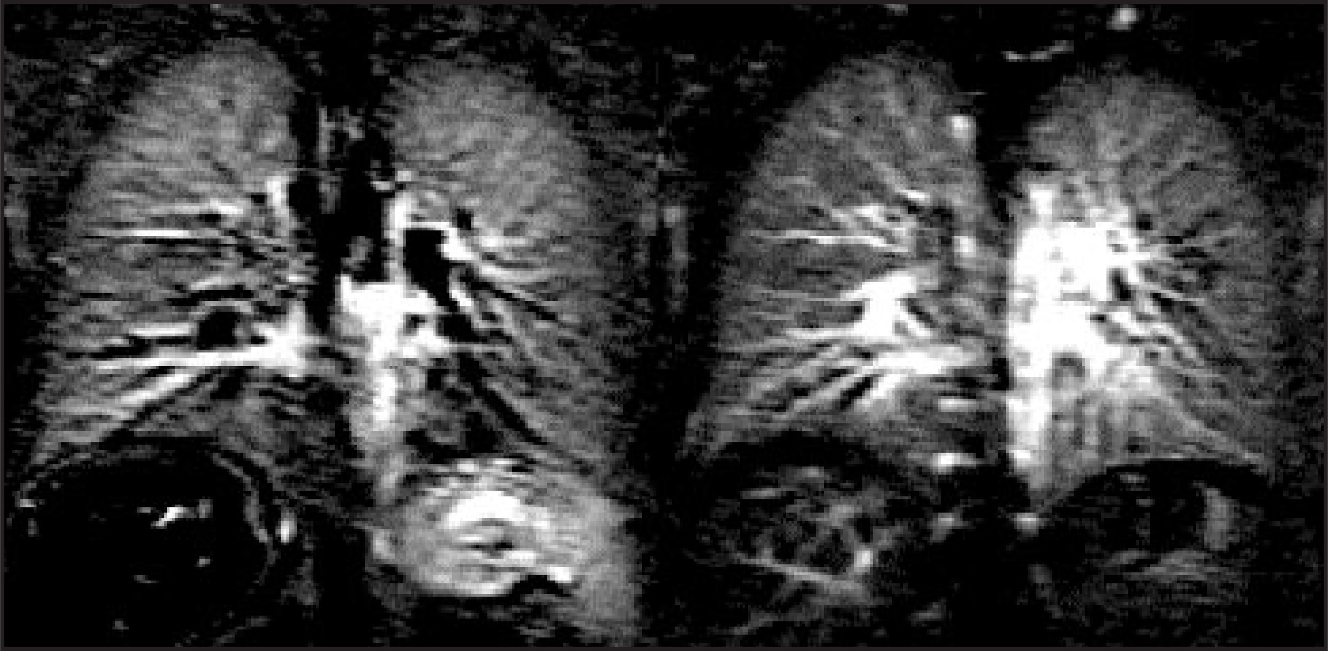
Oxygen-enhanced ventilation imaging has been successful in detecting regional ventilation defects in animal models and in patients with emphysema (Figure 10).38,42 Oxygen-enhanced MRI provides a means to study oxygen transfer from the pulmonary alveoli to the pulmonary vasculature because oxygen is a principal component in the functional gas exchange. Additionally, oxygen is readily available as part of the emergency equipment in most MR suites and is safe and inexpensive. By coupling oxygen ventilation MRI with ASL perfusion, a completely safe and noninvasive evaluation of regional V/Q may be performed (Figure 11).43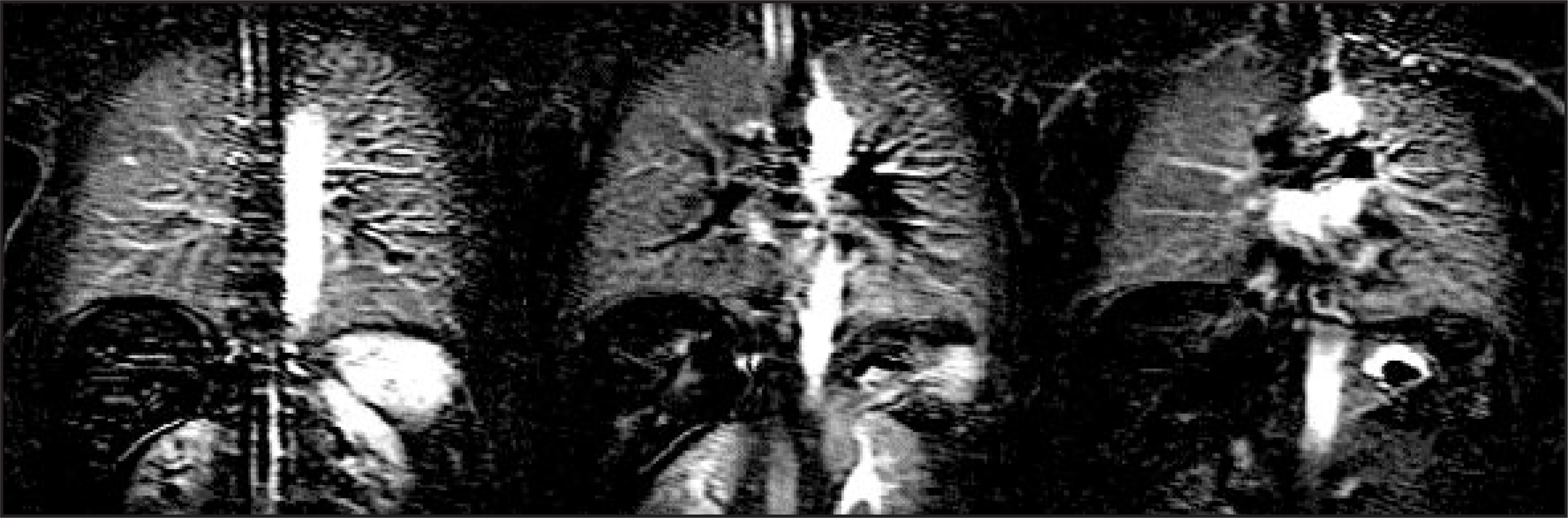
Sulfur hexafluoride
Pulmonary physiologists have used sulfur hexaflouride (SF6) to study gas distribution in the lung by taking advantage of its low blood solubility and high density.44 Additionally, high-resolution computed tomography (HRCT) imaging using helium and SF6 has been shown to be useful in predicting acinar gas distribution abnormalities in patients with pulmonary disease.45 Recently, SF6 has been described as a contrast agent for ventilation MRI.46 The most abundant isotope of fluorine is 19F, whose nucleus possesses a spin ½ and is therefore potentially visible in an MRI system. The resonance frequency and gyromagnetic ratio of 19F are close to those of the proton nucleus. In contrast to 3He, the fluorine signal from SF6 gas is generated from an equilibrium magnetization, just as for routine proton imaging. This is advantageous because the gas is less sensitive to the surrounding environment and there is no loss of magnetization with successive radiofrequency pulses. Imaging may therefore be performed during relatively long periods of time. Figure 12 shows a coronal image of SF6 gas obtained in rat lung at 4.7 T.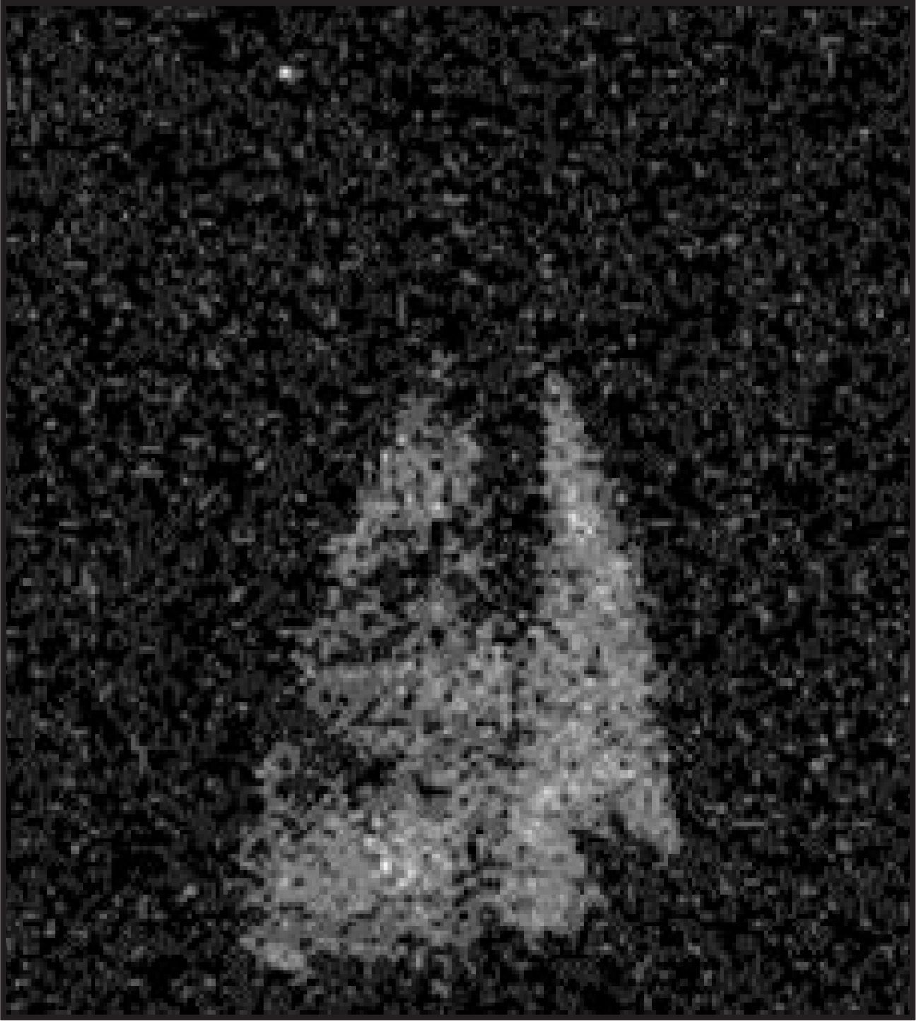
Discussion
Functional pulmonary MRI ventilation and perfusion imaging methods have several distinct advantages over those that employ radioactive tracers. In general, the resolution of ventilation and perfusion MRI techniques is superior to that of nuclear medicine techniques. In addition, the MRI system offers the capability to acquire spatially co-registered three-dimensional images of ventilation and perfusion and therefore makes feasible imaging of the V/Q ratio. The absence of ionizing radiation makes this a safe method for use in children.
There are many clinical areas in which the V/Q MRI methods may have an impact. They may be useful in improving the noninvasive diagnosis of pulmonary embolism, where the improved resolution may reduce the number of indeterminate functional scans. Due to its intrinsically high sensitivity, helium MRI may become useful as a means for early detection of rejection following lung transplantation36 or for following patients with asthma during therapy. 3He MRI has been shown to be sensitive to early functional changes occurring in smokers.47 Diffusion-weighted 3He MRI has been shown useful in the early detection of emphysema.25 As such, the fMRI methods may become an important tool in the evaluation of patients who undergo lung volume reduction surgery (LVRS), both to predict response preoperatively, and to evaluate outcome postoperatively.
Functional pulmonary MRI methods continue to evolve, and they are likely to improve in both spatial and temporal resolution as improvements in hardware occur. They have broad applicability to many important disease states, including emphysema, pulmonary embolism, and asthma. Given their accuracy and safety, these methods are likely to become an important clinical tool in the evaluation of patients with pulmonary disease in the future. AR
Acknowledgments
The authors thank Norman Butler, RT, Tanya Kurtz, RT, and Doris Cain, RT, Jaime F. Mata, John M. Christopher, RT (R)(MR), and Doris A. Harding, RN for valuable assistance.
Dr. Roberts is supported by an RSNA Scholar 2000 grant. Dr. Roberts and Dr. Rizi are supported by NIH grants RR02305 and RO1-HL64741. Dr. Mai is supported by a grant from the American Heart Association. Dr. Lipson is supported by NIH grant K23 HL 04486. Dr. Salerno is supported by NIH grants R44-HL059022 and RO1HL66479, and by the University of Virginia School of Medicine, Amersham Health, and Siemens Medical Systems.
References
- Alsop D, Hatabu H, Bonnet M. Multi-slice, breathhold imaging of the lung with submillisecond echo times.. Magn Reson Med.. 1995;33:678-682.
- Rosen B, Belliveau J, Chien D. Perfusion imaging by nuclear magnetic resonance.. Magn Reson Q.. 1989;5:263-281.
- Edelman R, Hatabu H, Tadamura E. Proceedings of the Society of Magnetic Resonance in Medicine, 12th Annual Scientific Meeting; 1996. 1996:1314.
- Hatabu H, Gaa J, Kim D. Pulmonary perfusion: Qualitative assessment with dynamic contrast-enhanced MRI using ultra-short TE and inversion recovery turbo FLASH.. Magn Reson Med.. 1996;36:503-508.
- Hatabu H, Gaa J, Kim D. Pulmonary perfusion and angiography: Evaluation with breath-hold enhanced three-dimensional fast imaging steady-state precession MR imaging with short TR and TE.. AJR Am J Roentgenol.. 1996;167:653-655.
- Hatabu H, Tadamura E, Prasad P. Noninvasive pulmonary perfusion imaging by STAR-HASTE sequence.. Magn Reson Med.. 2000;44:808-812.
- Detre J, Leigh J, Williams D, Koretsky A. Perfusion imaging.. Magn Reson Med.. 1991;22:1-9.
- Kwong K, Belliveau J, Chesler D. Dynamic magnetic resonance imaging of human brain activity during primary sensory stimulation.. Proc Natl Acad Sci USA.. 1992;89:5675-5679.
- Williams D, Detre J, Leigh J, Koretsky A. Magnetic resonance imaging of perfusion using spin inversion of arterial water.. Proc Natl Acad Sci USA. Proc Natl Acad Sci USA. 1992;89:212-216.
- Alsop D, Detre J. Reduced transit-time sensitivity in noninvasive magnetic resonance imaging of human cerebral blood flow.. J Cereb Blood Flow Metab.. 1996;16:1236-1249.
- Chen Q, Siewert B, Bly B. STAR-HASTE: Perfusion imaging without magnetic susceptibility artifact.. Magn Reson Med.. 1997;38:404-408.
- Roberts D, Gefter W, Hirsch J. Pulmonary perfusion: Respiratory-triggered three-dimensional MR imaging with arterial spin tagging—Preliminary results in healthy volunteers.. Radiology.. 1999;212:890-895.
- Mai V, Hagspiel K, Christopher J. Perfusion imaging of the human lung using flow-sensitive alternating inversion recovery with an extra radiofrequency pulse (FAIRER).. Magn Reson Imaging.. 1999;17:355-361.
- Mai V, Berr S. MR perfusion imaging of pulmonary parenchyma using pulsed arterial spin labeling techniques: FAIRER and FAIR.. J Magn Reson Imaging.. 1999;9:483-487.
- Williams D, Detre J, Zhang W, Koretsky A. Fast serial MRI of perfusion in the rat brain during bicuculline induced seizures using spin inversion of arterial water [abstract]. 1992:711.
- Roberts D, Rizi R, Lipson D. Dynamic observation of pulmonary perfusion using continuous arterial spin-labeling in a pig model.. J Magn Reson Imaging.. 2001;14:175-180.
- Middleton H, Black R, Saam B. MR imaging with hyperpolarized 3-He gas.. Magn Reson Med.. 1995;33:271-275.
- Kauczor H, Hofmann D, Kreitner K. Normal and abnormal pulmonary ventilation: Visualization at hyperpolarized He-3 MR imaging.. Radiology.. 1996;201:564-568.
- Mugler J, Driehuys B, Brookeman J. MR imaging and spectroscopy using hyperpolarizedXe gas: Preliminary human results. 129. Magn Reson Med.. 1997;37:809-815.
- Guenther D, Hanisch G, Kauczor H. Functional MR imaging of pulmonary ventilation using hyperpolarized noble gases.. Acta Radiol.. 2000;41:519-528.
- Roberts D, Rizi R, Lipson D. Detection and localization of pulmonary air leaks using laser-polarized (3)He MRI.. Magn Reson Med.. 2000;44:379-382.
- Kauczor H, Markstaller K, Puderbach M. Volumetry of ventilated airspaces byHe MRI: Preliminary results. 3. Invest Radiol.. 2001;36:110-114.
- Salerno M, Altes T, Mugler J. Hyperpolarized noble gas MR imaging of the lung: Potential clinical applications.. Eur J Radiol.. 2001;40:33-44.
- Saam B, Yablonskiy D, Kodibagkar V. MR imaging of diffusion of (3)He gas in healthy and diseased lungs.. Magn Reson Med.. 2000;44:174-179.
- Salerno M, de Lange E, Altes T. Emphysema: Hyperpolarized helium 3 diffusion MR imaging of the lungs compared with spirometric indexes—Initial experience.. Radiology.. 2002;222:252-260.
- Deninger A, Eberle B, Ebert M. Quantification of regional intrapulmonary oxygen partial pressure evolution during apnea by (3)He MRI.. J Magn Reson.. 1999;141:207-216.
- Deninger A, Eberle B, Ebert M. (3)he-MRI-based measurements of intrapulmonary p(O) and its time course during apnea in healthy volunteers: First results, reproducibility, and technical limitations. 2. NMR Biomed.. 2000;13:194-201.
- Deninger A, Eberle B, Bermuth J. Assessment of a single-acquisition imaging sequence for oxygen-sensitive (3)He-MRI.. Magn Reson Med.. 2002;47:105-114.
- Eberle B, Weiler N, Markstaller K. Analysis of intrapulmonary O(2) concentration by MR imaging of inhaled hyperpolarized helium-3.. J Appl Physiol.. 1999;87:2043-2052.
- Salerno M, Altes T, Brookeman J. Dynamic spiral MRI of pulmonary gas flow using hyperpolarizedHe: Preliminary studies in healthy and diseased lungs. 3. Magn Reson Med.. 2001;46:667-677.
- Saam B, Yablonskiy D, Gierada D, Conradi M. Rapid imaging of hyperpolarized gas using EPI.. Magn Reson Med.. 1999;42:507-514.
- Ruppert K, Brookeman J, Hagspiel K, Mugler J. Probing lung physiology with xenon polarization transfer contrast (XTC).. Magn Reson Med.. 2000;44:349-357.
- Kauczor H, Ebert M, Kreitner K. Imaging of the lungs usingHe MRI: Preliminary clinical experience in 18 patients with and without lung disease. 3. J Magn Reson Imaging.. 1997;7:538-543.
- de Lange E, Mugler J, Brookeman J. Lung air spaces: MR imaging evaluation with hyperpolarizedHe gas. 3. Radiology.. 1999;210:851-857.
- Altes T, Powers P, Knight-Scott J. HyperpolarizedHe MR lung ventilation imaging in asthmatics: Preliminary findings. 3. J Magn Reson Imaging.. 2001;13:378-384.
- McAdams H, Palmer S, Donnelly L. HyperpolarizedHe-enhanced MR imaging of lung transplant recipients: Preliminary results. 3. AJR Am J Roentgenol.. 1999;173:955-959.
- Donnelly L, MacFall J, McAdams H. Cystic fibrosis: Combined hyperpolarizedHe-enhanced and conventional proton MR imaging in the lung—Preliminary observations. 3. Radiology.. 1999;212:885-889.
- Edelman R, Hatabu H, Tadamura E. Noninvasive assessment of regional ventilation in the human lung using oxygen-enhanced magnetic resonance imaging.. Nat Med.. 1996;2:1236-1239.
- Mai V, Chen Q, Bankier A, Edelman R. Multiple inversion recovery MR subtraction imaging of human ventilation from inhalation of room air and pure oxygen.. Magn Reson Med.. 2000;43:913-916.
- Loffler R, Muller C, Peller M. Optimization and evaluation of the signal intensity change in multisection oxygen-enhanced MR lung imaging.. Magn Reson Med.. 2000;43:860-866.
- Mai V, Li W, Chen C. Application of functional magnetic resonance imaging analysis in oxygen-enhanced ventilation and arterial spin labeling perfusion imaging in the lung [abstract]. 2002:1972.
- Chen Q, Levin D, Kim D. Pulmonary disorders: Ventilation-perfusion MR imaging with animal models.. Radiology.. 1999;213:871-879.
- Mai V, Bankier A, Prasad P. MR ventilation-perfusion imaging of human lung using oxygen-enhanced and arterial spin labeling techniques.. J Magn Reson Imaging.. 2001;14:574-579.
- Yamaguchi K, Mori M, Kawai A. Effects of pH and SO2 on solubility coefficients of inert gases in human whole blood.. J Appl Physiol.. 1993;74:643-649.
- Yamaguchi K, Soejima K, Koda E, Sugiyama N. Inhaling gas with different CT densities allows detection of abnormalities in the lung periphery of patients with smoking-induced COPD.. Chest.. 2001;120:1907-1916.
- Schreiber W, Eberle B, Laukemper-Ostendorf S. Dynamic (19)F-MRI of pulmonary ventilation using sulfur hexafluoride (SF(6)) gas.. Magn Reson Med.. 2001;45:605-613.
- Guenther D, Eberle B, Hast J. (3)He MRI in healthy volunteers: preliminary correlation with smoking history and lung volumes.. NMR Biomed.. 2000;13:182-189.
Citation
. Functional magnetic resonance imaging of the lung. Applied Radiology. 2003;32(2):41-48. doi:10.37549/AR1160.