Glioneuronal Tumors: A Pictorial Review and Practical Approach
Applied Radiology — Vol. 55 , Issue 1
Published: December 22, 2025
1 Department of Radiology and Imaging Sciences, Emory University, Atlanta, Georgia
2 Emory University, Atlanta, Georgia
3 Philadelphia College of Osteopathic Medicine, Philadelphia, Pennsylvania
* Corresponding author: Zofia Lasiecka (zofia.maria.lasiecka@emory.edu)
Abstract
Many radiologists struggle to parse between glioneuronal tumors due to significant overlap in imaging, histology, and long-term behavior. However, most glioneuronal tumors have long, primarily benign courses, and therefore, it is not uncommon for imaging to be the primary source for clinical decision-making. In cases requiring resection, imaging features can inform preoperative risk and determine the need for adjuvant therapies. While these tumors are diagnostically challenging, specific imaging features have stronger diagnostic and prognostic associations. This review aims to provide an approach to navigating the glioneuronal tumor differential for practicing radiologists and radiology trainees. Through a set of representative cases, key imaging features are discussed that inform specific glioneuronal tumor diagnoses as well as subsequent clinical management.
Keywords
glioneuronal tumors, central nervous system tumors, neuroradiology, pediatric neuroradiology
Categories
Introduction
As their name indicates, glioneuronal tumors contain glial and neuronal cellular features. They typically exhibit low-grade behavior and do not fall into the molecular subtype of other brain tumors such as gliomas, oligodendrogliomas, and astrocytomas. 1,2 Their clinical symptoms usually stem from mass effect and/or medically refractory seizures, and while they can occur across all age groups, they are often diagnosed in childhood or early adulthood. 2
Advances in molecular pathophysiology have led the World Health Organization to update its diagnostic criteria for brain tumors, switching from an imaging or histopathological approach to genetic and molecular categorization. 1 Nevertheless, radiologists play a key role in diagnosis and prognosis. MRI is the modality of choice for tumor characterization, with occasional use of advanced techniques such as MR perfusion and spectroscopy (MRS) for indeterminate cases. 3
Here, we present an overview of glioneuronal tumors and typical imaging findings. We also present an approach to differentiating these tumors from other types based on key imaging features that can inform diagnosis and subsequent management.
Review of Tumors
Supratentorial: Cortical
Desmoplastic Infantile Astrocytoma and Ganglioglioma
Desmoplastic infantile gangliogliomas (DIGs) and astrocytomas (DIAs) occur most often in infants under age 24 months. 4 DIGs demonstrate a slight male predominance (~60%) while DIAs have no gender predilection. 4,5 In contrast to other glioneuronal tumors, DIG/DIA presents with seizures in a minority of cases. 4,6 DIGs/DIAs are large tumors most often located within the superficial cortex in the frontal or parietal lobes (Figure 1). 4,6 Cystic components are found in most cases (>90%) and often located deep to solid components (Figure 1). 6 The peripheral solid components enhance vividly. 4,5 While these tumors are considered benign, they can demonstrate metastatic behavior; one case series has documented metastasis at presentation in 11.7% of cases (Figure 1B). 7
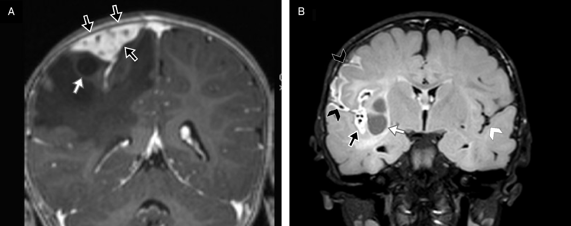
The main differentials are pleomorphic xanthoastrocytoma (PXA), ganglioglioma, dysembryoplastic neuroepithelial tumor (DNET), and atypical teratoid/rhabdoid tumor (AT/RT). DIAs/DIGs typically present at younger ages and have stronger T2 hyperintensity compared with PXAs and gangliogliomas. 4 In addition, DIAs and DIGs enhance more strongly than DNETs. 4,6 AT/RTs are more likely to calcify and hemorrhage than DIAs and DIGs; however, calcification and hemorrhage can be seen in DIA/DIG cases with acute clinical symptoms. 4,5 Total resection is uncommon (27-30% of cases), and adjuvant therapy is recommended only in cases of documented tumor progression. 6
Imaging offers a few key predictors of patient outcomes. 6 The leptomeninges are the most common site of metastasis (Figure 1), but intracranial and spinal metastases have also been reported. 6 Midline location is associated with more aggressive histological behavior, increased chance of recurrence, and increased intraoperative risk. 4,6 Specifically, involvement of the hypothalamus should be reported, as it is associated with increased mortality secondary to postoperative respiratory compromise. 4 Finally, the solid components of DIAs/DIGs usually encase vessels, which can help with diagnosis but can also limit surgical resection. 6
Dysembryoplastic Neuroepithelial Tumors
Slow-growing and cortical-based, DNETs most commonly affect patients <20 years and are found in the temporal and frontal lobes. 8
DNETs demonstrate a characteristic multicystic, “bubbly” lesion with high T2 and low T1 signal (Figure 2). 8 Multicystic morphology is associated more with DNETs than with other cortically based lesions such as oligodendrogliomas and gangliogliomas. 9 Most DNETs extend into the subcortical white matter, occasionally in a tapered fashion. 9 The FLAIR rim sign has been suggested as a specific finding for DNET (82% specificity in one case series) (Figure 2B). 10
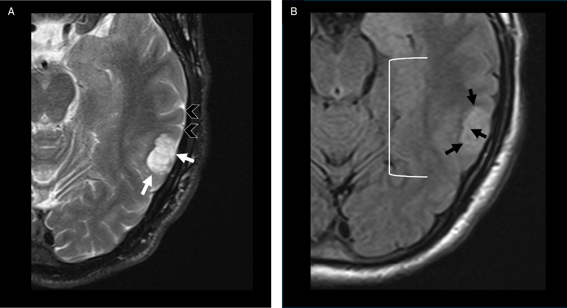
Some DNETs will demonstrate peripheral contrast enhancement, may contain macroscopic calcification (~20%), and rarely contain intratumoral hemorrhage. 11 They frequently present with multiple satellite lesions, preferentially occurring along the medial aspect of the primary lesion. 8 Additionally, focal cortical dysplasia can be associated with higher-grade DNETs. 12
Surgical resection provides effective seizure control. Persistent seizures have been seen in 46% of patients with satellite lesions but only 18% of patients without satellite lesions. 13
Polymorphous Low-Grade Neuroepithelial Tumors of the Young
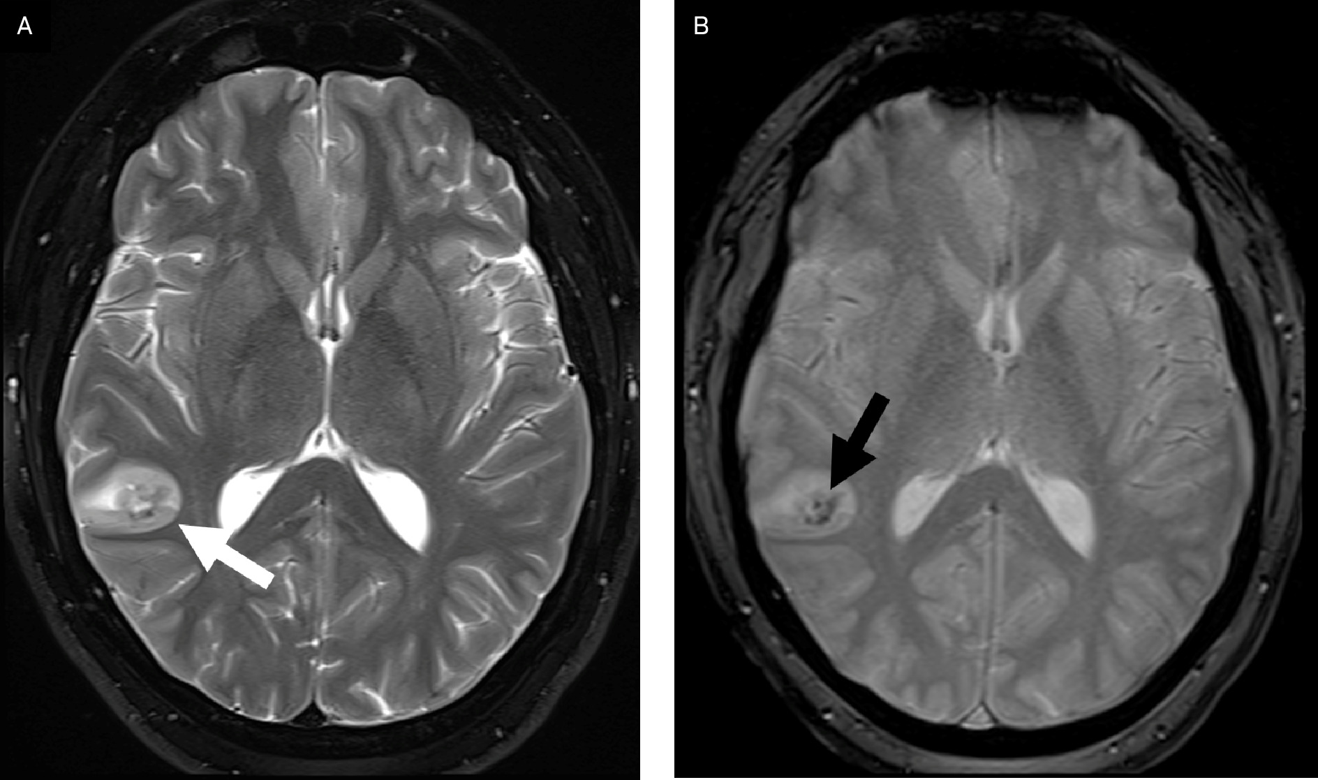
Polymorphous low-grade neuroepithelial tumors of the young (PLNTY) were previously considered a subtype of DNET, differentiated via mutations within the MAPK pathway. 14 They are primarily seen in children and young adults, 15 occurring only rarely in older adults. 16 Typically, PLNTYs are cortically based, heterogeneous cystic or mixed solid cystic masses (Figure 3) classically with central calcification 17 and mild, heterogeneous enhancement. 18 A “transmantle”-like signal abnormality (linear increased T2 signal in the deep white matter extending peripherally) can be seen, with some mixed evidence around its postresection persistence for predicting poorer seizure control. 18 Beyond seizure-associated morbidity, the tumor follows a benign course with minimal mass effect. There is low recurrence after total resection 17 ; however, a recent meta-analysis reported that the extent of resection was not significantly associated with postoperative seizure control. 19
Gangliogliomas
Gangliogliomas are the most common epilepsy-associated neoplasm, typically found in the temporal and frontal lobes. 20,21 They can occur infratentorially, including in the spinal column. They occur in both pediatric and adult populations, with most pediatric cases diagnosed in those over the age of 10 years 21 and adult cases at a median age of 32 years. 22
On imaging, gangliogliomas are typically superficial, well-circumscribed mixed cystic and solid tumors, but a diffuse infiltrative appearance is possible. Enhancement is variable and associated with the solid components (Figure 4); infratentorial gangliogliomas are more likely to demonstrate “paintbrush” enhancement (dorsal predominant) compared with supratentorial tumors. 22 Brainstem gangliogliomas are associated with older patient age (median of 30 years), peritumoral edema, higher solid proportion, and lower rates of calcification. 23
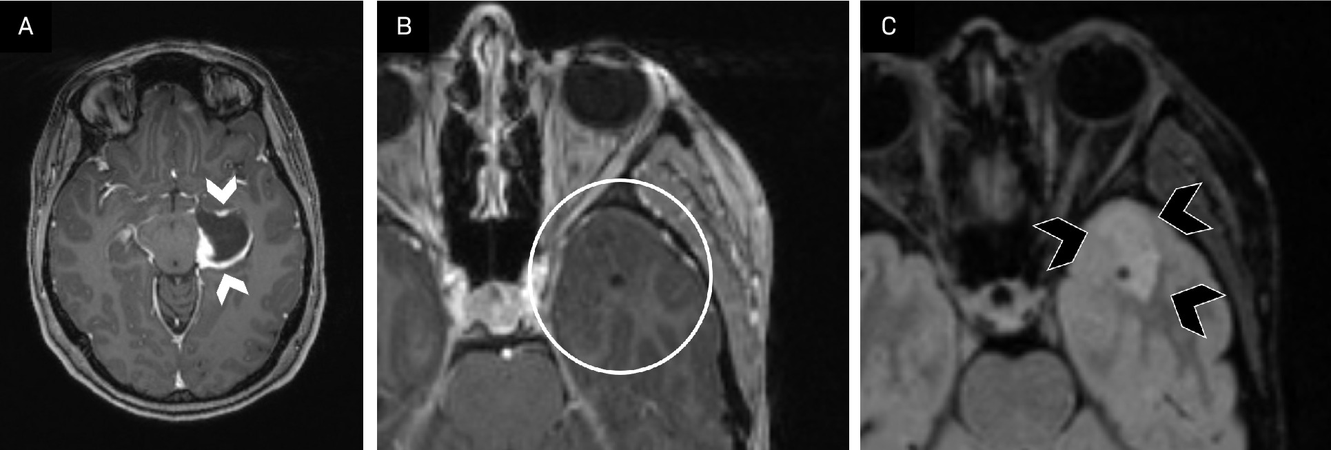
With their variable imaging appearance, the differential is wide, including PXA, PLNTY, and oligodendrogliomas. In general, PXAs enhance more vividly than gangliogliomas and very rarely calcify, while up to 50% of gangliogliomas calcify. 23 Finally, gangliogliomas have been reported to have higher normalized cerebral blood volume and lower normalized apparent diffusion coefficient compared with oligodendrogliomas. 24
Tumor locations associated with worse outcomes include the brainstem, cerebellopontine angle, spinal cord, and midline tumors. 22 Brainstem involvement is often the defining factor for total vs subtotal resection. 21 There is mixed evidence for the ability of resection extent to predict long-term outcomes. 22,25,26 Finally, there have been rare case reports of malignant degeneration; in these cases, leptomeningeal involvement was common and MRS showed anaplastic features (increased choline, lipid, and lactate). 22
Cerebral Gangliocytoma
Cerebral gangliocytomas (CGs) are rare (<3% of CNS tumors), occurring most frequently in children and young adults. 2 These tumors typically arise in the cerebral cortex, most often in the temporal lobe (~75%) (Figure S1), though occasional exophytic and rare extra-axial presentations have been described.
Sometimes exhibiting variable imaging features, CGs can be difficult to distinguish from other tumors, particularly gangliogliomas. Meningioma is in the differential, given that CGs can be extra-axial and demonstrate a dural tail. 2,27 MRS may be a helpful diagnostic adjunct, as CGs demonstrate a highly elevated choline signal and reduced N-acetylaspartate, a pattern more typical of high-grade gliomas than other glioneuronal tumors such as ganglioglioma. 27 Diagnosis is ultimately based on histopathology.
Supratentorial: Intraventricular
Myxoid Glioneuronal Tumors
Previously known as “DNET of the septum pellucidum,” 1,28 myxoid glioneuronal tumors (MGTs) are now defined by a gain-of-function mutation in the PDGFRA gene. 29 Given their similar histology and patient populations, MGTs and DNETs are difficult to distinguish from each other; therefore, imaging and genetic testing play an important diagnostic role.
The pathognomonic finding of MGT is location within the septum pellucidum (Figure 5), but they have also been reported in the midbrain. 29 Other imaging characteristics are similar to DNETs, with a multicystic morphology with well-defined margins and variable enhancement. 30 Unlike DNETs, no macrocalcifications have been reported in histologically proven MGTs. 29
![Myxoid glioneuronal tumor (formerly dysembryoplastic neuroepithelial tumor [DNET] of the septum pellucidum). Axial T2 FLAIR imaging (A) demonstrates a mass in the septum pellucidum (arrow) with imaging features similar to DNET: well-circumscribed with multiple hyperintense internal cystic structures. The mass is nonenhancing on axial postcontrast T1 image (B, arrowhead), consistent with the variable enhancement patterns of DNET. At biopsy, the mass was confirmed to contain histological characteristics of DNET of the septum pellucidum, which subsequently was classified as myxoid glioneuronal tumor. Genetic testing for PDGFRA was not performed for this patient owing to lack of testing availability but would be considered the gold standard for myxoid glioneuronal tumor diagnosis.](https://appliedradiologystorage.blob.core.windows.net/jats-replatform/10.37549-AR-D-25-0143/AR-D-25-0143-F005.jpeg)
Central Neurocytoma
Originally classified as oligodendrogliomas, central neurocytomas (CNs) are now recognized as a distinct entity. 31,32 These tumors present at a median age of 30-31 years and typically demonstrate no gender predilection. 31,32
Classically, CNs are found in the frontal horn of the lateral ventricles in close association with the foramen of Monro. 31,32 Most are unilateral at presentation, with bilateral involvement more common in larger tumors. 32 These tumors consist of mixed calcific, cystic, and solid tissue components, resulting in heterogeneous imaging characteristics (Figure 6). CNs may contain coarse “popcorn” calcifications on CT, 31,32 while cystic components produce a “Swiss-cheese” or “soap bubble” appearance on T2 sequences. 31,33 The solid tissue component dictates the degree of diffusion restriction and postcontrast enhancement, with the adjacent choroid plexus used for baseline enhancement comparison. 31,33,34
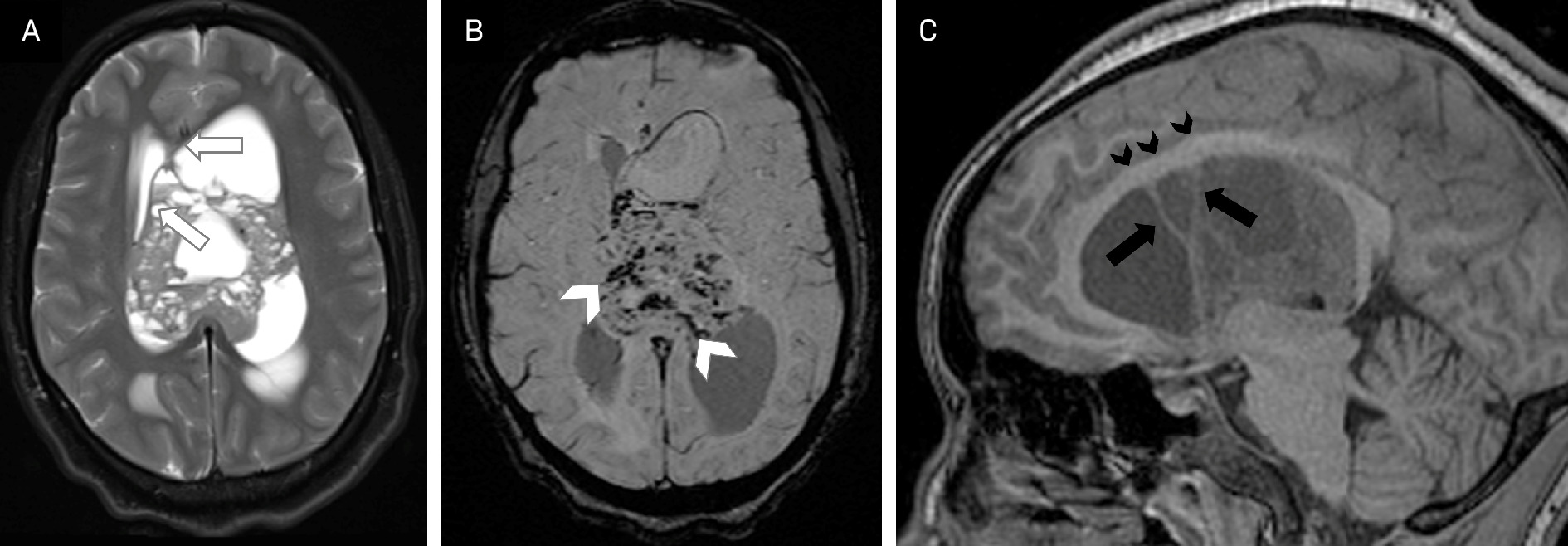
While the imaging appearance of CNs can vary, specific characteristics can assist with diagnosis and prognosis. These include a broad-based septum pellucidum attachment (Figure 6), peripheral cystic components, periventricular edema, and the “scalloping sign,” a wavy ventricular wall with adjacent rows of cysts. 12,15,16 On MRS, CNs typically demonstrate elevated choline and reduced creatinine and N-acetylaspartate 31,33 ; elevated glycine is considered a sensitive but nonspecific finding for CN. 32
Total resection is the gold standard treatment for CNs; however, this is limited by the degree of vascularity and potential neurologic complications, the rates of which range from 30% to 70%. 33 Adjuvant radiation is controversial in cases of subtotal resection but may be considered for tumors that demonstrate atypical and/or aggressive histologic features (~20% of CNs). 31,32,35 Imaging findings associated with aggressive histologic features include enhancement, larger size, and the presence of irregular vessels. 36 Radiation is also considered for recurrent disease, which typically occurs 3-4 years post-treatment. 31,35
Supratentorial: Subcortical Tumors
Multinodular and Vacuolating Neuronal Tumors
Primarily occurring in adults, multinodular and vacuolating tumors (MVNTs) present at mean and median ages ranging from 38 to 45 years. 37,38 They are benign tumors with very slow growth and do not typically require resection. 38
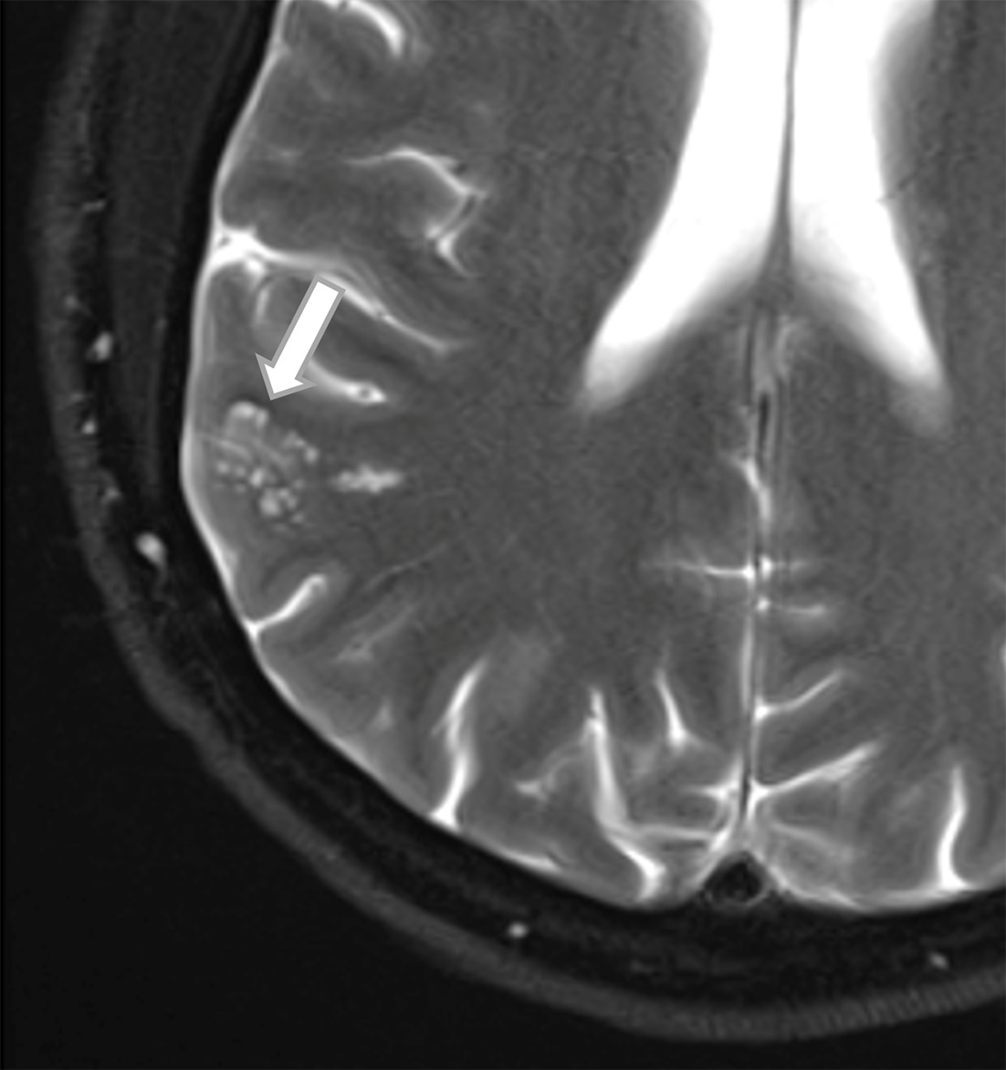
Their classic MRI appearance is a cluster of nonenhancing T2 and FLAIR hyperintense nodules centered at the cortical-subcortical junction, most commonly in the temporal lobe (Figure 7), without restriction. 37,38 The differential is wide and includes other glioneuronal tumors and non-neoplastic processes such as prominent perivascular spaces or focal cortical dysplasia. The most appropriate follow-up MRI protocol for MVNTs continues to be debated. 38
Papillary Glioneuronal Tumors
Extremely rare entities, papillary glioneuronal tumors (PGNTs) primarily affect adults in their second decade. 39 There is no gender-based predilection.
The imaging features of these tumors are similar to those of other glioneuronal tumors, with a mixed solid cystic appearance. The 2 features suggestive of PGNT are location—80% are within or adjacent to the lateral ventricles—and septated cystic components, though these are not specific. 40 PGNTs are generally diagnosed histologically, genetically differentiated by the fusion of SLC44A1-PRKCA proteins. 41 Treatment involves resection, which has a good prognosis. 2
Posterior Fossa
Rosette-Forming Glioneuronal Tumors
Rosette-forming glioneuronal tumors (RGNTs) are defined histologically 42 and typically present in adults at a mean of 23 years, with a slight female predilection (1.36:1). 43 The classic location is adjacent to the 4th ventricle, with local parenchymal extension (Figure 8). 2 Less common sites include the cerebellum and spine. 43 Given their mixed solid-cystic composition, their imaging appearance is variable; thus, tissue sampling is required for diagnosis.
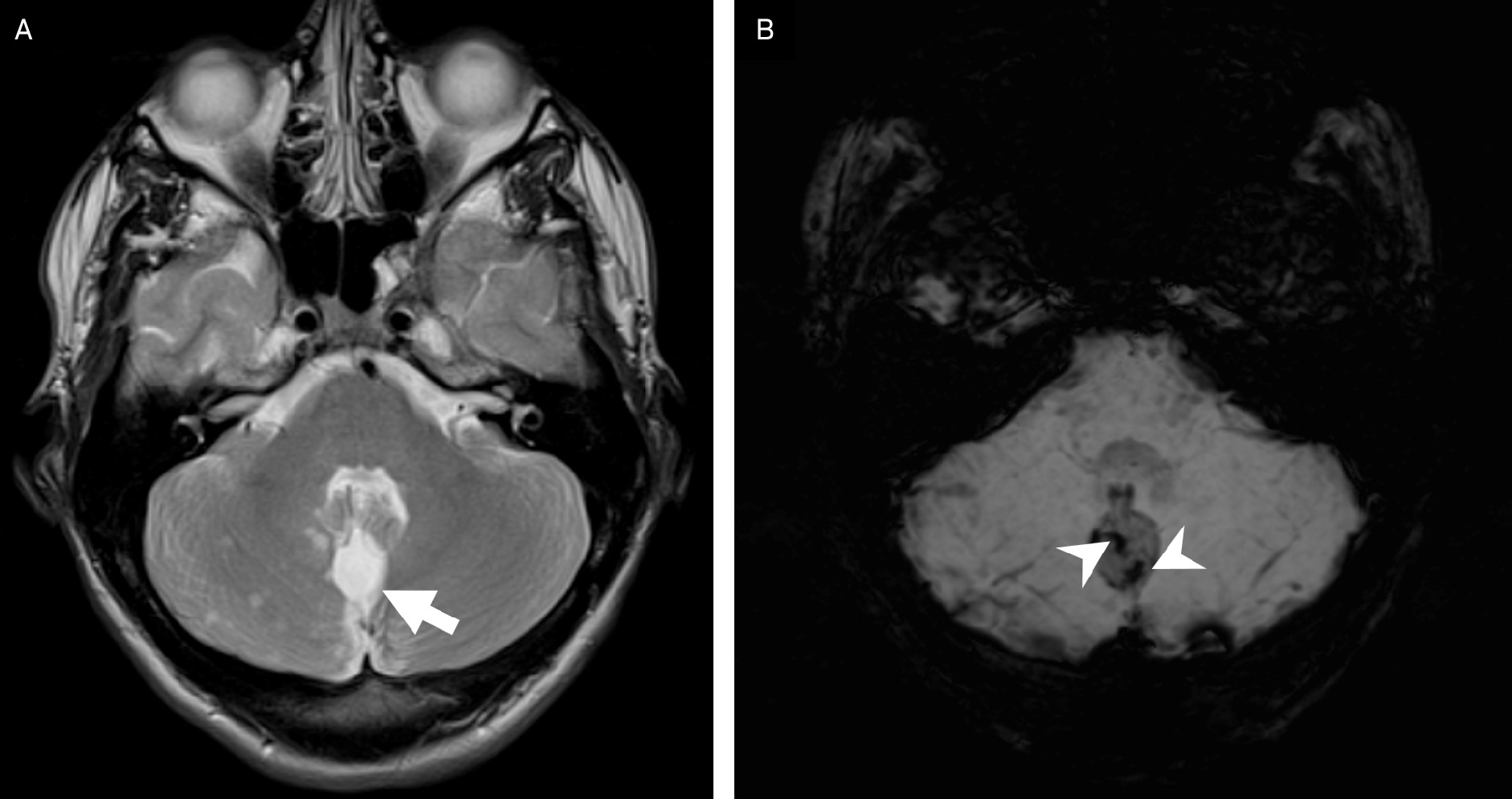
The main differential considerations are pilocytic astrocytoma and DNET. Most reported RGNTs are <3.0 cm, 43 with a diameter <3.5 cm being just over 3 times more likely for RGNT. 44 Calcifications in RGNTs are exceedingly rare, with only 2 cases reported, 43 while pilocytic astrocytomas and DNETs have reported calcification incidences of 17% 45 and 20%, 11 respectively. Microhemorrhage is common in RGNTs (Figure 8), and while pilocytic astrocytomas can hemorrhage, it is exceedingly rare for DNETs, reported in 3%. 46
While RGNTs are considered benign, 2 mass effect can be significant and require resection. 47 Pineal involvement is possible and should be reported, as it changes surgical approach. 48 Overall recurrence rates between total and subtotal resection of these tumors are similar, at about 10% 44 ; however, the recurrence/progression timeline for subtotal resection vs total resection is reported at an average of 2.9 and 6.2 years, respectively. 44 Additionally, there is evidence that a higher proportion of solid tissue within the tumor predicts higher recurrence risk and worse histological features. 43 Rarely, RGNTs can develop satellite lesions and CSF involvement, which can lead to drop metastases; thus, postcontrast series should be evaluated for leptomeningeal involvement. 42,43 Finally, MRS usually shows elevated choline, reduced N-acetylaspartate, and absent lactate and lipid peaks 2 ; deviations from this pattern should raise suspicion for malignant transformation.
Dysplastic Cerebellar Gangliocytomas
Also known as Lhermitte-Duclos disease, dysplastic cerebellar gangliocytomas are found primarily in patients with Cowden syndrome. 49 Arising in the cerebellum, they are benign, with no reported malignant transformation. 49 Surgical resection provides definitive treatment in acute cases; however, these tumors often do not require intervention. 50
MRI demonstrates a unilateral cerebellar parenchymal mass with T2 hyperintensity, no enhancement, and variable diffusion restriction. Widened cerebellar folia create the pathognomonic “tiger-stripe” pattern, 49,50 which is primarily seen in adults. 51 SWI and contrast-enhanced sequences can demonstrate prominent vascular structures within the folia (Figure S2), 52 while MRS may reveal reduced choline and an inverted lactate peak. 52,53 The differential is limited, but subacute infarct or cerebritis can be considered in patients with acute deterioration.
Discussion
Differentiating between glioneuronal tumors is challenging, owing to similarities in demographics, clinical presentation, and imaging appearance, as well as their overall low incidence, resulting in limited reports in the literature. In this paper, we propose a diagnostic approach to these tumors, present representative cases, and describe prognostic imaging features based on our review of the current literature.
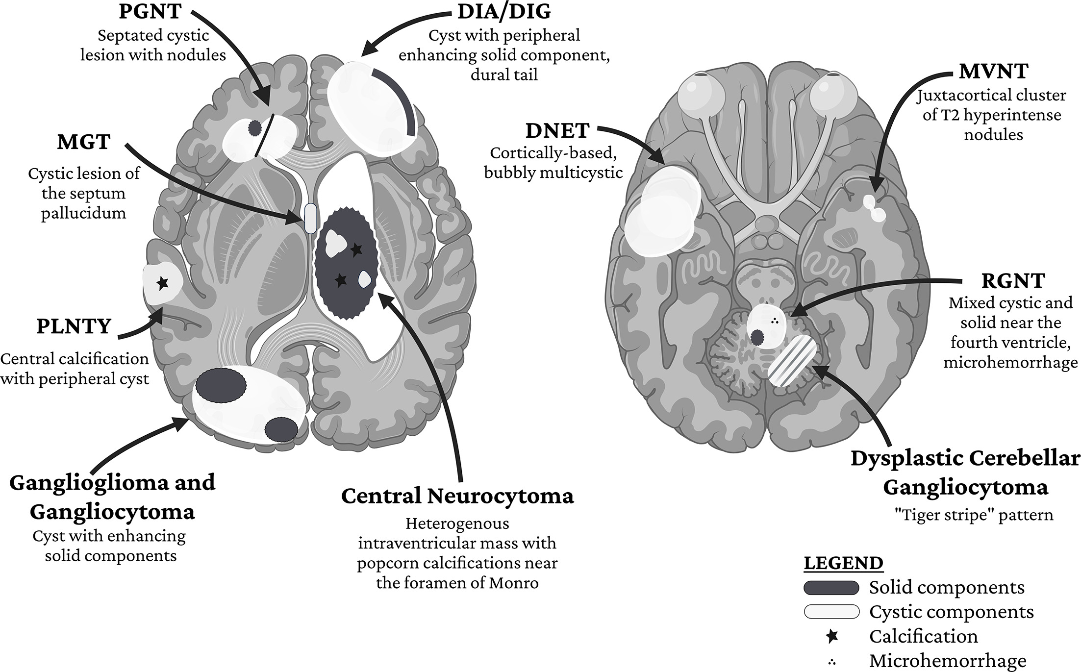
Owing to their shared cellular origin, most glioneuronal tumors have a mixed cystic-solid composition (Figure 9). Location and parenchymal compartment are most useful for initially narrowing the differential, followed by the morphology and arrangement of the specific tumor components (eg, peripheral vs septated cysts, calcification vs no calcification) (Figure 10). MR perfusion and spectroscopy may be helpful in some cases. Ultimately, many glioneuronal tumors require histopathology for definitive diagnosis, but some patients may choose to forgo biopsy, relying instead entirely on image-based diagnosis.
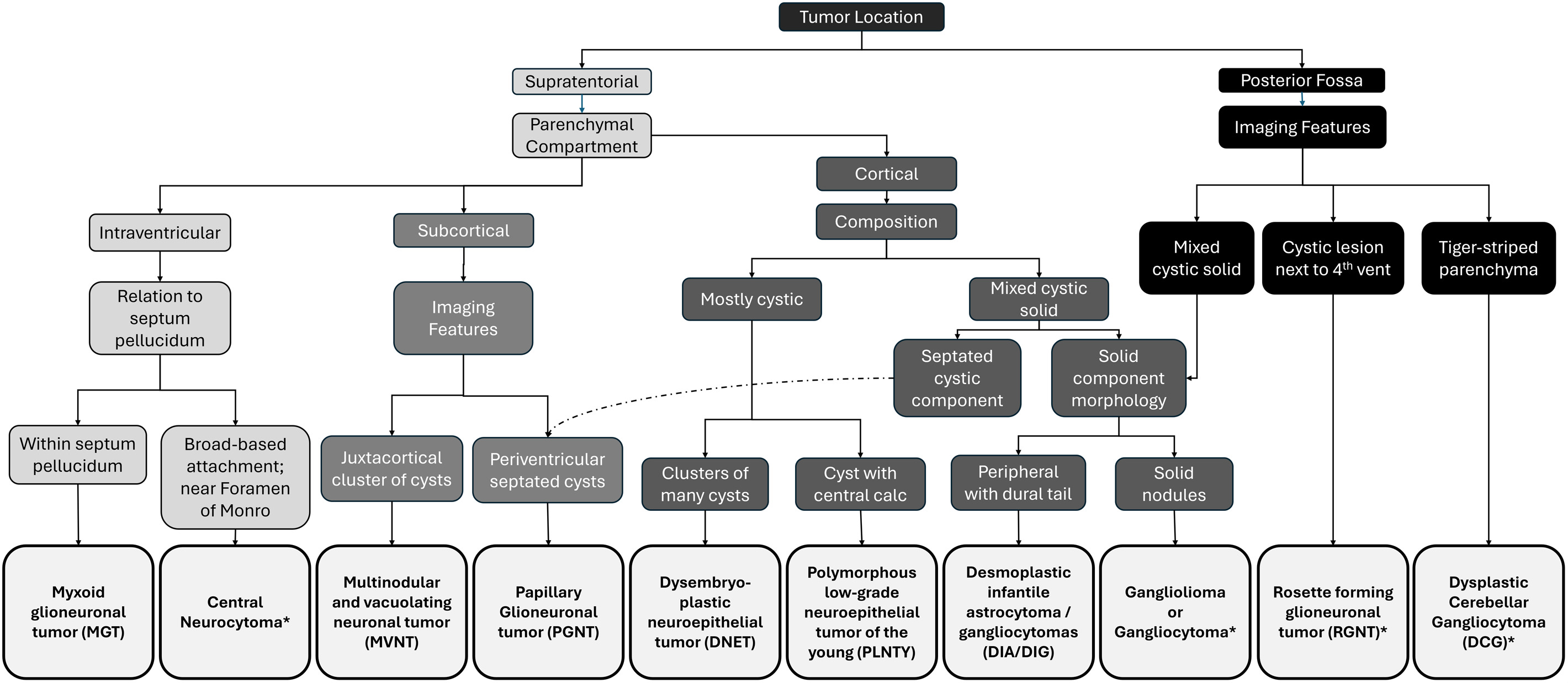
Radiologists have a great impact on the management of glioneuronal tumors, as the primary treatment consists of excision with continued surveillance. Preoperative reports should include features that could alter operative approach and risk, such as involvement of midline structures and their relationship to major vessels. Furthermore, specific imaging features are associated with more aggressive histopathology in certain glioneuronal tumors; these can inform care management discussions. While the evidence is mixed for several glioneuronal tumors on the relationship between resection extent and long-term outcomes, including seizure control, the presence of metastatic foci or recurrent tumor on imaging affects the decision whether to pursue adjuvant systemic therapy.
In conclusion, while differentiating between glioneuronal tumors is challenging, narrowing the differential diagnosis is possible with careful assessment of tumor location and relational morphology. Radiologists can provide useful evidence for clinical management, including surgical approach, operative risk, and the need for adjuvant treatment.
References
- Louis D, Perry A, Wesseling P. The 2021 WHO classification of tumors of the central nervous system: a summary. Neuro Oncol. 2021;23(8):1231-1251. doi:10.1093/neuonc/noab106.
- Vaz A, Cavalcanti M, da Silva Junior E, Ramina R, de Almeida Teixeira B. Uncommon glioneuronal tumors: a radiologic and pathologic synopsis. AJNR Am J Neuroradiol. 2022;43(8):1080-1089. doi:10.3174/ajnr.A7465.
- Park Y, Vollmuth P, Foltyn-Dumitru M. The 2021 WHO classification for gliomas and implications on imaging diagnosis: part 1-key points of the fifth edition and summary of imaging findings on adult-type diffuse gliomas. J Magn Reson Imaging. 2023;58(3):677-689. doi:10.1002/jmri.28743.
- Bianchi F, Tamburrini G, Massimi L, Caldarelli M. Supratentorial tumors typical of the infantile age: Desmoplastic Infantile Ganglioglioma (DIG) and Astrocytoma (DIA). A review. Childs Nerv Syst. 2016;32(10):1833-1838. doi:10.1007/s00381-016-3149-4.
- Ho C, Gener M, Bonnin J, Kralik S. Diffusion, perfusion, and histopathologic characteristics of desmoplastic infantile ganglioglioma. J Radiol Case Rep. 2016;10(7):1-13. doi:10.3941/jrcr.v10i7.2715.
- Imperato A, Spennato P, Mazio F. Desmoplastic infantile astrocytoma and ganglioglioma: a series of 12 patients treated at a single institution. Childs Nerv Syst. 2021;37(7):2187-2195. doi:10.1007/s00381-021-05057-3.
- Wang S, Sun M, Abecassis I. Predictors of mortality and tumor recurrence in desmoplastic infantile ganglioglioma and astrocytoma-and Individual Participant Data Meta-Analysis (IPDMA). J Neurooncol. 2021;155(2):155-163. doi:10.1007/s11060-021-03860-1.
- Phi J, Kim S. Dysembryoplastic neuroepithelial tumor: a benign but complex tumor of the cerebral cortex. Brain Tumor Res Treat. 2022;10(3):144-150. doi:10.14791/btrt.2022.0015.
- Paudel K, Borofsky S, Jones R, Levy L. Dysembryoplastic neuroepithelial tumor with atypical presentation: MRI and diffusion tensor characteristics. J Radiol Case Rep. 2013;7(11):7-14. doi:10.3941/jrcr.v7i11.1559.
- Parmar H, Hawkins C, Ozelame R. Fluid-attenuated inversion recovery ring sign as a marker of dysembryoplastic neuroepithelial tumors. J Comput Assist Tomogr. 2007;31(3):348-353. doi:10.1097/01.rct.0000243453.33610.9d.
- Liao D, Zheng X, Feng Q, Xia T. Association of CT and MRI manifestations with pathology in dysembryoplastic neuroepithelial tumors. J Belg Soc Radiol. 2022;106(1):135. doi:10.5334/jbsr.2940.
- Nolan M, Sakuta R, Chuang N. Dysembryoplastic neuroepithelial tumors in childhood: long-term outcome and prognostic features. Neurology. 2004;62(12):2270-2276. doi:10.1212/01.wnl.0000130495.69512.6f.
- Yang J, Kim S, Kim K. Satellite lesions of DNET: implications for seizure and tumor control after resection. J Neurooncol. 2019;143(3):437-445. doi:10.1007/s11060-019-03174-3.
- Singh D, Joshi V, Pattankar S. Polymorphous Low-grade Neuroepithelial Tumor of the Young (PLNTY): scoping review of case reports and case series. Asian J Neurosurg. 2024;19(2):126-136. doi:10.1055/s-0044-1786700.
- Huse J, Snuderl M, Jones D. Polymorphous Low-grade Neuroepithelial Tumor of the Young (PLNTY): an epileptogenic neoplasm with oligodendroglioma-like components, aberrant CD34 expression, and genetic alterations involving the MAP kinase pathway. Acta Neuropathol. 2017;133(3):417-429. doi:10.1007/s00401-016-1639-9.
- Xu C, Beers C, Lu J, Hann C, Ramos R. Case report: polymorphous low-grade neuroepithelial tumor of the young and supratentorial ependymoma diagnosed in an adult male. Front Neurol. 2024;15:1482832. doi:10.3389/fneur.2024.1482832.
- Johnson D, Giannini C, Jenkins R, Kim D, Kaufmann T. Plenty of calcification: imaging characterization of polymorphous low-grade neuroepithelial tumor of the young. Neuroradiology. 2019;61(11):1327-1332. doi:10.1007/s00234-019-02269-y.
- He G, Tan H, Li S. Polymorphic low-grade neuroepithelial tumors of the young: disease characteristics and treatment decisions from the epilepsy surgery perspective. Front Neurol. 2024;15:1454056. doi:10.3389/fneur.2024.1454056.
- Armocida D, Berra L, Frati A, Santoro A. Radiological and surgical aspects of Polymorphous Low-grade Neuroepithelial Tumor of the Young (PLNTY). Acta Neurol Belg. 2023;123(2):327-340. doi:10.1007/s13760-023-02231-z.
- Adachi Y, Yagishita A. Gangliogliomas: characteristic imaging findings and role in the temporal lobe epilepsy. Neuroradiology. 2008;50(10):829-834. doi:10.1007/s00234-008-0410-x.
- Dudley R, Torok M, Gallegos D. Pediatric low-grade ganglioglioma: epidemiology, treatments, and outcome analysis on 348 children from the surveillance, epidemiology, and end results database. Neurosurgery. 2015;76(3):313-319. doi:10.1227/NEU.0000000000000619.
- Gatto L, Franceschi E, Nunno V. Glioneuronal tumors: clinicopathological findings and treatment options. Future Neurol. 2020;15(3):FNL47. doi:10.2217/fnl-2020-0003.
- Zhang S, Wang X, Liu X, Ju Y, Hui X. Brainstem gangliogliomas: a retrospective series. J Neurosurg. 2013;118(4):884-888. doi:10.3171/2013.1.JNS121323.
- Lee S, Yun T, Kang K. Application of diffusion-weighted imaging and dynamic susceptibility contrast perfusion-weighted imaging for ganglioglioma in adults: comparison study with oligodendroglioma. J Neuroradiol. 2016;43(5):331-338. doi:10.1016/j.neurad.2016.06.001.
- Yahanda A, Patel B, Sutherland G. A multi-institutional analysis of factors influencing surgical outcomes for patients with newly diagnosed grade I gliomas. World Neurosurg. 2020;135. doi:10.1016/j.wneu.2019.12.156.
- Tandon V, Bansal S, Chandra P. Ganglioglioma: single-institutional experience of 24 cases with review of literature. Asian J Neurosurg. 2016;11(4):407-411. doi:10.4103/1793-5482.153500.
- Alarifi N, Del Bigio M, Beiko J. Adult gangliocytoma arising within the lateral ventricle: a case report and review of the literature. Surg Neurol Int. 2022;13:11. doi:10.25259/SNI_814_2021.
- Baisden B, Brat D, Melhem E. Dysembryoplastic neuroepithelial tumor-like neoplasm of the septum pellucidum: a lesion often misdiagnosed as glioma: report of 10 cases. Am J Surg Pathol. 2001;25(4):494-499. doi:10.1097/00000478-200104000-00009.
- Caporalini C, Scagnet M, Giunti L. Myxoid glioneuronal tumor: histopathologic, neuroradiologic, and molecular features in a single center series. Neoplasia. 2023;37:100885. doi:10.1016/j.neo.2023.100885.
- Zamora C, Castillo M. From dysembryoplastic neuroepithelial tumors to myxoid glioneuronal tumors, a new entity. AJNR Am J Neuroradiol. 2021;42(11). doi:10.3174/ajnr.A7273.
- Ramsahye H, He H, Feng X, Li S, Xiong J. Central neurocytoma: radiological and clinico-pathological findings in 18 patients and one additional MRS case. J Neuroradiol. 2013;40(2):101-111. doi:10.1016/j.neurad.2012.05.007.
- Pawar D, Chatterjee A, Epari S. Clinico-radiological characteristics, histo-pathological features and long-term survival outcomes in central neurocytoma: a single-institutional audit. J Clin Neurosci. 2021;84:91-96. doi:10.1016/j.jocn.2020.11.051.
- Sachani P, Dhande R, Parihar P, Saifi I, Kasat P. Central neurocytoma: a case report with literature review. Cureus. 2024;16(5). doi:10.7759/cureus.60969.
- Zhang Y, Liu L, Li W, Ran C, Luo Y. Imaging characteristics of central neurocytomas according to ki-67 proliferation index. Front Oncol. 2025;15:1601817. doi:10.3389/fonc.2025.1601817.
- Michel A, Rodemerk J, Rauschenbach L. Treatment of central neurocytoma. Cancers. 2025;17(12):2005. doi:10.3390/cancers17122005.
- Li X, Guo L, Sheng S. Diagnostic value of six MRI features for central neurocytoma. Eur Radiol. 2018;28(10):4306-4313. doi:10.1007/s00330-018-5442-y.
- Nunes R, Hsu C, da Rocha A. Multinodular and vacuolating neuronal tumor of the cerebrum: a new “leave me alone” lesion with a characteristic imaging pattern. AJNR Am J Neuroradiol. 2017;38(10):1899-1904. doi:10.3174/ajnr.A5281.
- Dogra S, Zagzag D, Young M. Long-term follow-up of multinodular and vacuolating neuronal tumors and implications for surveillance imaging. AJNR Am J Neuroradiol. 2023;44(9):1032-1038. doi:10.3174/ajnr.A7946.
- Myung J, Byeon S, Kim B. Papillary glioneuronal tumors: a review of clinicopathologic and molecular genetic studies. Am J Surg Pathol. 2011;35(12):1794-1805. doi:10.1097/PAS.0b013e31823456e6.
- Du X, He Y, Li F. Imaging manifestations of papillary glioneuronal tumors. Neurosurg Rev. 2024;47(1):179. doi:10.1007/s10143-024-02393-1.
- Nagaishi M, Nobusawa S, Matsumura N. SLC44A1-PRKCA fusion in papillary and rosette-forming glioneuronal tumors. J Clin Neurosci. 2016;23:73-75. doi:10.1016/j.jocn.2015.04.021.
- Yang Z, Zhang X. The clinical and molecular landscape of rosette-forming glioneuronal tumors. Biomedicines. 2024;12(10):2325. doi:10.3390/biomedicines12102325.
- Anyanwu C, Robinson T, Huang J. Rosette-forming glioneuronal tumor: an update. Clin Transl Oncol. 2020;22(5):623-630. doi:10.1007/s12094-019-02179-8.
- Wilson C, Chakraborty A, Pelargos P. Rosette-forming glioneuronal tumor: an illustrative case and a systematic review. Neurooncol Adv. 2020;2(1). doi:10.1093/noajnl/vdaa116.
- Knight J, Karsonovich T, Jesus O. In: StatPearls. 2025.
- Singh S, Kedia S, Garg A, Kumar H, Singh G. DNET presenting with bleed: an infrequent event – histopatho-radio-surgical report. Interdiscip Neurosurg. 2021;23:100890. doi:10.1016/j.inat.2020.100890.
- Huang X, Zhang H, Feng Y, Lei Y, Lin F. Rosette-forming glioneuronal tumours: two case reports and a review of the literature. BJR Case Rep. 2022;8(1):20210125. doi:10.1259/bjrcr.20210125.
- Michel A, Dinger T, Jabbarli R. Treatment of pineal region Rosette-Forming Glioneuronal Tumors (RGNT). Cancers. 2022;14(19):4634. doi:10.3390/cancers14194634.
- Joo G, Doumanian J. Radiographic findings of dysplastic cerebellar gangliocytoma (Lhermitte-Duclos disease) in a woman with Cowden syndrome: a case study and literature review. J Radiol Case Rep. 2020;14(3):1-6. doi:10.3941/jrcr.v14i3.3814.
- Nowak D, Trost H. Lhermitte-Duclos disease (dysplastic cerebellar gangliocytoma): a malformation, hamartoma or neoplasm?. Acta Neurol Scand. 2002;105(3):137-145. doi:10.1034/j.1600-0404.2002.1r127.x.
- Ma J, Jia G, Chen S, Jia W. Clinical perspective on dysplastic gangliocytoma of the cerebellum (Lhermitte-Duclos disease). World Neurosur. 2019;122:16-23. doi:10.1016/j.wneu.2018.10.085.
- Moenninghoff C, Kraff O, Schlamann M. Assessing a dysplastic cerebellar gangliocytoma (Lhermitte-Duclos disease) with 7T MR imaging. Korean J Radiol. 2010;11(2):244-248. doi:10.3348/kjr.2010.11.2.244.
- Fauria-Robinson C, Meyers R, Castillo-Jorge S, Nguyen J, Palacios E. Dysplastic cerebellar gangliocytoma Lhermitte-Duclos disease imaging and magnetic resonance spectroscopy. J La State Med Soc. 2014;166(5):193-196.
Disclosures
The authors have no conflicts of interest to disclose. None of the authors received outside funding for the production of this original manuscript and no part of this article has been previously published elsewhere.
Citation
. Glioneuronal Tumors: A Pictorial Review and Practical Approach. Applied Radiology. 2025;55(1). doi:10.37549/AR-D-25-0143.