Hepatic Cirrhosis
Journal of Pediatric Case Reports — Vol. 1 , Issue 7
Published: October 1, 2026
1 SUNY Upstate Medical University, Syracuse, New York
2 Department of Radiology, Phoenix Children’s Hospital, Phoenix, Arizona
3 Department of Radiology, Cincinnati Children’s Hospital and University of Cincinnati College of Medicine, Ohio
* Corresponding author: Richard B. Towbin (rtowbin@gmail.com)
Abstract
Hepatic cirrhosis is a progressive disease that results from chronic liver injury and leads to fibrosis, architectural distortion, and ultimately liver failure. In children, biliary atresia is the leading cause, while metabolic and infectious etiologies are less common. Imaging plays a central role in diagnosis and follow-up. Key features include surface nodularity, heterogeneous parenchymal echotexture, altered hepatic volume distribution, and secondary signs of portal hypertension. Elastography provides a noninvasive method for assessing liver stiffness, with MR elastography offering the highest accuracy for detecting and staging fibrosis.
Keywords
gastrointestinal, liver, chronic injury, abdomen, metabolic
Categories
Case Summary
A child with a history of biliary atresia and prior Kasai procedure received surveillance imaging after blood tests revealed elevated alpha-fetoprotein levels.
Imaging Findings
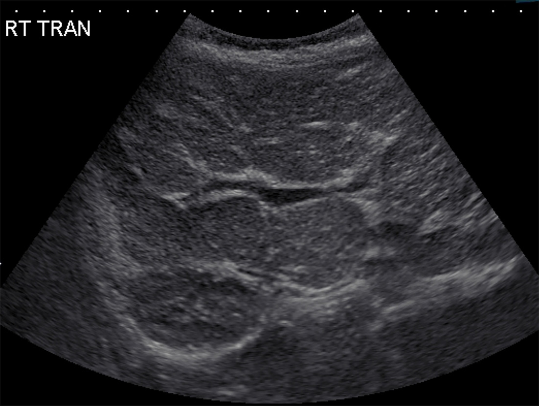
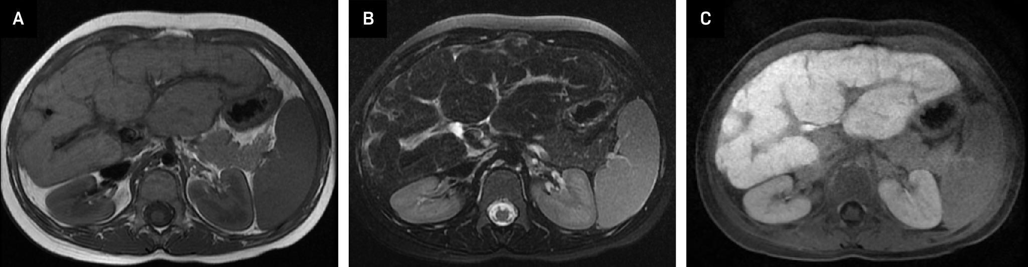
US (Figure 1) and MRI (Figure 2) showed the liver to be small, with a nodular contour and lobular band of apparent fibrosis. There were findings of portal hypertension with an enlarged main portal vein and splenomegaly.
Diagnosis
Hepatic cirrhosis caused by biliary atresia
Discussion
Hepatic cirrhosis is a progressive liver disease caused by chronic hepatocellular injury that leads to the replacement of normal parenchyma with collagenous scar tissue. The prevalence and causes of pediatric cirrhosis vary worldwide. In the United States, biliary atresia is the leading cause and the most common indication for pediatric liver transplantation. Less common causes include genetic metabolic disorders such as alpha-1 antitrypsin deficiency and Wilson disease. Chronic viral hepatitis, now rare in the United States due to routine vaccination, remains a leading cause of pediatric cirrhosis in Africa and Southeast Asia, where the prevalence of hepatitis B seropositivity among young children can reach 4.7%.1,2
Patients with early, compensated cirrhosis may be asymptomatic or present with nonspecific symptoms such as loss of appetite, nausea, or vomiting. Symptoms typically develop as liver fibrosis increases and blood flow through the cirrhotic tissue becomes impaired. In the later, decompensated stage, patients may show extrahepatic manifestations of liver dysfunction, including jaundice, ascites, hepatosplenomegaly, and variceal bleeding secondary to portal hypertension.2 Although biopsy remains the gold standard for diagnosis and staging, cirrhosis can also be identified through a combination of liver function testing and US imaging, with reported diagnostic accuracy of approximately 86%.3 Imaging findings such as surface nodularity, hepatic atrophy, altered parenchymal elasticity, and signs of portal hypertension are similar in both children and adults.4
Abdominal US is the preferred initial noninvasive imaging method for assessing cirrhosis in children and adolescents. Findings may include surface nodularity and heterogeneous parenchymal echotexture.2 In cases of neonatal hepatitis, US is used to evaluate for biliary atresia. Potential findings include a hyperechoic band of tissue anterior to the portal vein (the triangular cord sign) and an atretic gallbladder.4 As hepatic cirrhosis progresses, portal hypertension may develop, leading to secondary findings such as splenomegaly, ascites, varices, and portosystemic shunts.4 MRI and CT are typically used as second-line modalities to further characterize parenchymal changes and hepatic nodules, and to evaluate abdominal vasculature.5 Early cirrhotic changes may include altered hepatic volume distribution, with decreased right lobe volume and compensatory hypertrophy of the left and caudate lobes.4 In advanced disease, parenchymal atrophy results in overall reduction in liver size, widening of the hepatic fissures, and enlargement of the gallbladder fossa.
Metabolic dysfunction-associated fatty liver disease is the most common cause of liver disease in children. In this condition, excess fat accumulates within hepatocytes, resulting in hepatic steatosis. In some patients, inflammation and fibrosis develop, leading to metabolic dysfunction-associated steatohepatitis (MASH). A small subset of patients with MASH will progress to cirrhosis. US shear-wave elastography and MR elastography (MRE) measure liver stiffness and serve as noninvasive surrogate markers for fibrosis. These techniques are increasingly used instead of liver biopsy for diagnosis, staging, and monitoring of pediatric liver disease. While US offers the advantages of portability and ease of use, current evidence suggests that MRE provides greater accuracy for assessing fibrotic changes.6,7 In children, MRE stiffness thresholds generally align with adult criteria, with values above 2.8 kPa considered abnormal and values above 4.0 kPa typically associated with advanced fibrosis.8-10
Management of pediatric cirrhosis focuses on treating the underlying cause to prevent further liver damage, minimizing complications, and managing symptoms.2 Prognosis varies depending on etiology, disease severity, associated complications, and comorbid conditions. Patients who develop complications such as variceal hemorrhage or ascites generally have a poorer prognosis. Ascites are managed with sodium restriction and diuretics, with paracentesis or shunting reserved for refractory cases. Regular endoscopic surveillance and prophylactic band ligation are recommended for varices, particularly when high-risk features are present.2 In children with decompensated chronic liver disease or severe complications unresponsive to medical therapy, liver transplantation is the treatment of choice.2
Conclusion
Hepatic cirrhosis is a progressive disease that results from chronic liver injury and leads to fibrosis, architectural distortion, and ultimately liver failure. In children, biliary atresia is the leading cause, while metabolic and infectious etiologies are less common. Imaging plays a central role in diagnosis and follow-up. Key features include surface nodularity, heterogeneous parenchymal echotexture, altered hepatic volume distribution, and secondary signs of portal hypertension. Elastography provides a noninvasive method for assessing liver stiffness, with MRE offering the highest accuracy for detecting and staging fibrosis.
References
- Olave M, Gurung A, Mistry P. Etiology of cirrhosis in the young. Hum Pathol. 2020;96:96-103. doi:10.1016/j.humpath.2019.09.015.
- Forna L, Bozomitu L, Lupu V. Pediatric perspectives on liver cirrhosis: unravelling clinical patterns and therapeutic challenges. J Clin Med. 2024;13(14). doi:10.3390/jcm13144275.
- Luo H, Peng S, Ouyang W. Assessment of liver fibrosis by transient elastography and multi-parameters model in young children with chronic hepatitis B virus infection. BMC Infect Dis. 2022;22(1). doi:10.1186/s12879-022-07142-7.
- Herrmann J, Petit P, Grabhorn E. Liver cirrhosis in children - the role of imaging in the diagnostic pathway. Pediatr Radiol. 2023;53(4):714-726. doi:10.1007/s00247-022-05480-x.
- Hetland L, Kronborg T, Thing M. Suboptimal diagnostic accuracy of ultrasound and CT for compensated cirrhosis: evidence from prospective cohort studies. Hepatol Commun. 2023;7(9). doi:10.1097/HC9.0000000000000231.
- Catucci D, Hrycyk J, Lange N. Liver segmental volumes and their relationship with 5-year prognostication. Abdom Radiol. September 10, 2024;50(3):1133-1142. doi:10.1007/s00261-024-04552-w.
- Ito K, Mitchell D, Kim M. Right posterior hepatic notch sign: a simple diagnostic MR finding of cirrhosis. J Magn Reson Imaging. 2003;18(5):561-566. doi:10.1002/jmri.10387.
- Ludwig D, Fraum T, Ballard D, Narra V, Shetty A. Imaging biomarkers of hepatic fibrosis: reliability and accuracy of hepatic periportal space widening and other morphologic features on MRI. AJR Am J Roentgenol. 2021;216(5):1229-1239. doi:10.2214/AJR.20.23099.
- Ito K, Mitchell D, Gabata T. Enlargement of hilar periportal space: a sign of early cirrhosis at MR imaging. J Magn Reson Imaging. 2000;11(2):136-140. doi:10.1002/(sici)1522-2586(200002)11:2<136::aid-jmri9>3.0.co;2-b.
- Serai S, Franchi-Abella S, Syed A. MR and ultrasound elastography for fibrosis assessment in children: practical implementation and supporting evidence-AJR expert panel narrative review. AJR Am J Roentgenol. January 3, 2024;223(5). doi:10.2214/AJR.23.30506.
Disclosures
The authors have no conflicts of interest to disclose. None of the authors received outside funding for the production of this original manuscript and no part of this article has been previously published elsewhere.
Citation
. Hepatic Cirrhosis. Journal of Pediatric Case Reports. 2026;1(7). doi:10.37549/JPCR-26-0119.