Hepatic Hydatid Disease
Applied Radiology — Vol. 55 , Issue 2
Published: January 29, 2026
1 University of New Mexico, Albuquerque, New Mexico
* Corresponding author: William Thompson (WThompson@salud.unm.edu)
Categories
Case Summary
A previously healthy, middle-aged patient presented to the emergency department for chronic, intermittent worsening abdominal pain in the right upper quadrant with associated intermittent nausea, vomiting, diarrhea, and fatigue, without subjective fever. US and CT of the abdomen showed cystic structures within the liver. The patient was prescribed a 2-week course of oral doxycycline and discharged with a plan for outpatient follow-up. The patient returned to the emergency department on day 15 of the antibiotic course, reporting initial improvement but the return of symptoms the day after finishing the antibiotic course. At this time, it was noted that the patient worked as a sheep herder, butcher, and wool weaver. The patient also had a long history of work-related travel, including visits to international slaughterhouses. A CT of the abdomen again showed cystic disease within the liver. A complete blood count with differential showed eosinophilia of 2.0. The patient was started on albendazole (ABZ) and admitted for pain control. Serum Echinococcus antibody titer was drawn and later showed elevated titer of 6.14. The patient was discharged with an outpatient appointment for surgical evaluation. Later, the patient received outpatient surgery for resection of hepatic segments 2 and 3, along with elective cholecystectomy. A calcified segment 7 lesion was determined to be inactive without the need for surgical intervention.
Imaging Findings
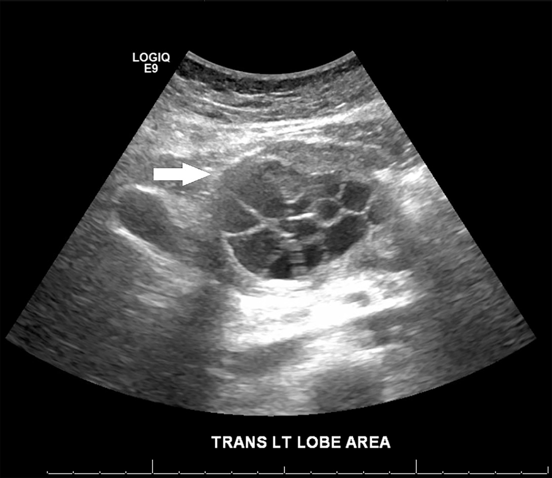
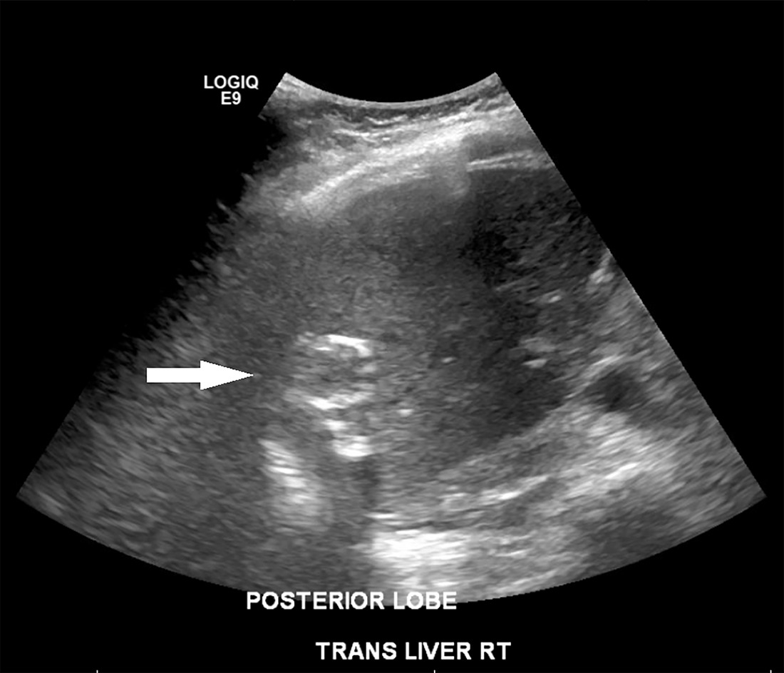
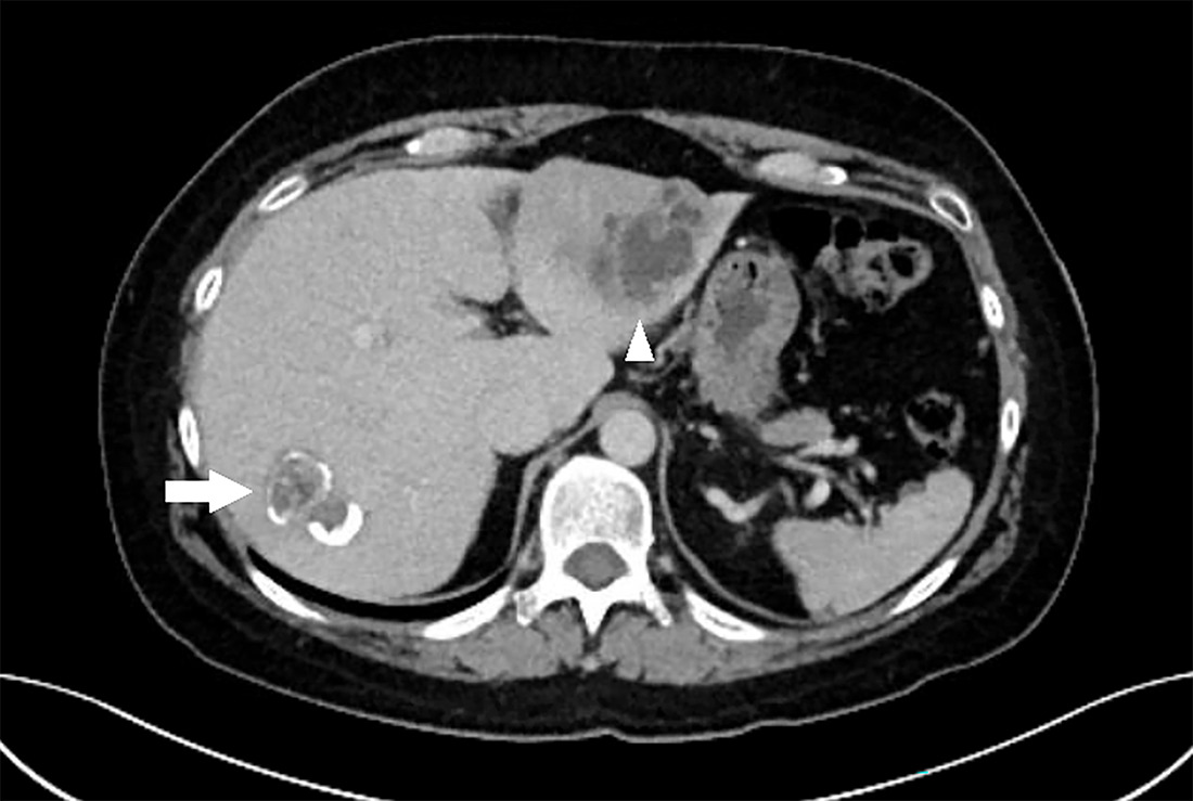
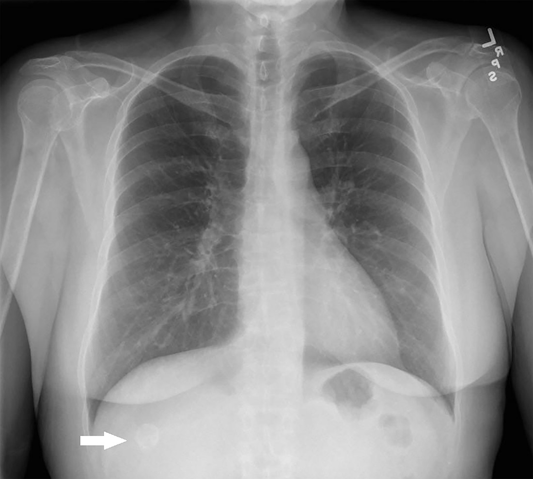
Abdominal US of the right upper quadrant showed a multiloculated cystic lesion in the left lobe of the liver (Figure 1, arrow). US of the posterior right liver showed multiple circular hyperechoic shadowing calcified structures (Figure 2, arrow). Axial contrast-enhanced abdominal CT through the liver showed a multiloculated cystic lesion in the left lobe of the liver (Figure 3, arrowhead), as well as a calcified cystic lesion in the posterior right liver (Figure 4, arrow).
Diagnosis
Hepatic hydatid disease.
Aside from hydatid disease, the main differential considerations for multifocal cystic hepatic lesions are hepatic abscesses from other etiologies or neoplasms.
Discussion
The tapeworm Echinococcus granulosus and its genetic variants are responsible for cystic hydatid disease. Humans are considered accidental intermediate hosts after they acquire infection similarly to other intermediate hosts through contaminated food and water. 1 The majority of human infection involves a dog-sheep-dog cycle, but other domestic animals may also be involved. 1 Most human cases of infection in the United States are diagnosed in immigrants from countries where cystic echinococcosis is endemic, who acquired infection in their country of origin. 2,3 Primary infections in the United States mainly occur in California, Utah, Arizona, and New Mexico. 2 Hospital records indicate an estimated incidence of 1-4 cases per year. 2
Clinical presentation is usually in the 3rd to 5th decade of life, but primary infection usually occurs in childhood. 4 Most cysts are asymptomatic and are detected incidentally on imaging obtained for unrelated reasons or at autopsy. When patients are symptomatic, presentation depends on the sites involved, and symptoms are usually related to expanding mass pressure on adjacent structures or infection from rupture of cyst contents into surrounding areas. 4 Cyst rupture into the hollow abdominal organs is rare, even in endemic areas, and can result in fever and nonspecific symptoms, such as abdominal pain. 5
Imaging findings, along with a concurrent history of potential exposure, can be key to diagnosis. On US, cysts are typically well visualized, with primary cysts appearing as well-defined margins containing fluid, and daughter cysts appearing as multivesicular cystic structures. 4 CT can reveal the cystic structures as well as the location of cysts that are not easily seen with US. 4 Differential diagnosis of cystic hepatic lesions is broad, including benign and malignant etiologies such as simple hepatic cysts, polycystic liver disease, hepatic abscess, primary or metastatic neoplasms, biloma, and hepatic hydatid cyst, among others. 6 Diagnosis with aspiration or biopsy is rarely performed due to the risk of anaphylaxis with cyst rupture. 4 Diagnosis is further supported by serological detection of serum antibodies or detection of specific antigens. 4
Cystic echinococcosis disease is treated using observation, benzimidazoles, drainage, and/or surgery depending on the patient’s symptoms, the stage, size, and location of the cyst, and the risk of complications. 5 Medical treatments are mainly limited to benzimidazole-carbamate compounds, which include ABZ and mebendazole. 7 Prognosis is variable and dependent on each cystic lesion. Follow-up repeat imaging with CT or MRI, or with serologies, should be considered as local recurrence has been reported up to 10 years after apparently successful treatment. 8
Conclusion
Hydatid disease is caused by the tapeworms of the genus Echinococcus, with typical clinical presentation in the 3rd to 5th decade of life, although primary infection usually occurs in childhood. Classic imaging features on US and CT show the characteristic primary cystic structure containing multilocular daughter cysts. Calcification of the cysts can occur with long-standing chronic infection. If the classic daughter cysts are not seen or if there are alternative diagnoses, such as pyogenic abscess or neoplasm, diagnosis can be supported by appropriate history, including exposure to livestock and laboratory analysis showing eosinophilia and positive serum antibody titer.
References
- Echinococcosis. 2021.
- Moro P, Schantz P. Cystic echinococcosis in the Americas. Parasitol Int. 2006;55 Suppl:S181-S186. doi:10.1016/j.parint.2005.11.048.
- Parasites–Echinococcosis biology. 2019.
- Khuroo M. Hydatid disease: current status and recent advances. Ann Saudi Med. 2002;22(1-2):56-64. doi:10.5144/0256-4947.2002.56.
- Stojković M, Weber T, Junghanss T. Clinical management of cystic echinococcosis: state of the art and perspectives. Curr Opin Infect Dis. 2018;31(5):383-392. doi:10.1097/QCO.0000000000000485.
- Knipe H. Cystic lesions of the liver (differential). 2020.
- Siles-Lucas M, Casulli A, Cirilli R, Carmena D. Progress in the pharmacological treatment of human cystic and alveolar echinococcosis: compounds and therapeutic targets. PLOS Negl Trop Dis. 2018;12(4). doi:10.1371/journal.pntd.0006422.
- Rinaldi F, Brunetti E, Neumayr A. Cystic echinococcosis of the liver: a primer for hepatologists. World J Hepatol. 2014;6(5):293-305. doi:10.4254/wjh.v6.i5.293.
Disclosures
The authors have no conflicts of interest to disclose and have not received outside funding for the production of this original manuscript. No part of this article has been previously published elsewhere.
Citation
. Hepatic Hydatid Disease. Applied Radiology. 2026;55(2). doi:10.37549/AR-D-25-0110.