Imaging of intracranial hemorrhage: Subarachnoid hemorrhage and its sequelae
Applied Radiology — Vol. 44 , Issue 11 , pp. 9 -20
DOI: 10.37549/AR2224
Published: November 1, 2015
Categories
Subarachnoid hemorrhage (SAH) is a medical emergency in which radiologists play an important role in diagnosis and characterization to optimize treatment. Incidence varies geographically, with reported rates ranging from 2 to 32 per 100,000 individuals annually.1,2 SAH can inflict considerable morbidity and mortality, and the burden imposed on society is significant given the relatively young age of many affected individuals compared to other neurological pathologies.3,4 Prompt diagnosis is crucial, and knowledge of underlying pathologic processes and potential complications guides the diagnostic workup. This article will review imaging features and relevant clinical characteristics.
Diagnosis and initial imaging
Patients with SAH present with severe headaches. While most patients with a headache will not have SAH, computed tomography (CT) is typically used to exclude SAH in the setting of severe headache.1-3 Headaches from SAH are classically described as the most severe of one’s life, but acute onset within seconds is a more specific feature.3 CT is widely available, has short acquisition times, and is very accurate for diagnosis of SAH.1 CT correctly demonstrates hyperdense material within the subarachnoid space in the setting of acute SAH 95% of the time (Figure 1).2 As cerebrospinal fluid (CSF) is resorbed by arachnoid granulations, blood contents are also resorbed, causing dilution of the SAH and resultant diminution of the density seen on CT and reduced sensitivity in the subacute period.2 Given the potential for false negative CTs, lumbar puncture must be utilized to exclude occult hemorrhage after a negative CT.1,2
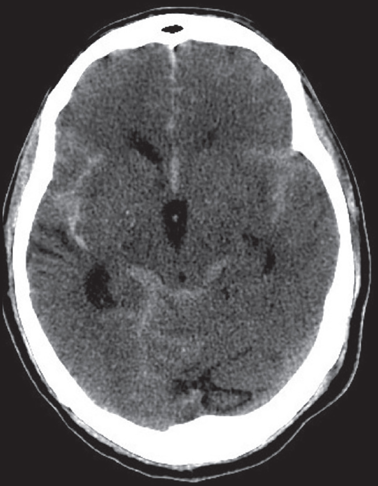
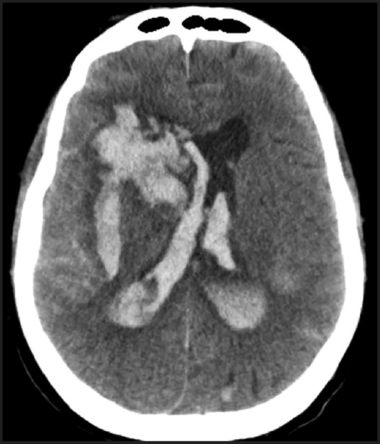
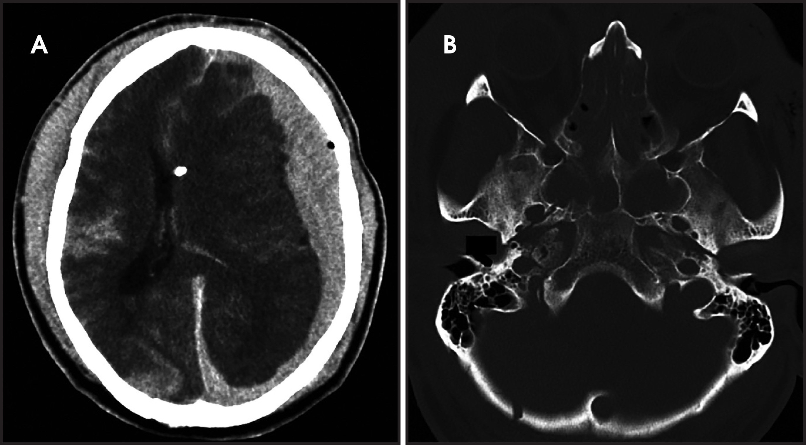
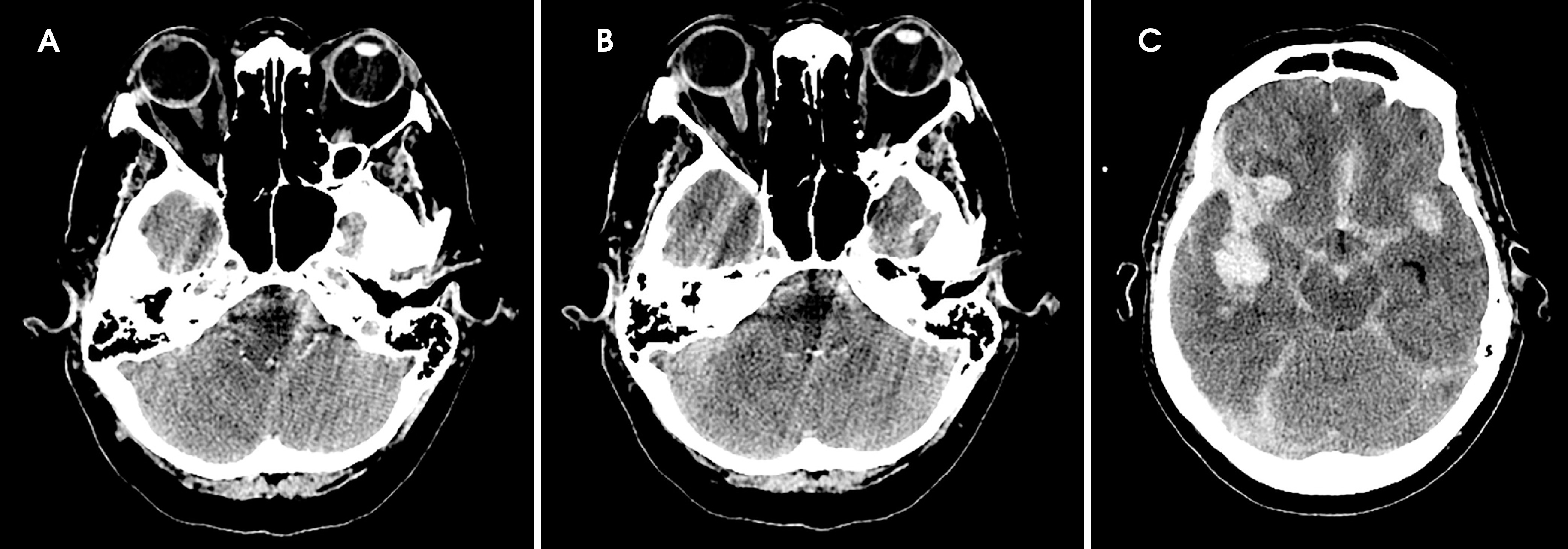
Computed tomography at diagnosis can also provide useful information to guide treatment and determine prognosis. The Fisher scale is widely used to grade SAH and is based on CT findings.5 Modifications have occurred as SAH thickness and presence of IVH were found to be additive in risk for ischemia.6 The Fisher and modified Fisher scales are summarized in Table 1. Hemorrhage may be present in other intracranial compartments. Intraventricular hemorrhage (IVH) may be found with varying severity; as time passes blood is more likely to be found within the ventricular system due to its contiguity with the subarachnoid space and the mobile nature of CSF (Figure 2).2 However, larger hemorrhage volumes can extend into the ventricles at the time of the initial insult, and outcomes are likely to be poor when IVH is massive (Figure 3).2,7 Such a description is important prognostically because CSF diversion in the setting of massive IVH has demonstrated no benefit, although some centers have reported improved outcomes when used in conjunction with fibrinolytic therapy.2,8,9 Epidural hemorrhage (EDH) and intraparenchymal hemorrhage (IPH) can be seen, with severities varying according to the underlying pathology (Figure 4).2 Subdural hemorrhage (SDH) rarely occurs but can be severe when present (Figure 5).2,10,11 CT can also identify concomitant soft tissue or osseous injuries of the head and neck (Figure 5 ).1 One in 7 patients with SAH will develop intraocular hemorrhage, known as Terson’s syndrome, which can be seen on CT, MR or fundoscopy and is a sign of poor prognosis (Figure 6).12-14
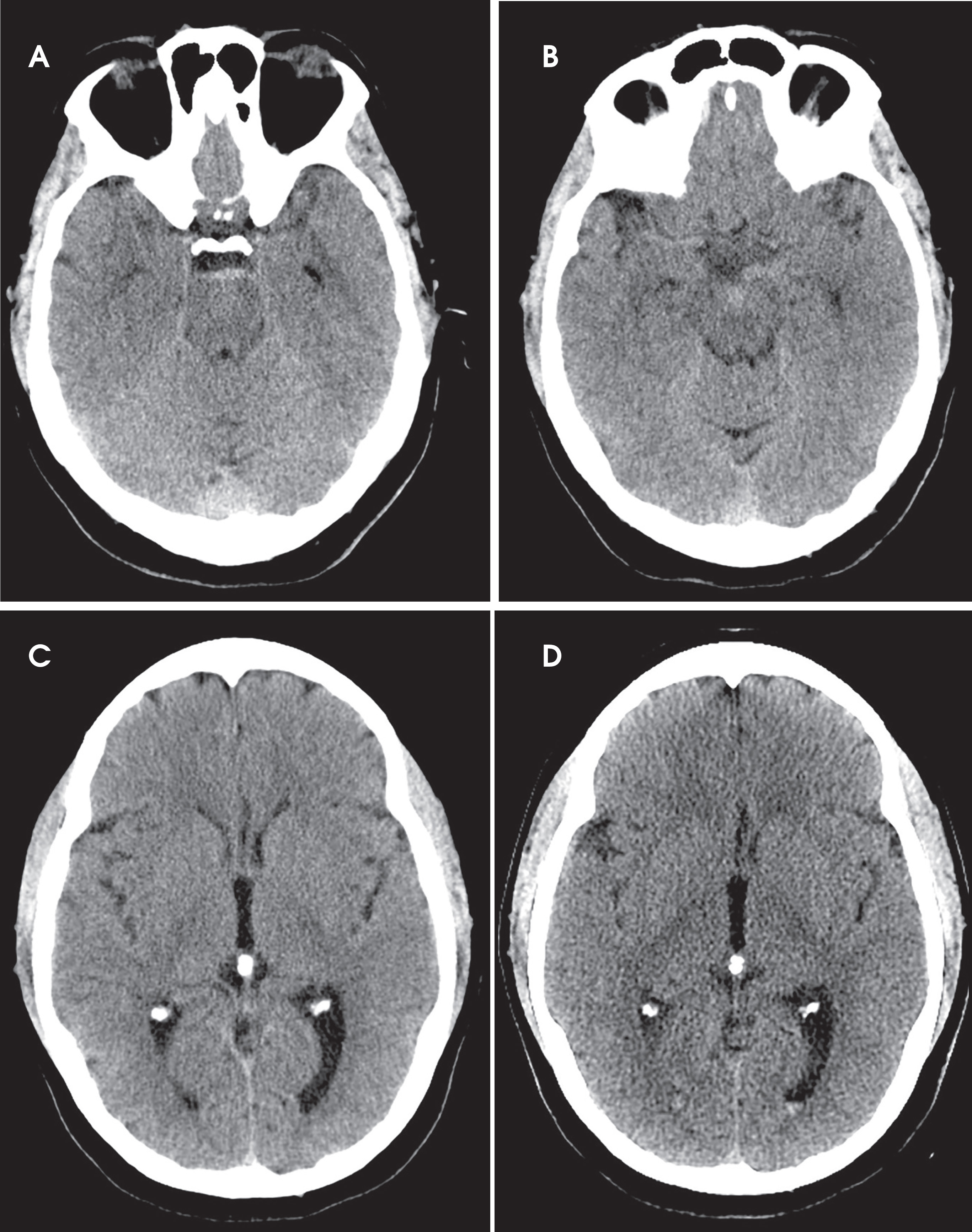
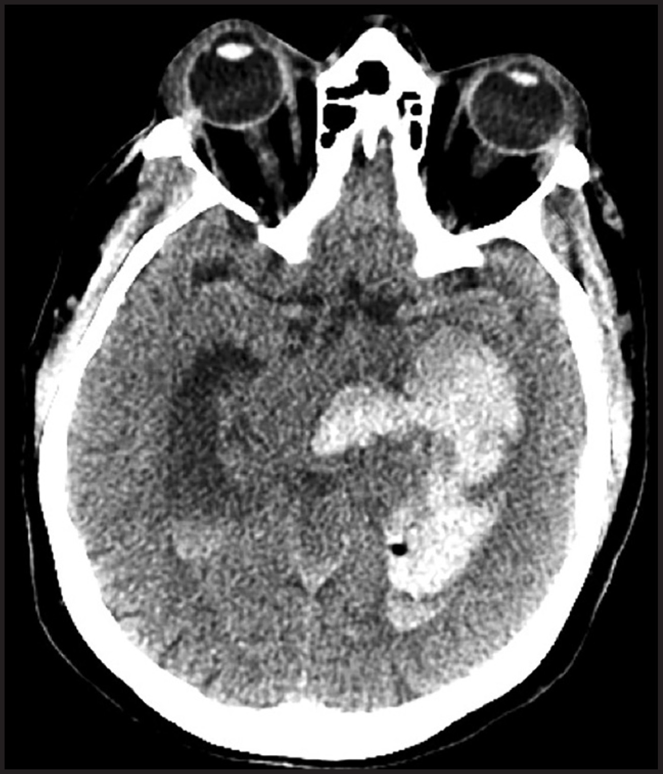
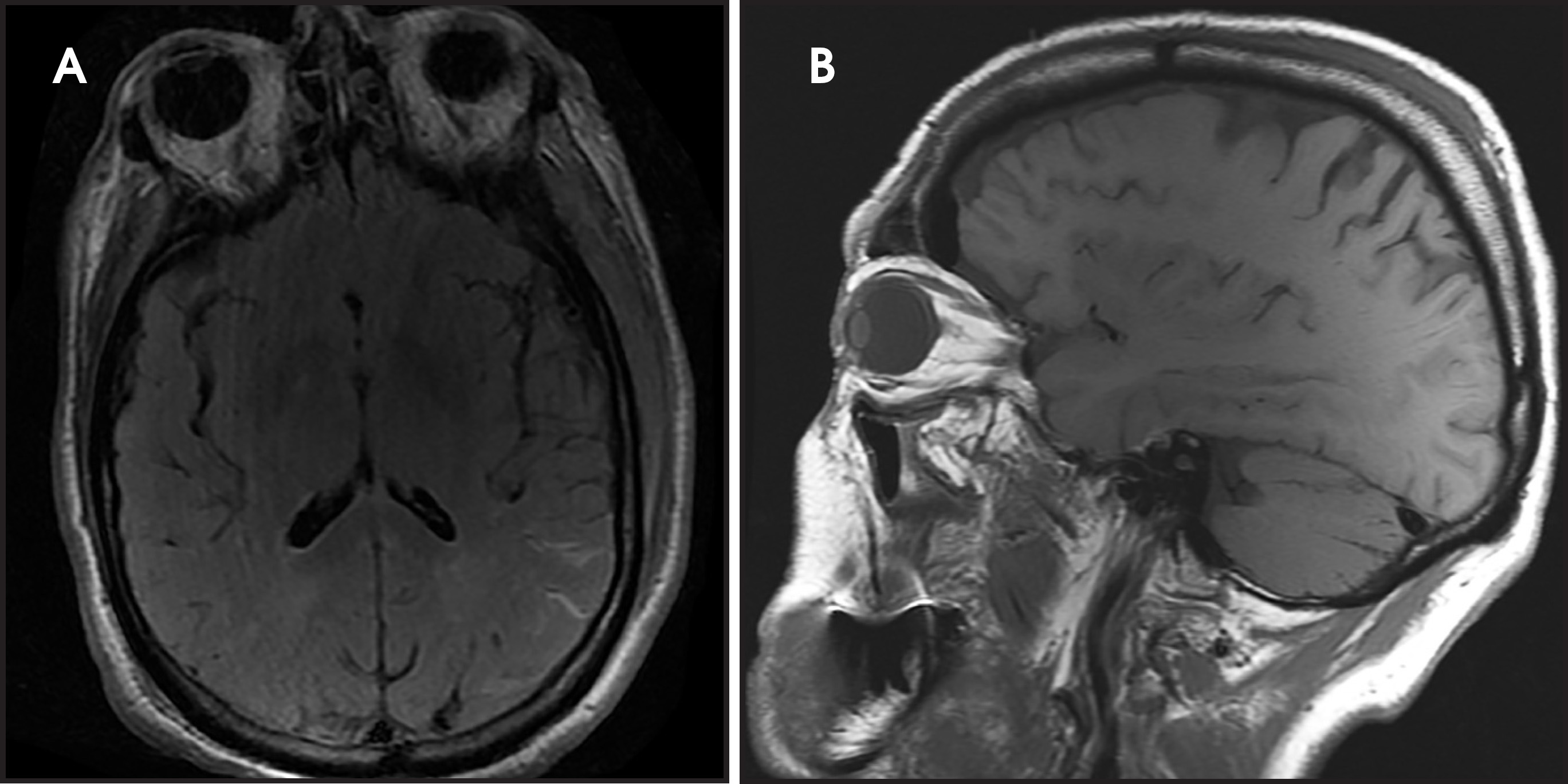
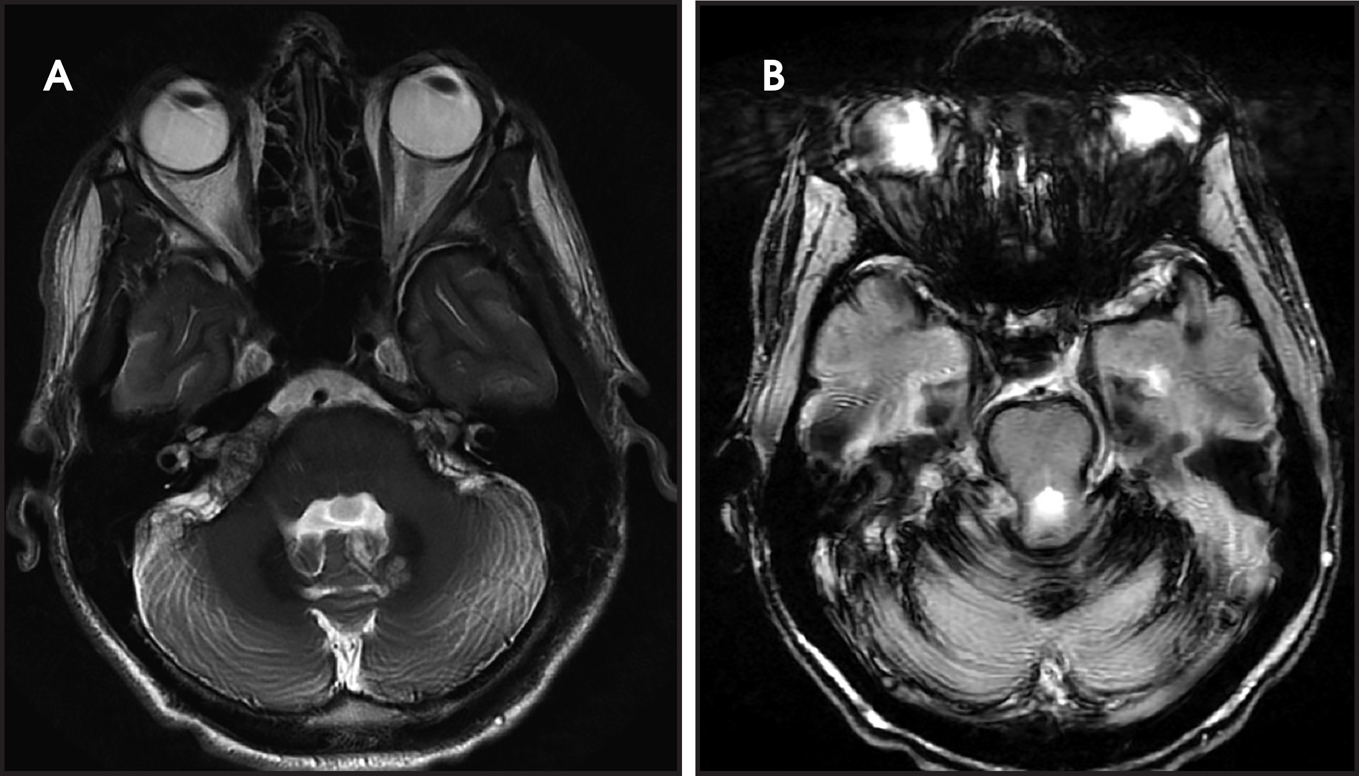
CT is utilized to assess for SAH largely because of its accuracy, efficiency and relative cost effectiveness. However, magnetic resonance (MR) imaging offers comparable accuracy, and familiarity with SAH appearances on these studies is crucial.2,15 MR characteristics of blood all relate to the paramagnetic properties of hemoglobin and the products of its degradation.16-18 As intracranial hemoglobin is degraded, it undergoes a well-described sequence from oxygenated to deoxygenated states and then conversion to methemoglobin, which can be present both intracellularly and extracellularly as cells are lysed.16-18 Imaging characteristics of chronic blood products are due to ferritin and hemosiderin.16-19 Most understanding of the appearance and timeframe for the degradation of intracranial blood is based on intraparenchymal hemorrhage.17 Due to higher levels of oxygen and free water in CSF, as well as protein with which hemorrhage may interact, SAH has unique MR characteristics.16,17 Early SAH is best visualized on fluid attenuation inversion recovery (FLAIR) imaging, on which it appears hyperintense; T1-weighted imaging may also demonstrate hyperintensity at this stage but is frequently less well seen (Figure 7).2,15-17 Over the ensuing days, SAH remains visible on FLAIR, but gradient echo (GRE) imaging becomes the best sequence for visualizing SAH (Figure 8).20,21 Degradation of hemoglobin progresses over a longer time period compared to other intracranial compartments, and resorption of products by arachnoid granulations may occur before methemoglobin or hemosiderin accumulate.17 However, any of the above-described degradation products can be seen, with typical signal characteristics as seen elsewhere in the brain and summarized in Table 2.16,17 Temporal descriptors such as hyperacute, acute, and subacute are accepted based on understanding IPH degradation. Given differences in temporal changes, such descriptors should be avoided in describing SAH to prevent confusion. With repeated SAH, hemosiderin may accumulate on the surface of the brain and cranial nerves, a condition known as superficial siderosis, which appears hypointense on T2 weighted images and GRE (Figure 8).17,22,23
Etiologies of subarachnoid hemorrhage
Numerous processes can cause subarachnoid hemorrhage, but a ruptured aneurysm is the origin in 85% of cases.2 Given the high likelihood of an aneurysm, further investigation is warranted upon the diagnosis of SAH, particularly given the substantial morbidity and mortality associated with them. Ten to thirteen percent of patients with aneurysmal SAH die before reaching the hospital, and overall mortality approaches 50%.3,24-32 Diagnostic cerebral angiography (DSA) has long been considered the gold standard for detection of cerebral aneurysms. (Figure 9) While techniques have been optimized to maximize safety of cerebral DSA, risks still remain.33-37 Additionally, these procedures can require considerable resources and coordination that may prohibit emergent performance in some centers. For these reasons utilization of noninvasive CT or MR angiography has increased, with sensitivities and specificities reported up to 97% and 100%, respectively (Figure 10).38-43 CT angiography is typically preferred to MR angiography due to the time constraints and clinical stability requirements of the latter.25 Diagnostic accuracy declines for aneurysms measuring less than 3mm, so DSA remains the gold standard.5,32,39-46 In addition to diagnosis of an aneurysm, high quality imaging is necessary to plan appropriate treatment, with best characterization occurring with both two- and three-dimensional DSA.2,3,25,47-49 Characteristics important to report include size, ratio of maximal depth to neck width, morphology, direction of aneurysm projection, any arteries arising from the aneurysm, and presence of an apical bleb (Figures 9, 10).50,51
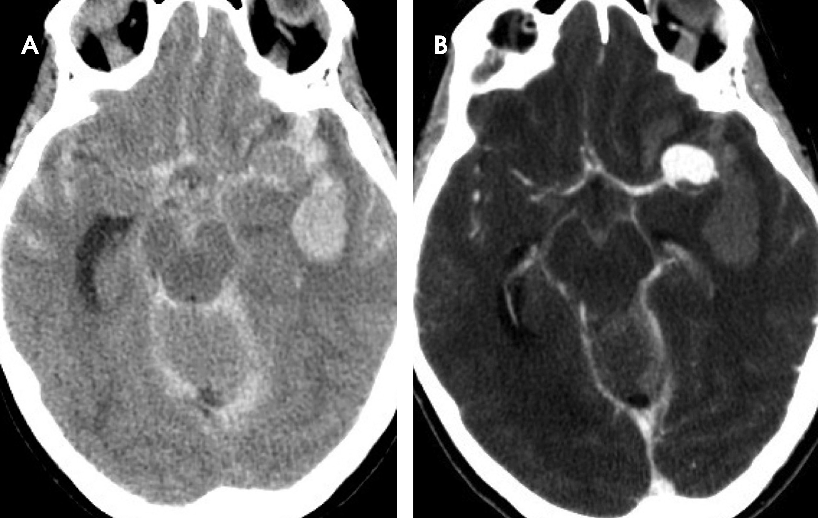
Aneurysms predominantly occur at arterial branching points, with the majority occurring in the anterior circulation. They most commonly arise from the anterior communicating (AComm, 30%), posterior communicating (PComm, 25%), middle cerebral (MCA, 20%), and distal internal carotid arteries (ICA, 7%). Seven percent of aneurysms occur at the distal basilar artery, and 3% arise from the posterior inferior cerebellar artery (PICA).25 Prevalence of cerebral aneurysms in the general population is 2%.52 In those individuals with a diagnosed aneurysm, an additional aneurysm is present in up to 35%.53-59 In the setting of SAH and multiple aneurysms, it is important to identify the aneurysm that has ruptured. Certain characteristics are suggestive of rupture, including length-to-neck ratio greater than 1.6, increased volume to surface area, aneurysm angulation, and presence of an apical bleb.60-63 Hemorrhage itself may aid identification of culprit aneurysms, although such clues are only reliable in the acute setting.64 Lateralized SAH typically indicates MCA, ICA, or PComm aneurysms, with degree of lateralization of SAH corresponding to degree of lateralization of aneurysms (Figure 11).64 Midline SAH occurs with basilar or AComm aneurysms (Figure 9).64 Posterior fossa SAH is associated with PComm and posterior circulation aneurysms, whereas anterior circulation aneurysms typically cause supratentorial SAH.64 When parenchymal hemorrhage occurs, the aneurysm typically points at it, with AComm aneurysms bleeding into the orbitofrontal gyrus or gyrus rectus and MCA aneurysms bleeding into the operculum (Figure 12).64 Aneurysms causing compression symptoms are more likely to rupture, and symptom localization can help identify the offending aneurysm.50 Prompt treatment of the ruptured aneurysm is imperative. 2-4% of aneurysms will rupture again within the first 24 hours, and there is a 1-2% risk of rupture for each day during the first month following initial rupture if the aneurysm is not secured.2,3,25,65
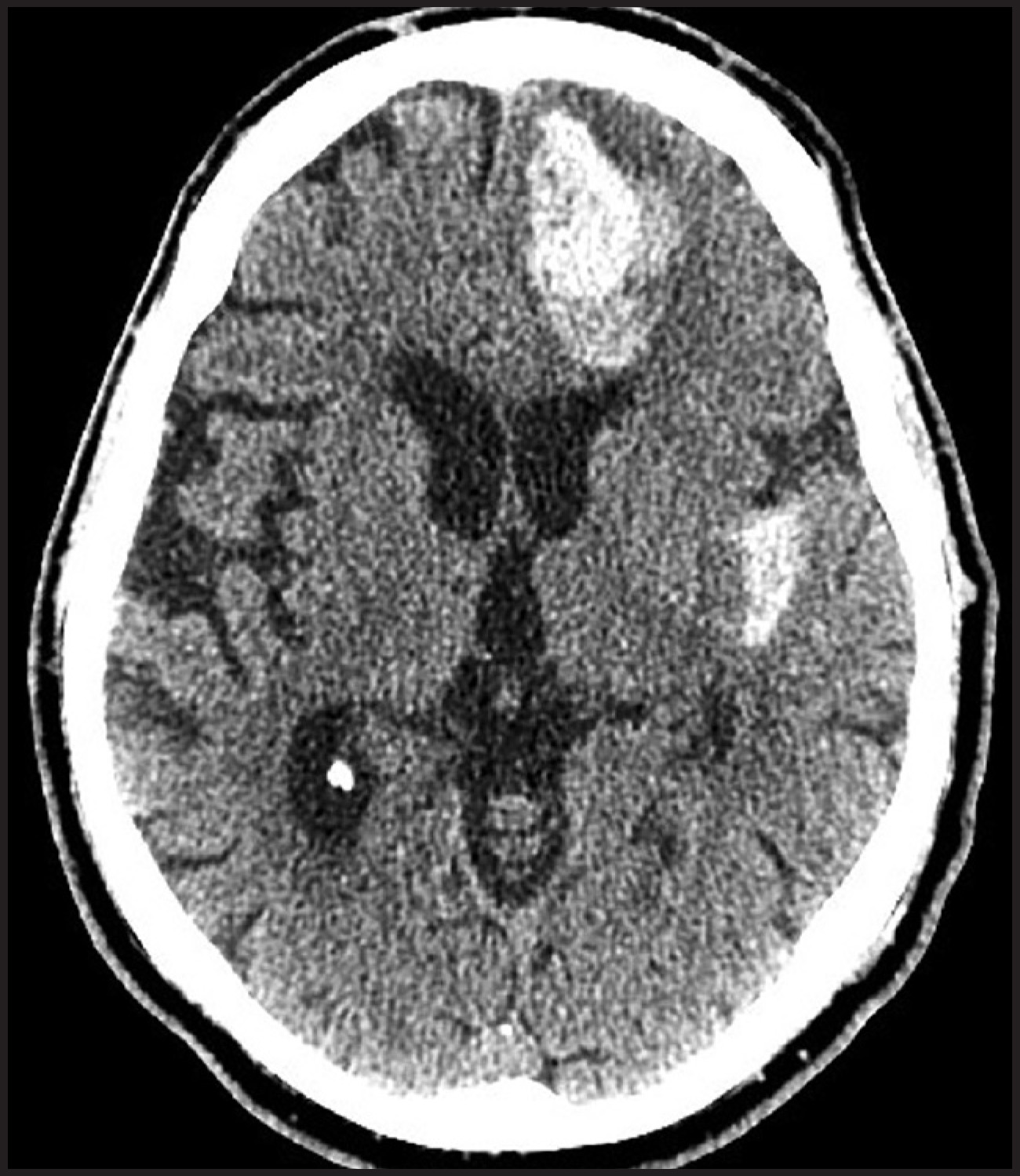
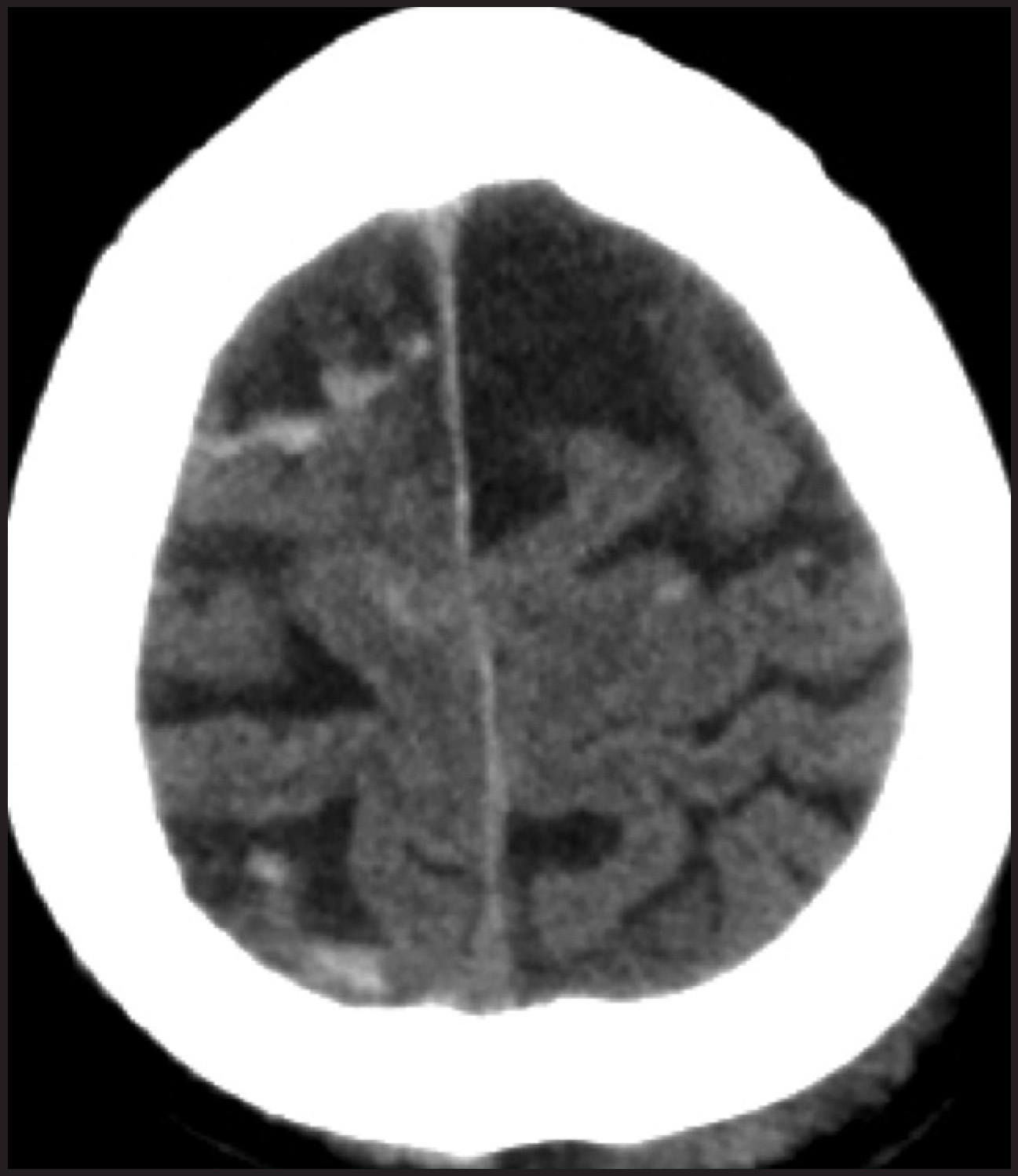
SAH frequently occurs following trauma and can have multiple appearances. Such SAH tends to be more peripheral and localized to the site of injury (Figure 13).64 Hemorrhage often occurs in other intracranial compartments, and important associated soft tissue or osseous injuries can be seen as well (Figure 5).1 Worse outcomes are associated with poor initial clinical state, larger volumes of hemorrhage, EDH, midline shift, or obliteration of basal cisterns.1,66,67 Numerous other pathologic processes can cause SAH, including nonaneurysmal vascular anomalies like arteriovenous malformations and dural arteriovenous fistulae, dissection, inflammatory vasculitides, idiopathic vasculopathy, reversible cerebral vasoconstriction syndrome, coagulopathy, neoplasms, and illicit drugs, among many others.2,64,68
Approximately 10% of SAH cases will yield no clear diagnosis. Within this group is a benign entity known as nonaneurysmal perimesencephalic SAH (NAPSAH).2 This is a diagnosis of exclusion and has well-described characteristics that are important for radiologists to know well.69-71 NAPSAH is believed to result from venous rupture in the region of the mesencephalon.72 Its clinical presentation is distinctively different from most cases of SAH from aneurysm rupture and other etiologies.2 Headaches are less sudden in onset with development over minutes rather than seconds, consciousness is never more than minimally altered, and seizures do not occur with NAPSAH.2,64,69,73-79 This entity demonstrates characteristic appearance on CT with hemorrhage isolated in the cisterns anterior to and near the midbrain, at times located solely in the quadrigeminal plate cistern (Figure 2).2,69,77,79-81 Trace hemorrhage layering dependently in the ventricles is allowable for this diagnosis, but frank IVH excludes NAPSAH.2,26,69,77,79 All patients with suspected NAPSAH must undergo evaluation with DSA since small aneurysms or other etiologies not visible on noninvasive angiography may be the source of SAH.2 2-5% of patients with a perimesencephalic SAH pattern on CT will subsequently be diagnosed with an aneurysm on DSA.2,69-71 Thrombosed aneurysms or very small aneurysms can elude detection on DSA, so repeat DSA has historically been recommended several weeks after an initial study.2,82-84 Some have questioned the utility of repeat DSA, although no studies have been published demonstrating the safety of foregoing a repeat study.82-85 NAPSAH does not carry risk of repeat hemorrhage or ischemia, so patients given this diagnosis do not require further surveillance beyond the time frame for potential hydrocephalus.79,86 As such, it is important to strictly follow requirements for this diagnosis of exclusion to avoid false negatives and unwarranted cessation of surveillance following SAH.
Complications
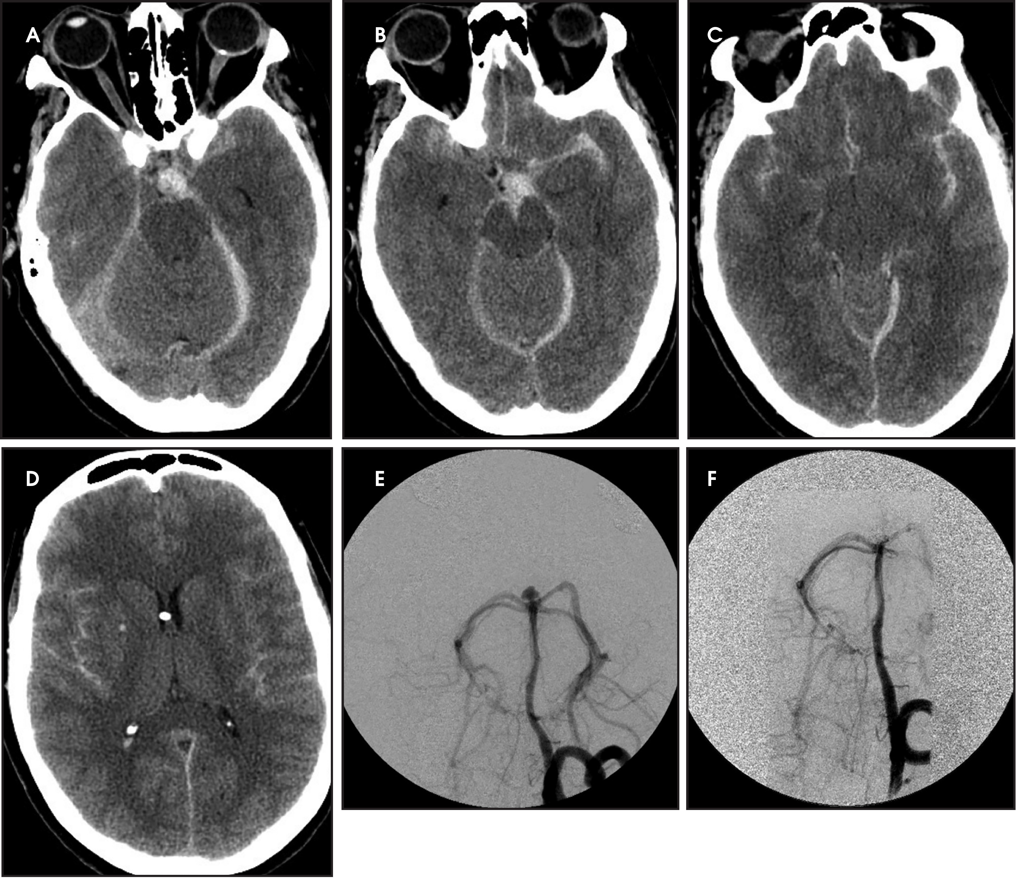
Morbidity from SAH can arise from several complications that can occur at the time of ictus or in the ensuing days and weeks. The most pressing complication can be mass effect from hemorrhage. Increased intracranial pressure from any source causes distinctive herniation syndromes.87,88 Subfalcine herniation involves displacement of the cingulate gyrus under the falx, with midline shift and medial displacement of a compressed ipsilateral ventricle (Figure 14).87 Descending transtentorial herniation involves medial displacement of the temporal lobe into the incisura and effacement and eventual obliteration of the basal cisterns, usually starting with the suprasellar cistern.87 Ascending transtentorial herniation occurs with increased pressure in the posterior fossa, and herniation of the cerebellum effaces the quadrigeminal plate cistern.87 Finally, in tonsillar herniation the cisterna magna is obliterated by cerebellar tonsils descending into the foramen magnum.87 Mass effect is more common in cases of traumatic SAH with additional intracranial hemorrhage, although herniation may be present with isolated SAH and may be clinically unapparent.1,89,90 Herniation is typically a surgical emergency as ischemic damage occurs from both physical compression of parenchyma and from reduced perfusion due to arterial compression.88 Additionally, parenchymal hemorrhage in the brainstem can occur in the setting of herniation, a process termed Duret hemorrhage that can be seen on CT and MR and is associated with poor outcomes (Figure 15).91
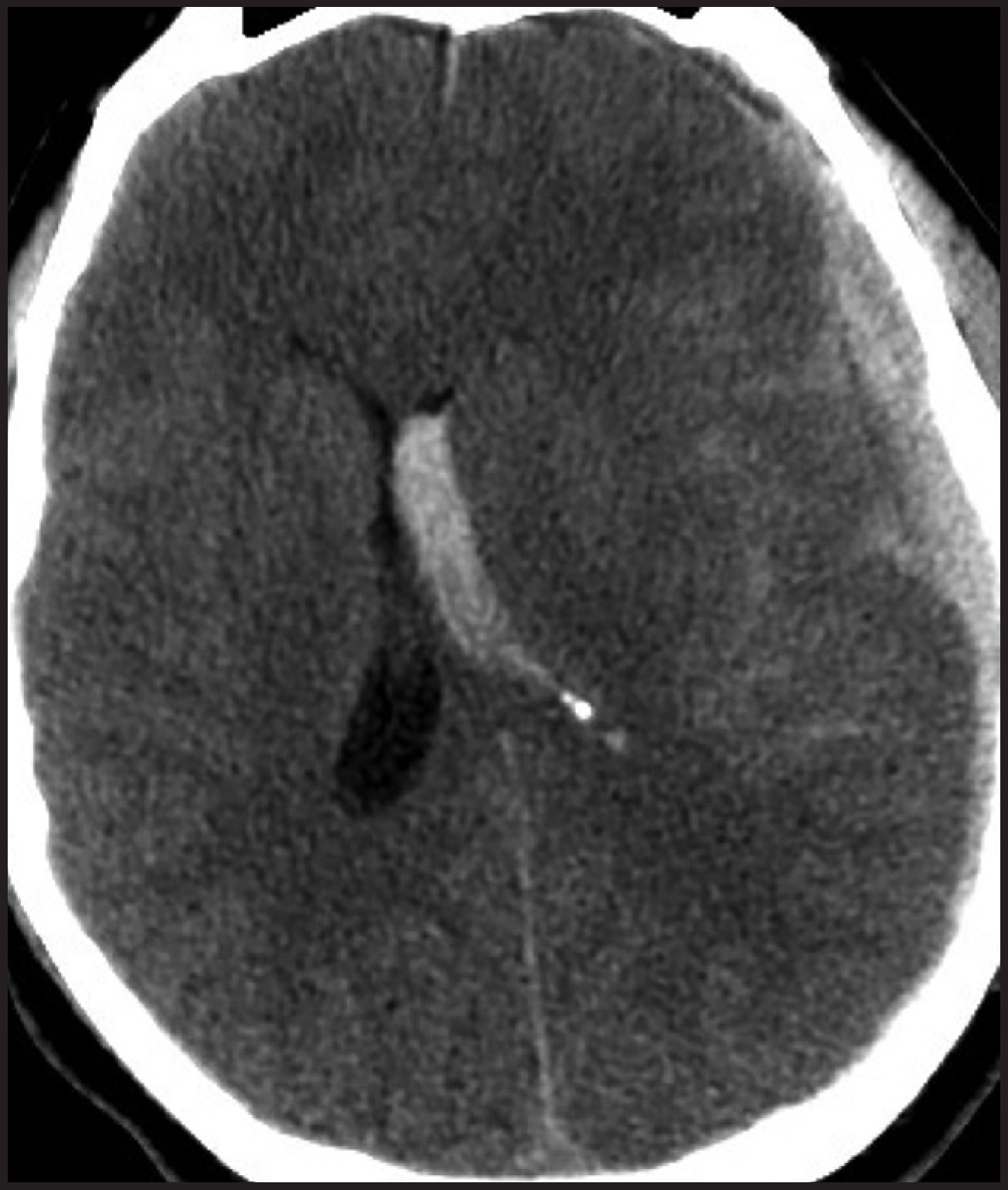
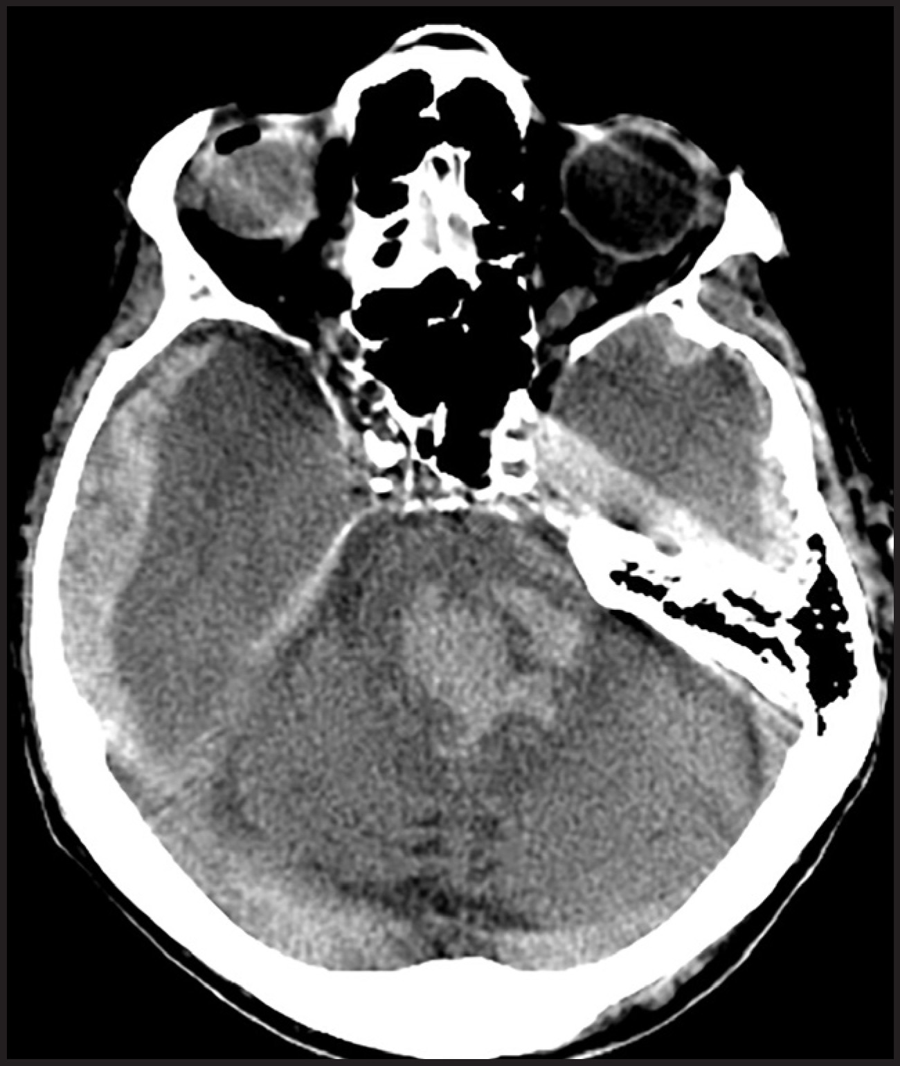
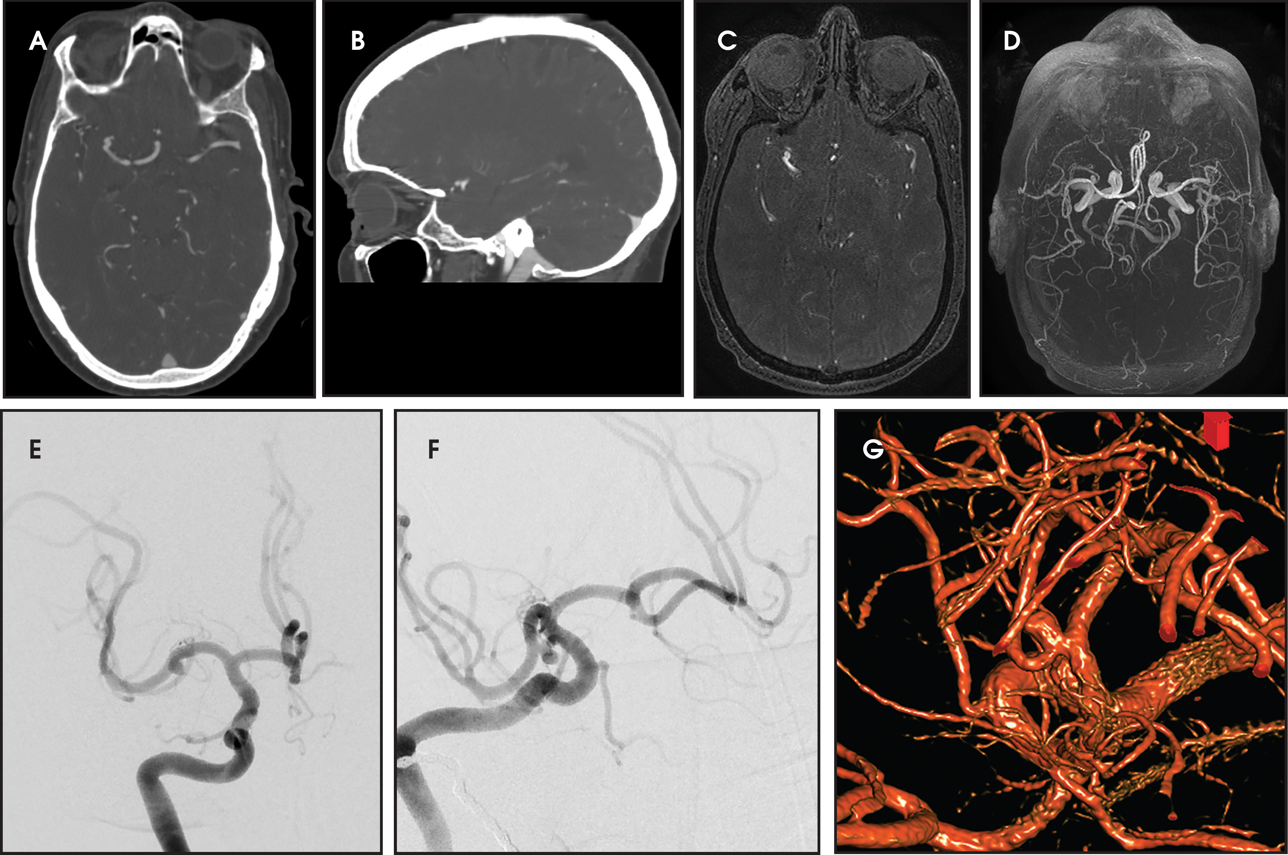
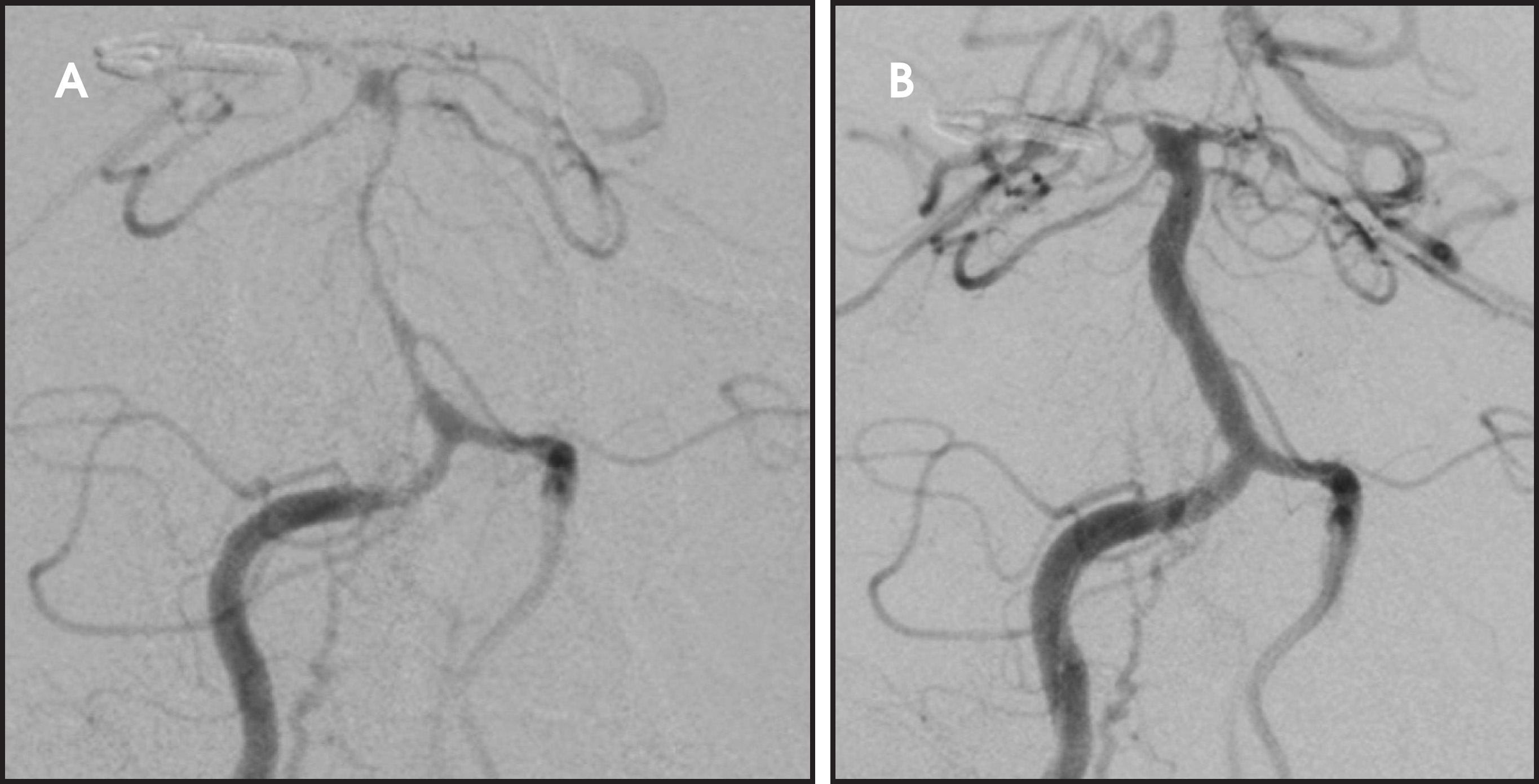
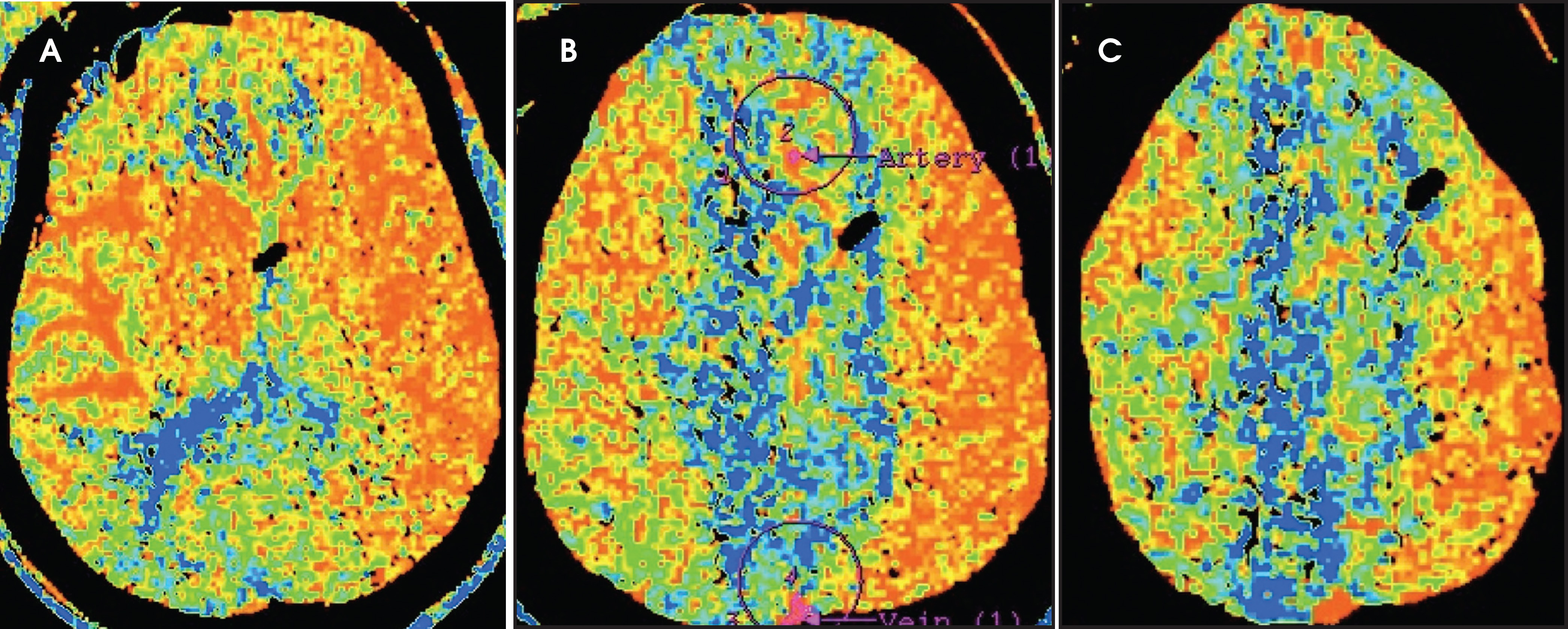
Ischemia can also occur following SAH in the absence of mass effect.2 This often occurs in the setting of vasospasm, although this is neither necessary nor sufficient for development of ischemia.2,92 Vasospasm occurs for unclear reasons on days 3 through 12 after SAH with risk peaking on day 7.25,65,92,93 Both vasospasm and infarct are more likely to occur with diffusely distributed SAH.6,94-96 If SAH has an arterial source, larger volumes of blood predict subsequent ischemia, as does loss of consciousness at the time of ictus.2,92,97,98 These factors can prepare clinicians to have appropriate levels of clinical suspicion in addition to providing recommended prophylactic treatment with nimodipine and maintenance of euvolemia.99 Measures to prevent vasospasm are important because no clearly superior means of screening have been identified, and treatment can be difficult.3 Neurological deficits progress slowly and typically refer to multiple arterial territories.92 Transcranial Doppler is employed for vasospasm screening at many centers, but investigational results have been mixed, and no randomized trials have been conducted.2,93,99,100 CT angiography is sensitive and specific for severe vasospasm in proximal arteries, but diagnostic accuracy plummets for mild to moderate vasospasm and distal involvement.45,101 DSA is the best modality for diagnosing vasospasm, and catheter-directed intra-arterial administration of calcium channel blockers and angioplasty can be performed for vasospasm refractory to noninvasive treatments (Figure 16).45,99,102 Treatment approaches vary between centers, but angioplasty is reserved for vasospasm in proximal arteries.102 Some treatment algorithms call for angioplasty only after failure of intra-arterial calcium channel blocker infusion, while others primarily treat proximal vasospasm with angioplasty primarily.102 Regardless of the presence of vasospasm, evaluation for ischemia and infarct can be performed with CT or MR perfusion studies or diffusion weighted MR imaging.99,101,103 More specifically, CT perfusion studies have demonstrated excellent value of mean transit time in the prediction of vasospasm on DSA and promise from blood-brain barrier permeability imaging as a physiologic biomarker that may guide treatments in the future (Figure 17).104,105
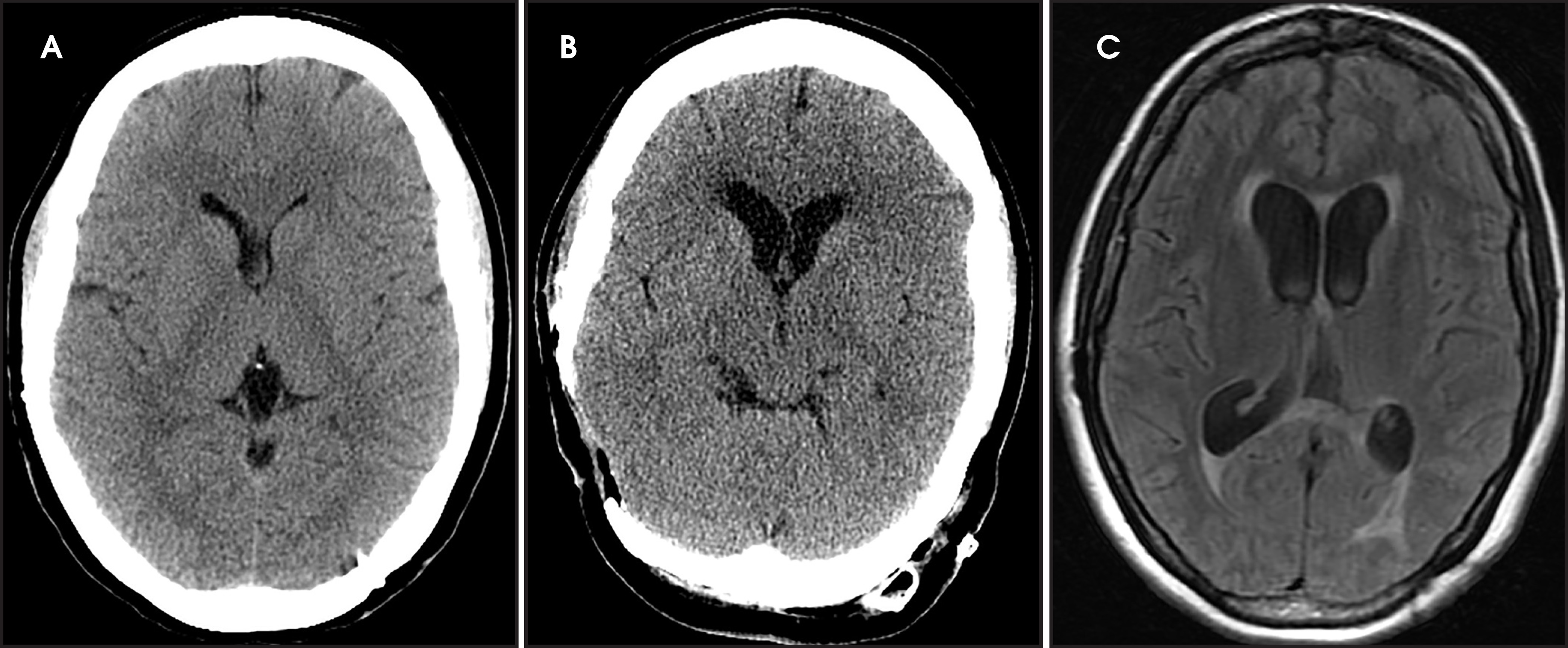
Hydrocephalus can occur with SAH of any etiology; it presents in up to 45% of SAH patients.25,65,86,106-110 It can be either acute or chronic and must be diverted when symptomatic.3 Symptoms are often subtle in onset with gradual progression, most commonly manifesting as a depressed level of consciousness.77 Hydrocephalus is more likely to occur in older patients, with diffuse distribution of SAH, when SAH measures more than 5 mm thick, and when IVH is present.106,111,112 Ventricular size on CT and MR is variable between individuals and has poor accuracy for diagnosis of hydrocephalus, although changes in size between different studies on the same patient correlate with level of consciousness (Figure 18).111,112 Periventricular edema consistent with transependymal flow is a marker of hydrocephalus, and this is better seen on MR than CT (Figure 18).113
Conclusion
SAH can occur from a variety of etiologies and result in a wide range of outcomes. Radiologists play a key role in identifying the source of SAH and providing information for planning the most appropriate treatment, SAH features with implications for prognosis and complications of the hemorrhage.
References
- Maas A, Steyerberg E, Butcher I. Prognostic value of computerized tomography scan characteristics in traumatic brain injury: results from the IMPACT study.. J Neurotrauma.. 2007;24:303-314.
- van Gijn J, Kerr R, Rinkel G. Subarachnoid haemorrhage. Lancet.. 2007;369:306-318.
- Bederson J, Connolly E, Batjer H. Guidelines for the management of aneurysmal subarachnoid hemorrhage: a statement for healthcare professionals from a special writing group of the Stroke Council, American Heart Association.. Stroke.. 2009;40:994-1025.
- Johnston S, Selvin S, Gress D. The burden, trends, and demographics of mortality from subarachnoid hemorrhage.. Neurology.. 1998;50:1413-1418.
- Fisher C, Kistler J, Davis J. Relation of cerebral vasospasm to subarachnoid hemorrhage visualized by computerized tomographic scanning.. Neurosurgery.. 1980;6:1-9.
- Claassen J, Bernardini G, Kreiter K. Effect of cisternal and ventricular blood on risk of delayed cerebral ischemia after subarachnoid hemorrhage: the Fisher scale revisited.. Stroke.. 2001;32:2020-2012.
- Roos Y, Hasan D, Vermeulen M. Outcome in patients with large intraventricular haemorrhages: a volumetric study.. J Neurol Neurosurg Psychiatry.. 1995;58:622-624.
- Nieuwkamp D, de Gans K, Rinkel G, Algra A. Treatment and outcome of severe intraventricular extension in patients with subarachnoid or intracerebral hemorrhage: a systematic review of the literature.. J Neurol.. 2000;247:117-121.
- Naff N, Carhuapoma J, Williams M. Treatment of intraventricular hemorrhage with urokinase : effects on 30-Day survival.. Stroke.. 2000;31:841-847.
- Inamasu J, Saito R, Nakamura Y. Acute subdural hematoma caused by ruptured cerebral aneurysms: diagnostic and therapeutic pitfalls.. Resuscitation.. 2002;52:71-76.
- Sasaki T, Sato M, Oinuma M. Management of poor-grade patients with aneurysmal subarachnoid hemorrhage in the acute stage: Importance of close monitoring for neurological grade changes.. Surg Neurol.. 2004;62.
- Hassan A, Lanzino G, Wijdicks E. Terson’s syndrome.. Neurocrit Care.. 2011;15:554-558.
- McCarron M, Alberts M, McCarron P. A systematic review of Terson’s syndrome: frequency and prognosis after subarachnoid haemorrhage.. J Neurol Neurosurg Psychiatry.. 2004;75:491-493.
- Sung W, Arnaldo B, Sergio C. Terson’s syndrome as a prognostic factor for mortality of spontaneous subarachnoid haemorrhage.. Acta Ophthalmol.. 2011;89:544-547.
- Fiebach J, Schellinger P, Gass A. Stroke magnetic resonance imaging is accurate in hyperacute intracerebral hemorrhage: a multicenter study on the validity of stroke imaging.. Stroke.. 2004;35:502-506.
- Bradley W, Schmidt P. Effect of methemoglobin formation on the MR appearance of subarachnoid hemorrhage.. Radiology.. 1985;156:99-103.
- Bradley W. MR appearance of hemorrhage in the brain.. Radiology.. 1993;189:15-26.
- Gomori J, Grossman R. Mechanisms responsible for the MR appearance and evolution of intracranial hemorrhage.. Radiographics.. 1988;8:427-440.
- Thulborn K, Sorensen A, Kowall N. The role of ferritin and hemosiderin in the MR appearance of cerebral hemorrhage: a histopathologic biochemical study in rats.. AJR Am J Roentgenol.. 1990;154:1053-1059.
- Mitchell P, Wilkinson I, Hoggard N. Detection of subarachnoid haemorrhage with magnetic resonance imaging.. J Neurol Neurosurg Psychiatry.. 2001;70:205-211.
- Atlas S, Mark A, Grossman R, Gomori J. Intracranial hemorrhage: gradient-echo MR imaging at 1.5 T. Comparison with spin-echo imaging and clinical applications.. Radiology.. 1988;168:803-807.
- Gomori J, Grossman R, Bilaniuk L. High-field MR imaging of superficial siderosis of the central nervous system.. J Comput Assist Tomogr.. 1985;9:972-975.
- Mamourian A. MR of superficial siderosis.. AJNR Am J Neuroradiol.. 1993;14:1445-1448.
- Wijdicks E, Kallmes D, Manno E. Subarachnoid hemorrhage: neurointensive care and aneurysm repair.. Mayo Clin Proc.. 2005;80:550-559.
- Brisman J, Song J, Newell D. Cerebral aneurysms.. N Engl J Med.. 2006;355:928-939.
- van Gijn J, Rinkel G. Subarachnoid haemorrhage: diagnosis, causes and management.. Brain.. 2001;124:249-278.
- Hijdra A, van Gijn J, Nagelkerke N. Prediction of delayed cerebral ischemia, rebleeding, and outcome after aneurysmal subarachnoid hemorrhage.. Stroke.. 1988;19:1250-1256.
- Hijdra A, Braakman R, van Gijn J. Aneurysmal subarachnoid hemorrhage. Complications and outcome in a hospital population.. Stroke.. 1987;18:1061-1067.
- Hop J, Rinkel G, Algra A, van Gijn J. Changes in functional outcome and quality of life in patients and caregivers after aneurysmal subarachnoid hemorrhage.. J Neurosurg.. 2001;95:957-963.
- Broderick J, Brott T, Duldner J. Initial and recurrent bleeding are the major causes of death following subarachnoid hemorrhage.. Stroke.. 1994;25:1342-1347.
- Sundt T, Kobayashi S, Fode N, Whisnant J. Results and complications of surgical management of 809 intracranial aneurysms in 722 cases. Related and unrelated to grade of patient, type of aneurysm, and timing of surgery.. J Neurosurg.. 1982;56:753-765.
- Wang H, Li W, He H. 320-Detector row CT angiography for detection and evaluation of intracranial aneurysms: Comparison with conventional digital subtraction angiography.. Clin Radiol.. 2013;68:15-20.
- Leonardi M, Cenni P, Simonetti L. Retrospective study of complications arising during cerebral and spinal diagnostic angiography from 1998 to 2003.. Interv Neuroradiol.. 2005;11:213-221.
- Hoh B, Cheung A, Rabinov J. Results of a prospective protocol of computed tomographic angiography in place of catheter angiography as the only diagnostic and pretreatment planning study for cerebral aneurysms by a combined neurovascular team.. Neurosurgery.. 2004;54.
- Heiserman J, Dean B, Hodak J. Neurologic complications of cerebral angiography.. AJNR Am J Neuroradiol.. 1994;15.
- Dion J, Gates P, Fox A. Clinical events following neuroangiography: a prospective study.. Stroke.. 1987;18:997-1004.
- Connors J, Sacks D, Furlan A. Training, competency, and credentialing standards for diagnostic cervicocerebral angiography, carotid stenting, and cerebrovascular intervention: a joint statement from the American Academy of Neurology, the American Association of Neurological Surgeons, the American Society of Interventional and Therapeutic Neuroradiology, the American Society of Neuroradiology, the Congress of Neurological Surgeons, the AANS/CNS Cerebrovascular Section, and the Society of Interventional Radiology.. Neurology.. 2005;64:190-198.
- Bederson J, Awad I, Wiebers D. Recommendations for the management of patients with unruptured intracranial aneurysms: A Statement for healthcare professionals from the Stroke Council of the American Heart Association.. Stroke.. 2000;31:2742-2750.
- Dammert S, Krings T, Moller-Hartmann W. Detection of intracranial aneurysms with multislice CT: comparison with conventional angiography.. Neuroradiology.. 2004;46:427-434.
- Chappell E, Moure F, Good M. Comparison of computed tomographic angiography with digital subtraction angiography in the diagnosis of cerebral aneurysms: a meta-analysis.. Neurosurgery.. 2003;52.
- White P, Teasdale E, Wardlaw J, Easton V. Intracranial aneurysms: CT angiography and MR angiography for detection prospective blinded comparison in a large patient cohort.. Radiology.. 2001;219:739-749.
- Harrison M, Johnson B, Gardner G, Welling B. Preliminary results on the management of unruptured intracranial aneurysms with magnetic resonance angiography and computed tomographic angiography.. Neurosurgery.. 1997;40.
- Okahara M, Kiyosue H, Yamashita M. Diagnostic accuracy of magnetic resonance angiography for cerebral aneurysms in correlation with 3D-digital subtraction angiographic images: a study of 133 aneurysms.. Stroke.. 2002;33:1808-1803.
- Tipper G, Price S. Detection and evaluation of intracranial aneurysms with 16-row multislice CT angiography.. Clin Radiol.. 2005;60:565-572.
- Anderson G, Ashforth R, Steinke D, Findlay J. CT angiography for the detection of cerebral vasospasm in patients with acute subarachnoid hemorrhage.. AJNR Am J Neuroradiol.. 2000;21:1011-1015.
- McKinney A, Palmer C, Truwit C. Detection of aneurysms by 64-section multidetector CT angiography in patients acutely suspected of having an intracranial aneurysm and comparison with digital subtraction and 3D rotational angiography.. AJNR Am J Neuroradiol.. 2008;29:594-602.
- Brinjikji W, Cloft H, Lanzino G, Kallmes D. Comparison of 2D digital subtraction angiography and 3D rotational angiography in the evaluation of dome-to-neck ratio.. AJNR Am J Neuroradiol.. 2009;30:831-834.
- Anxionnat R, Bracard S, Ducrocq X. Intracranial aneurysms: clinical value of 3D digital subtraction angiography in the therapeutic decision and endovascular treatment.. Radiology.. 2001;218:799-808.
- Tanoue S, Kiyosue H, Kenai H. Three-dimensional reconstructed images after rotational angiography in the evaluation of intracranial aneurysms: surgical correlation.. Neurosurgery.. 2000;47:866-871.
- Wiebers D, Whisnant J, Sundt T, O’Fallon W. The significance of unruptured intracranial saccular aneurysms.. J Neurosurg.. 1987;66:23-29.
- Wardlaw J, White P. The detection and management of unruptured intracranial aneurysms.. Brain.. 2000;123:205-221.
- Rinkel G, Djibuti M, Algra A, van Gijn J. Prevalence and risk of rupture of intracranial aneurysms: a systematic review.. Stroke.. 1998;29:251-256.
- Kaminogo M, Yonekura M, Shibata S. Incidence and outcome of multiple intracranial aneurysms in a defined population.. Stroke.. 2003;34:16-21.
- Ostergaard J, Hog E. Incidence of multiple intracranial aneurysms. Influence of arterial hypertension and gender.. J Neurosurg.. 1985;63:49-55.
- Inagawa T. Multiple intracranial aneurysms in elderly patients.. Acta Neurochir (Wien).. 1990;106:119-126.
- Rinne J, Hernesniemi J, Puranen M, Saari T. Multiple intracranial aneurysms in a defined population: prospective angiographic and clinical study.. Neurosurgery.. 1994;35:803-808.
- Qureshi A, Suarez J, Parekh P. Risk factors for multiple intracranial aneurysms.. Neurosurgery.. 1998;43.
- Juvela S. Risk factors for multiple intracranial aneurysms.. Stroke.. 2000;31:392-397.
- Ellamushi H, Grieve J, Jager H, Kitchen N. Risk factors for the formation of multiple intracranial aneurysms.. J Neurosurg.. 2001;94:728-732.
- Ujiie H, Tamano Y, Sasaki K, Hori T. Is the aspect ratio a reliable index for predicting the rupture of a saccular aneurysm?. Neurosurgery.. 2001;48.
- Lauric A, Baharoglu M, Malek A. Size ratio performance in detecting cerebral aneurysm rupture status is insensitive to small vessel removal.. Neurosurgery.. 2013;72:547-554.
- Chien A, Sayre J, Vinuela F. Comparative morphological analysis of the geometry of ruptured and unruptured aneurysms.. Neurosurgery.. 2011;69:349-356.
- Dhar S, Tremmel M, Mocco J. Morphology parameters for intracranial aneurysm rupture risk assessment.. Neurosurgery.. 2008;63.
- Krings T, Geibprasert S. Brugge KGt. Case-based interventional neuroradiology.. 2011.
- Greenberg M. Handbook of neurosurgery.. 2010.
- Compagnone C, d’Avella D, Servadei F. Patients with moderate head injury: a prospective multicenter study of 315 patients.. Neurosurgery.. 2009;64.
- Steyerberg E, Mushkudiani N, Perel P. Predicting outcome after traumatic brain injury: development and international validation of prognostic scores based on admission characteristics.. PLoS Med.. 2008;5.
- Singhal A, Hajj-Ali R, Topcuoglu M. Reversible cerebral vasoconstriction syndromes: Analysis of 139 cases.. Arch Neurol.. 2011;68:1012-1005.
- Rinkel G, Wijdicks E, Vermeulen M. Nonaneurysmal perimesencephalic subarachnoid hemorrhage: CT and MR patterns that differ from aneurysmal rupture.. AJNR Am J Neuroradiol.. 1991;12:829-834.
- Pinto A, Ferro J, Canhao P, Campos J. How often is a perimesencephalic subarachnoid haemorrhage CT pattern caused by ruptured aneurysms?. Acta Neurochir (Wien).. 1993;124:79-81.
- Van Calenbergh F, Plets C, Goffin J, Velghe L. Nonaneurysmal subarachnoid hemorrhage: prevalence of perimesencephalic hemorrhage in a consecutive series.. Surg Neurol.. 1993;39:320-323.
- Ronkainen A, Hernesniemi J, Ryynanen M. Familial subarachnoid hemorrhage in east Finland, 1977-1990.. Neurosurgery.. 1993;33.
- Brilstra E, Rinkel G, Algra A, van Gijn J. Rebleeding, secondary ischemia, and timing of operation in patients with subarachnoid hemorrhage.. Neurology.. 2000;55:1656-1660.
- Linn F, Rinkel G, Algra A, van Gijn J. Headache characteristics in subarachnoid haemorrhage and benign thunderclap headache.. J Neurol Neurosurg Psychiatry.. 1998;65:791-793.
- Pinto A, Canhao P, Ferro J. Seizures at the onset of subarachnoid haemorrhage.. J Neurol.. 1996;243:161-164.
- Butzkueven H, Evans A, Pitman A. Onset seizures independently predict poor outcome after subarachnoid hemorrhage.. Neurology.. 2000;55:1315-1320.
- van Gijn J, Hijdra A, Wijdicks E. Acute hydrocephalus after aneurysmal subarachnoid hemorrhage.. J Neurosurg.. 1985;63:355-362.
- Schwartz T, Farkas J. Quadrigeminal non-aneurysmal subarachnoid hemorrhage–a true variant of perimesencephalic subarachnoid hemorrhage. Case report.. Clin Neurol Neurosurg.. 2003;105:95-98.
- Schwartz T, Solomon R. Perimesencephalic nonaneurysmal subarachnoid hemorrhage: review of the literature.. Neurosurgery.. 1996;39:433-440.
- Zentner J, Solymosi L, Lorenz M. Subarachnoid hemorrhage of unknown etiology.. Neurol Res.. 1996;18:220-226.
- Schievink W, Wijdicks E. Pretruncal subarachnoid hemorrhage: an anatomically correct description of the perimesencephalic subarachnoid hemorrhage.. Stroke.;28.
- Jung J, Kim Y, Lee J. Spontaneous subarachnoid haemorrhage with negative initial angiography: a review of 143 cases.. J Clin Neurosci.. 2006;13:1011-1017.
- Little A, Garrett M, Germain R. Evaluation of patients with spontaneous subarachnoid hemorrhage and negative angiography.. Neurosurgery.. 2007;61.
- Delgado Almandoz J, Jagadeesan B, Refai D. Diagnostic yield of repeat catheter angiography in patients with catheter and computed tomography angiography negative subarachnoid hemorrhage.. Neurosurgery.. 2012;70:1135-1142.
- Alexander M, McTaggart R, Marks M. Initial hemorrhage pattern affects utility of repeat angiography following negative initial angiogram for non-traumatic subarachnoid Hemorrhage.. J NeuroIntervent Surg. 2012;4:A58-A59.
- Rinkel G, Wijdicks E, Vermeulen M. Acute hydrocephalus in nonaneurysmal peri-mesencephalic hemorrhage: evidence of CSF block at the tentorial hiatus.. Neurology.. 1992;42:1807-1805.
- Laine F, Shedden A, Dunn M, Ghatak N. Acquired intracranial herniations: MR imaging findings.. AJR Am J Roentgenol.. 1995;165:967-973.
- Hussain S, Cordero-Tumangday C, Goldenberg F. Brainstem ischemia in acute herniation syndrome.. J Neurol Sci.. 2008;268:190-192.
- Juul N, Morris G, Marshall S, Marshall L. Intracranial hypertension and cerebral perfusion pressure: influence on neurological deterioration and outcome in severe head injury. The Executive Committee of the International Selfotel Trial.. J Neurosurg.. 2000;92:1-6.
- Baraff L, Byyny R, Probst M. Prevalence of herniation and intracranial shift on cranial tomography in patients with subarachnoid hemorrhage and a normal neurologic examination.. Acad Emerg Med.. 2010;17:423-428.
- Parizel P, Makkat S, Jorens P. Brainstem hemorrhage in descending transtentorial herniation (Duret hemorrhage).. Intensive Care Med.. 2002;28:85-88.
- Rabinstein A, Weigand S, Atkinson J, Wijdicks E. Patterns of cerebral infarction in aneurysmal subarachnoid hemorrhage.. Stroke.. 2005;36:992-997.
- Rabinstein A, Friedman J, Weigand S. Predictors of cerebral infarction in aneurysmal subarachnoid hemorrhage.. Stroke.. 2004;35:1866-1862.
- Taneda M, Kataoka K, Akai F. Traumatic subarachnoid hemorrhage as a predictable indicator of delayed ischemic symptoms.. J Neurosurg.. 1996;84:762-768.
- Wilkins R, Odom G. Intracranial arterial spasm associated with craniocerebral trauma.. J Neurosurg.. 1970;32:626-633.
- Zubkov A, Pilkington A, Bernanke D. Posttraumatic cerebral vasospasm: clinical and morphological presentations.. J Neurotrauma.. 1999;16:763-770.
- Brouwers P, Wijdicks E, Van Gijn J. Infarction after aneurysm rupture does not depend on distribution or clearance rate of blood.. Stroke.. 1992;23:374-379.
- Hop J, Rinkel G, Algra A, van Gijn J. Initial loss of consciousness and risk of delayed cerebral ischemia after aneurysmal subarachnoid hemorrhage.. Stroke.. 1999;30:2268-2271.
- Connolly E, Rabinstein A, Carhuapoma J. Guidelines for the management of aneurysmal subarachnoid hemorrhage: a guideline for healthcare professionals from the American Heart Association/American Stroke Association.. Stroke.. 2012;43:1711-1737.
- Lysakowski C, Walder B, Costanza M, Tramer M. Transcranial Doppler versus angiography in patients with vasospasm due to a ruptured cerebral aneurysm: A systematic review.. Stroke.. 2001;32:2292-2298.
- Greenberg E, Gold R, Reichman M. Diagnostic accuracy of CT angiography and CT perfusion for cerebral vasospasm: a meta-analysis.. AJNR Am J Neuroradiol.. 2010;31:1860-1853.
- Jun P, Ko N, English J. Endovascular treatment of medically refractory cerebral vasospasm following aneurysmal subarachnoid hemorrhage.. AJNR Am J Neuroradiol.. 2010;31:1916-1911.
- Phan T, Huston J, Campeau N. Value of diffusion-weighted imaging in patients with a nonlocalizing examination and vasospasm from subarachnoid hemorrhage.. Cerebrovasc Dis.. 2003;15:177-181.
- Wintermark M, Dillon W, Smith W. Visual grading system for vasospasm based on perfusion CT imaging: comparisons with conventional angiography and quantitative perfusion CT.. Cerebrovasc Dis.. 2008;26:163-170.
- Kishore S, Ko N, Soares B. Perfusion-CT assessment of blood-brain barrier permeability in patients with aneurysmal subarachnoid hemorrhage.. J neuroradiol.. 2012;39:317-325.
- Tian H, Xu T, Hu J. Risk factors related to hydrocephalus after traumatic subarachnoid hemorrhage.. Surg Neurol.. 2008;69:241-246.
- Cardoso E, Galbraith S. Posttraumatic hydrocephalus–a retrospective review.. Surg Neurol.. 1985;23:261-264.
- Mazzini L, Campini R, Angelino E. Posttraumatic hydrocephalus: a clinical, neuroradiologic, and neuropsychologic assessment of long-term outcome.. Arch Phys Med Rehabil.;84:1637-1641.
- Phuenpathom N, Ratanalert S, Saeheng S, Sripairojkul B. Post-traumatic hydrocephalus: experience in 17 consecutive cases.. J Med Assoc Thai.. 1999;82:46-53.
- Poca M, Sahuquillo J, Mataro M. Ventricular enlargement after moderate or severe head injury: a frequent and neglected problem.. J Neurotrauma.. 2005;22:1303-1310.
- van Gijn J, van Dongen K, Vermeulen M, Hijdra A. Perimesencephalic hemorrhage: a nonaneurysmal and benign form of subarachnoid hemorrhage.. Neurology.. 1985;35:493-497.
- Hasan D, Vermeulen M, Wijdicks E. Management problems in acute hydrocephalus after subarachnoid hemorrhage.. Stroke.. 1989;20:747-753.
- Bradley W. Diagnostic tools in hydrocephalus.. Neurosurg Clin N Am.. 2001;12:661-684.
Citation
. Imaging of intracranial hemorrhage: Subarachnoid hemorrhage and its sequelae. Applied Radiology. 2015;44(11):9-20. doi:10.37549/AR2224.