Imaging of upper cervical spine injuries—Part II: The dens
Applied Radiology — Vol. 32 , Issue 2 , pp. 30 -38
DOI: 10.37549/AR1165
Published: February 1, 2003
Categories
[Editor’s note: The first article in this series was: West OC. Imaging upper cervical spine injuries—Part I: CO–C1. Appl Radiol. 2002;31(2):23-32. It is also available online at www.appliedradiology.com.]
This is the second in a series of articles that emphasizes the radiography and computed tomography (CT) of spine injuries. It is hoped that the subject will be covered with a depth and breadth rarely possible in print media. To achieve this goal, this article will focus exclusively on dens fractures. Throughout this series, our motto will be “Few words, many pictures.”
Dens fractures are common, accounting for 7% to 17% of all cervical spine fractures. While displaced dens fractures are readily apparent, less displaced fractures are easily overlooked. To avoid diagnostic error, the radiologist must pay careful attention to the cortical margins of the dens, the spinolaminar line in the craniocervical region, the C2 rings, and the prevertebral soft tissues. Each of these signs will be illustrated in this pictorial essay.
Dens fractures of C2 are classified into three types.1 A type I fracture represents an avulsion at the insertion of the alar ligament and appears as an obliquely oriented fracture at the rostral end of the dens (Figure 1). An isolated type I fracture is rare—we have never encountered one in our practice—and is considered to be stable. We do occasionally find type I fractures in association with an unstable craniocervical dislocation or subluxation.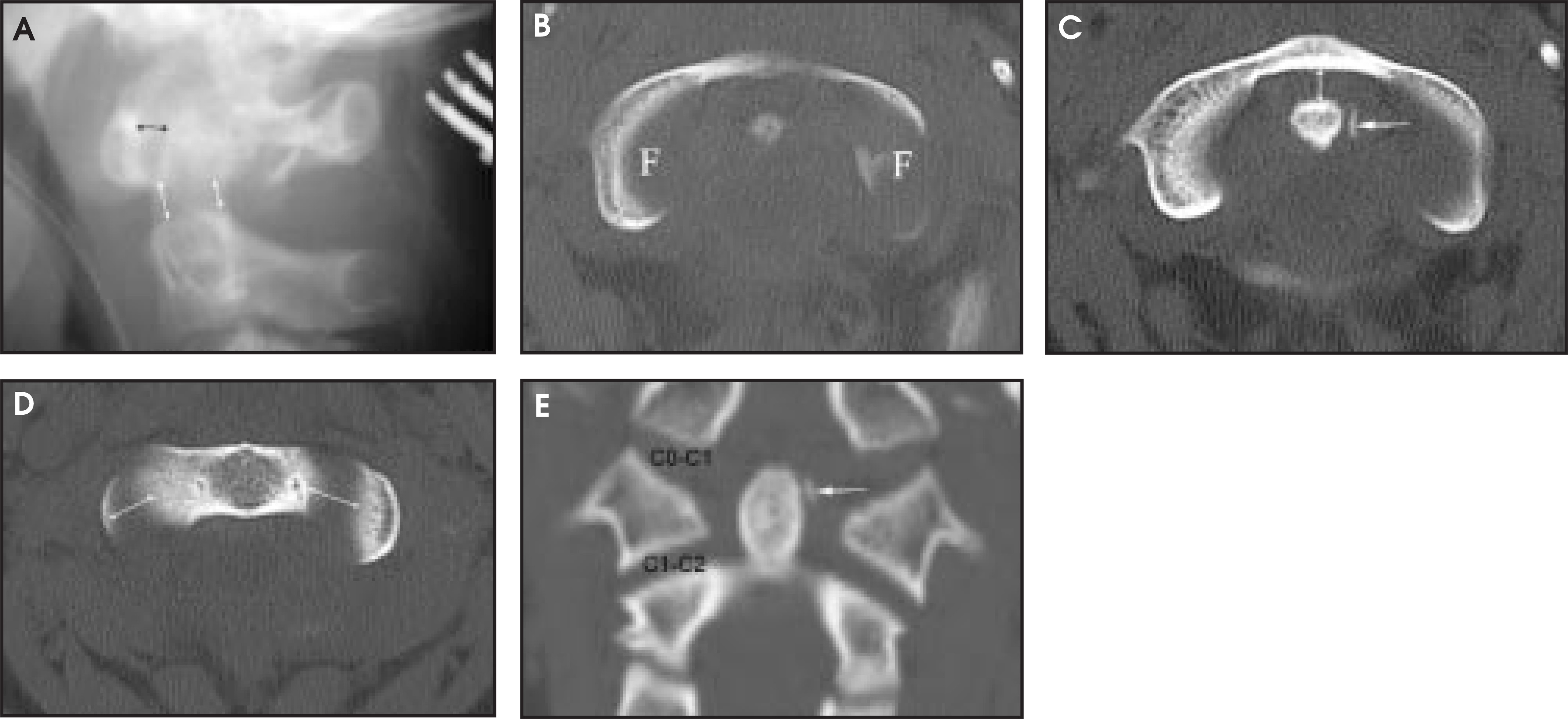
A type II fracture is the most common type. This horizontally oriented fracture occurs at the base of the dens, an area of diminished bone density compared with the rest of C2.2 Type II fractures are the least stable dens fracture and are most likely to develop nonunion when treated nonoperatively.
Minimally displaced type II dens fractures may be difficult or impossible to see on lateral radiographs (Figure 2). The open-mouth odontoid view may depict a fracture that is not otherwise visible, either as a fracture line (Figure 3) or a tilted dens (Figure 4).3 The presence of prevertebral soft-tissue swelling may prompt CT scanning of the upper cervical spine, allowing detection of otherwise radiographically occult fractures.4 However, not all dens fractures have abnormal prevertebral soft tissues (Figure 2). The existence of radiographically occult dens fractures in the absence of soft-tissue swelling is a compelling reason to perform screening CT in high-risk patients.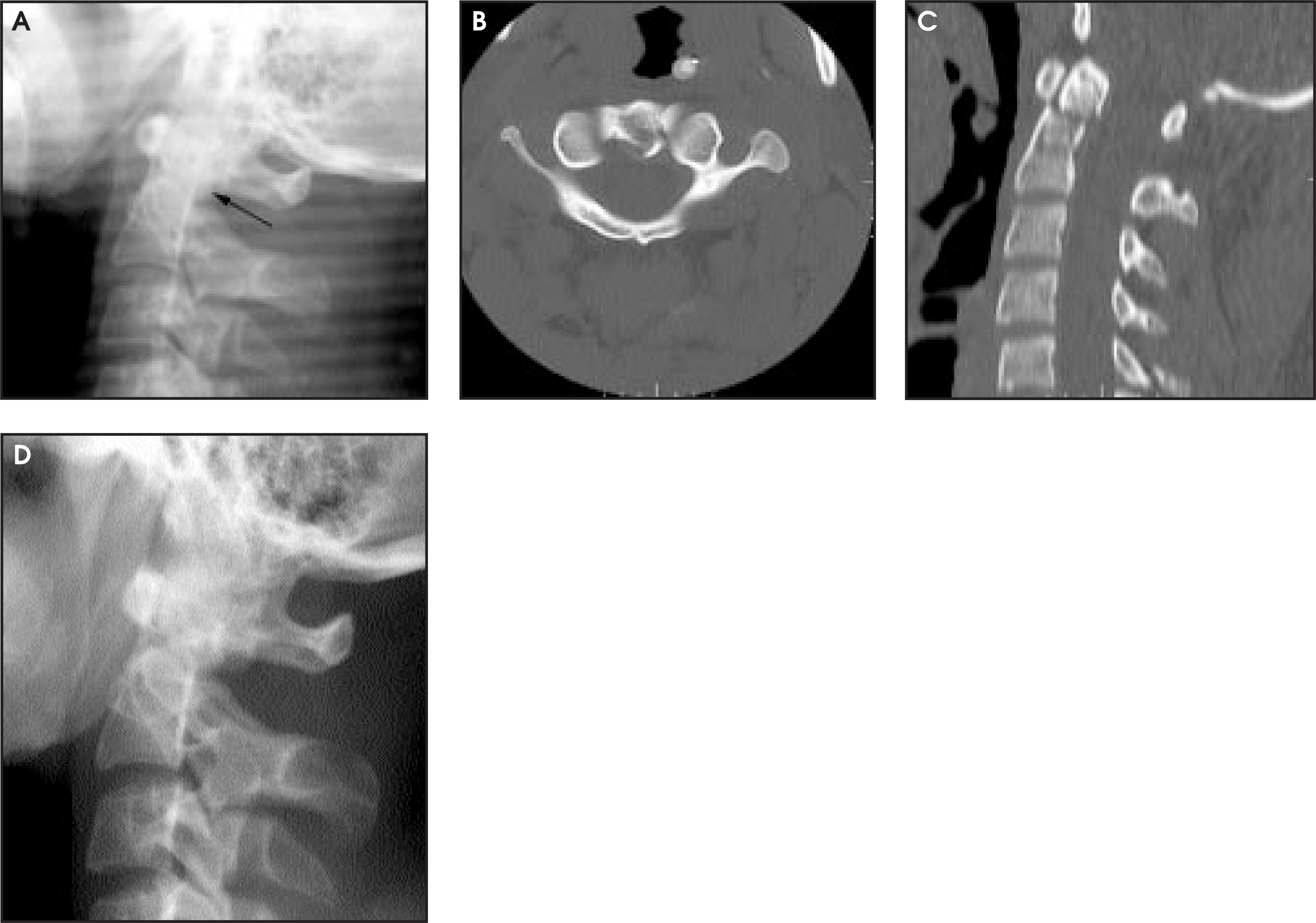
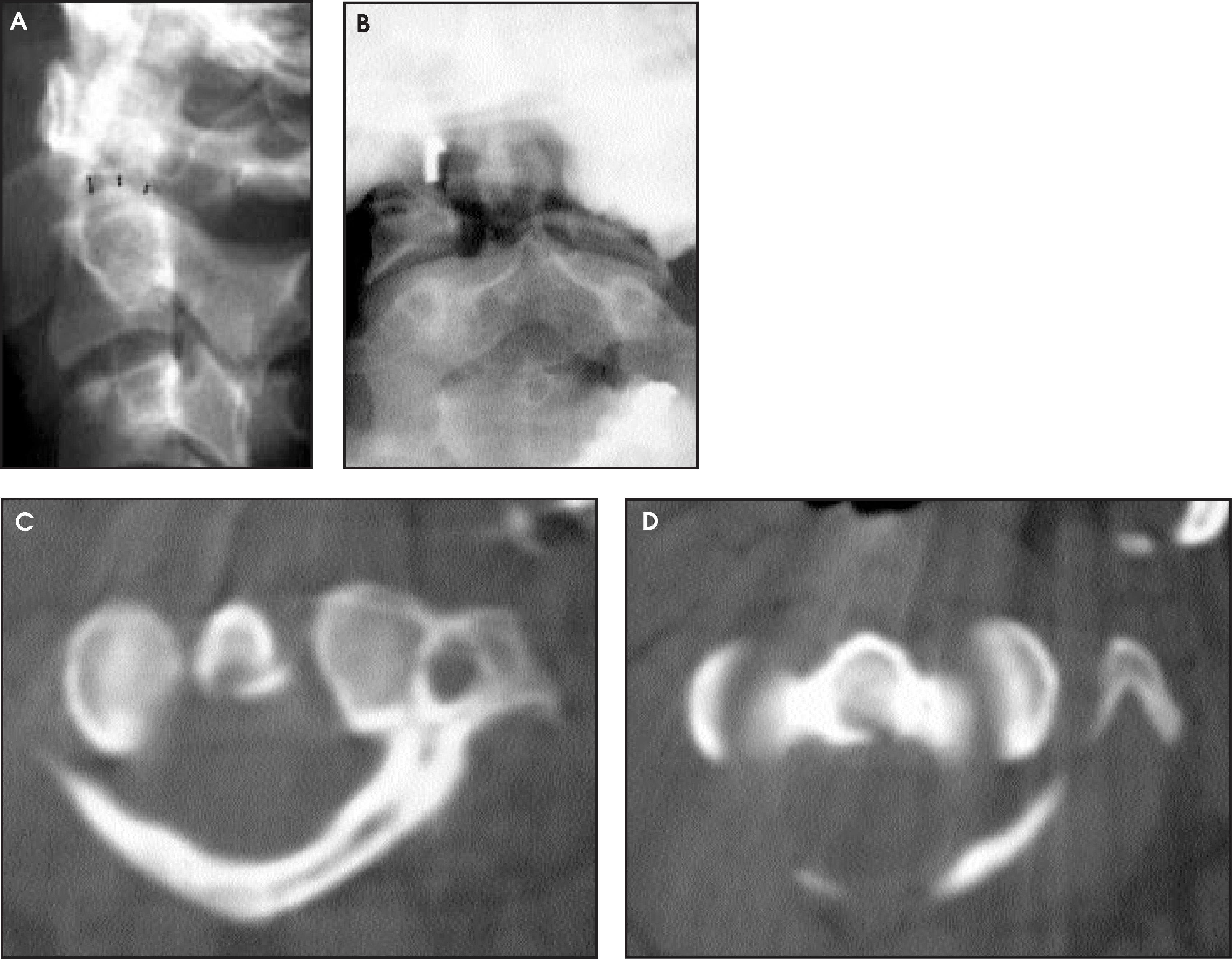
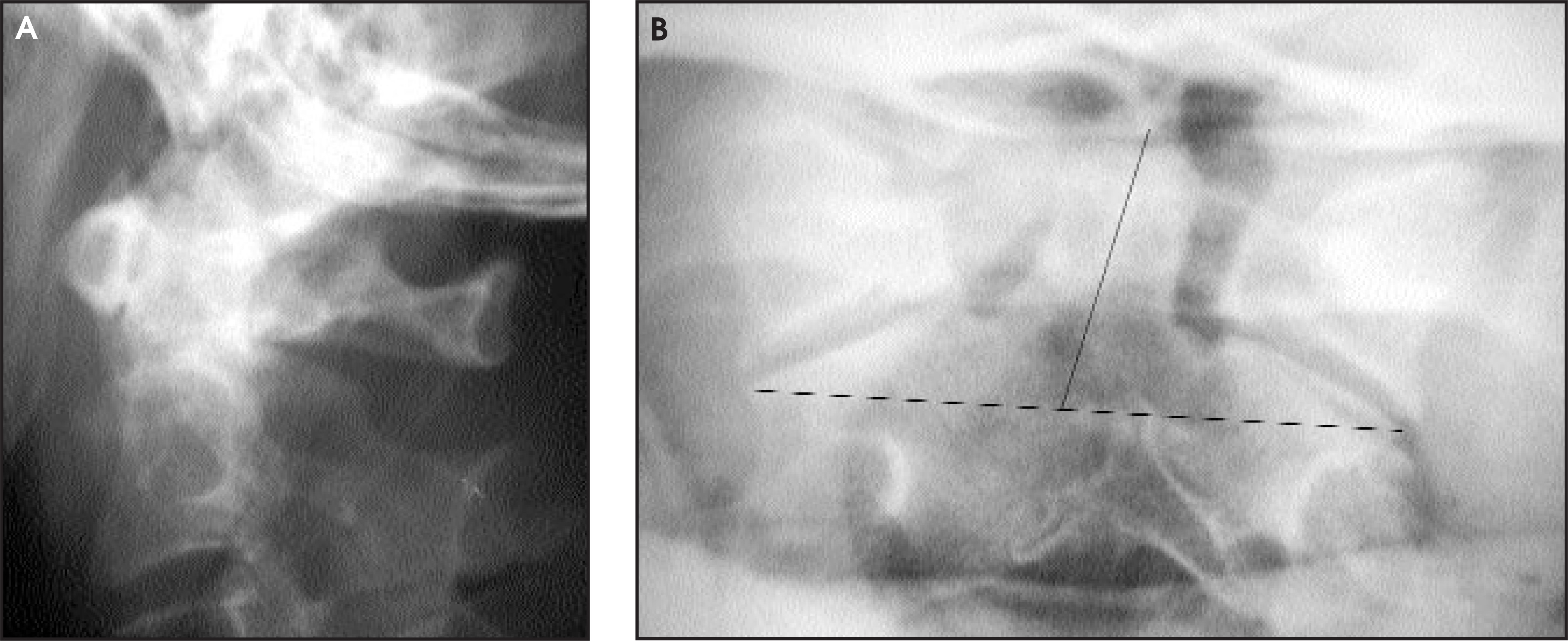
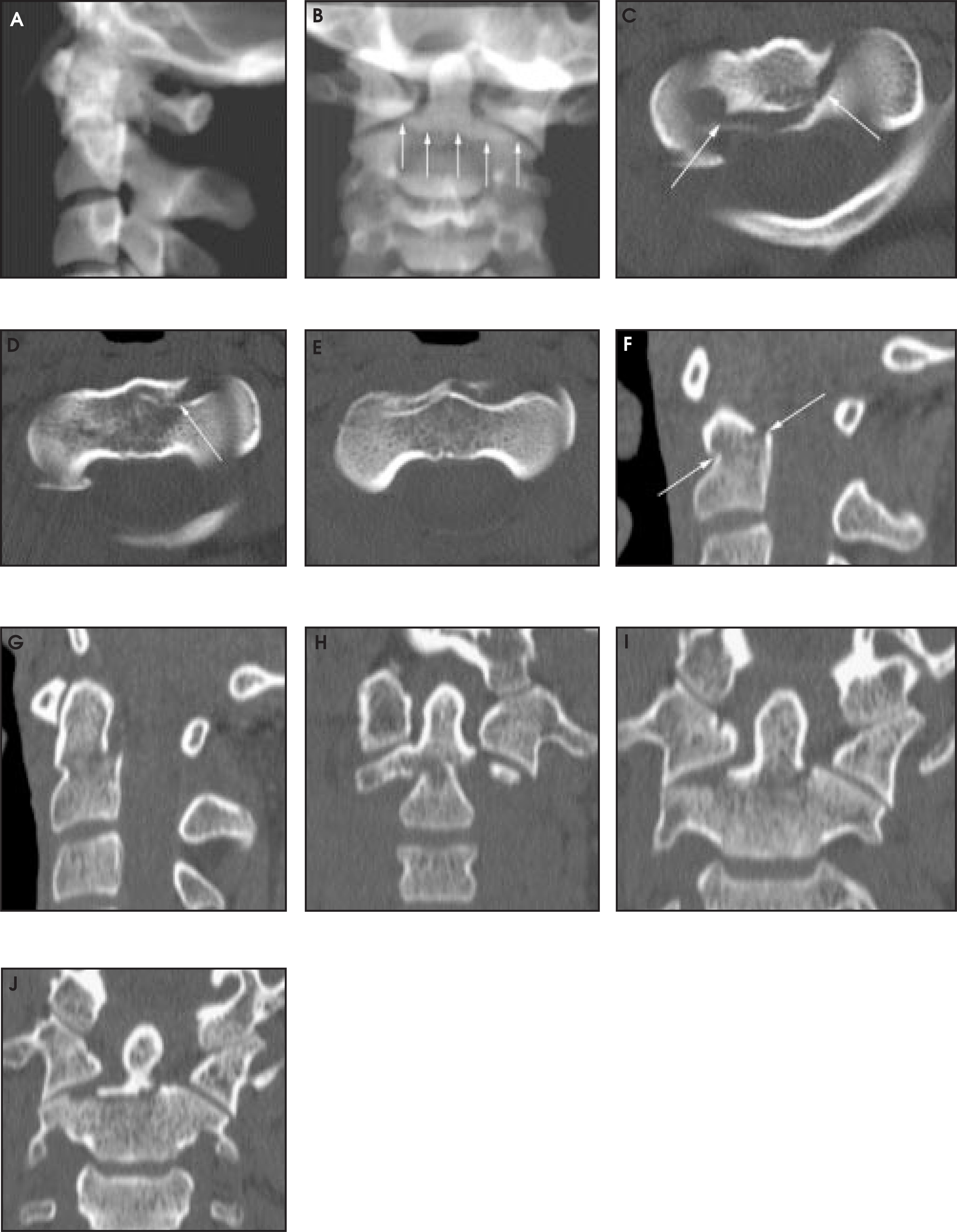
Most type II dens fractures are horizontally oriented and occur at or below the level of the inferior margin of the anterior arch of C1 (Figures 2 to 8). On occasion, the fracture line is oriented obliquely, from rostral anteriorly to caudal posteriorly (Figure 9).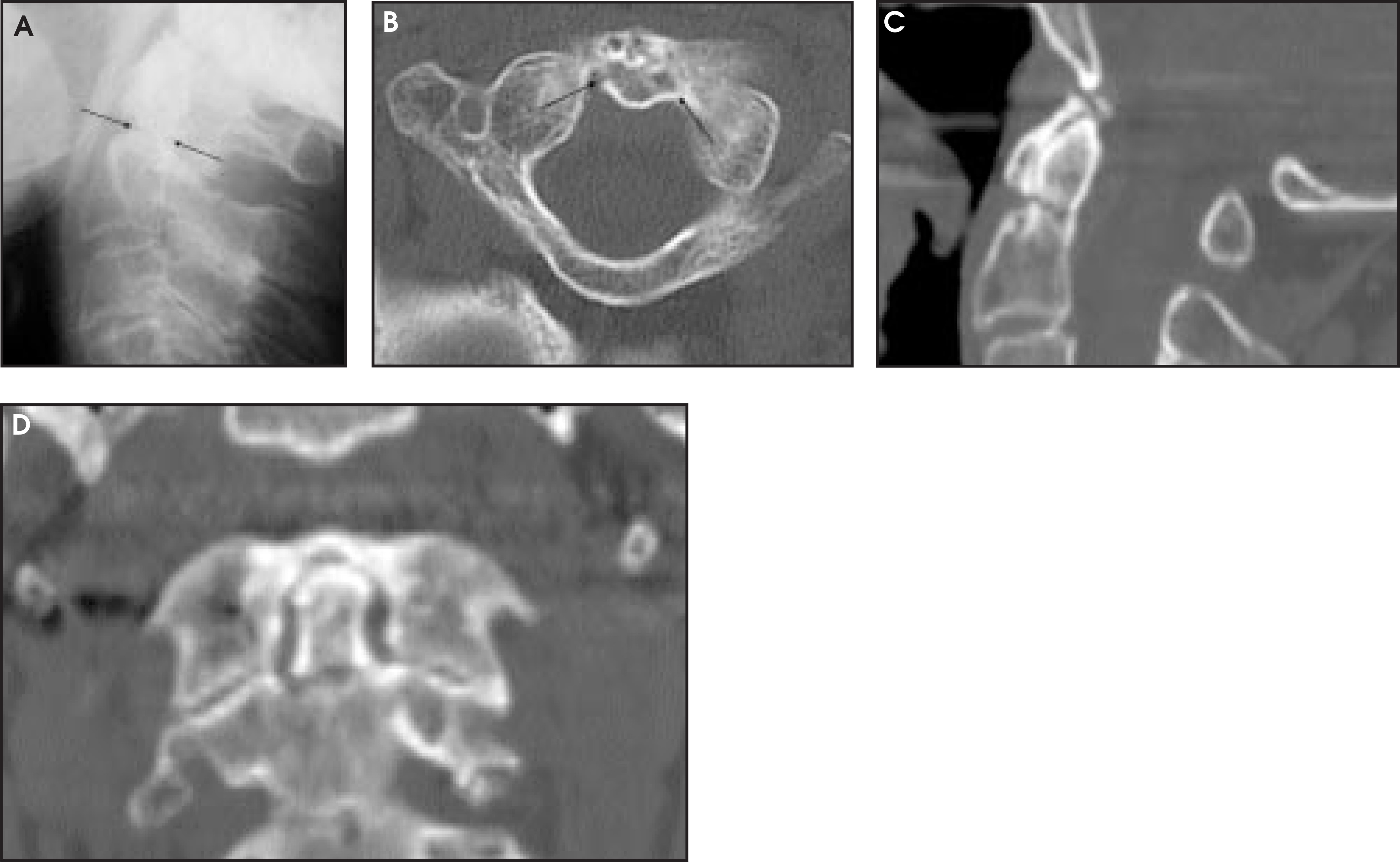
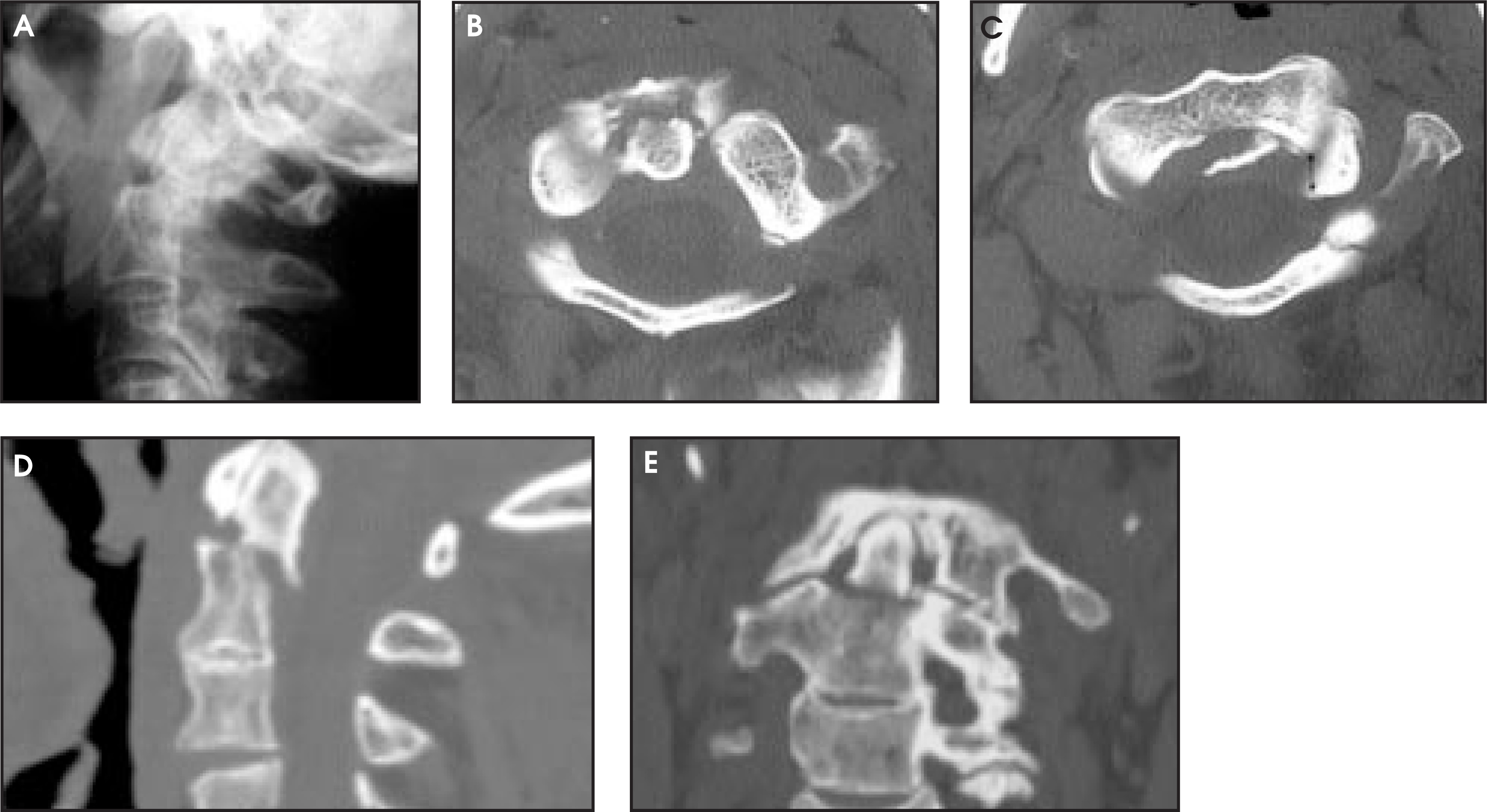
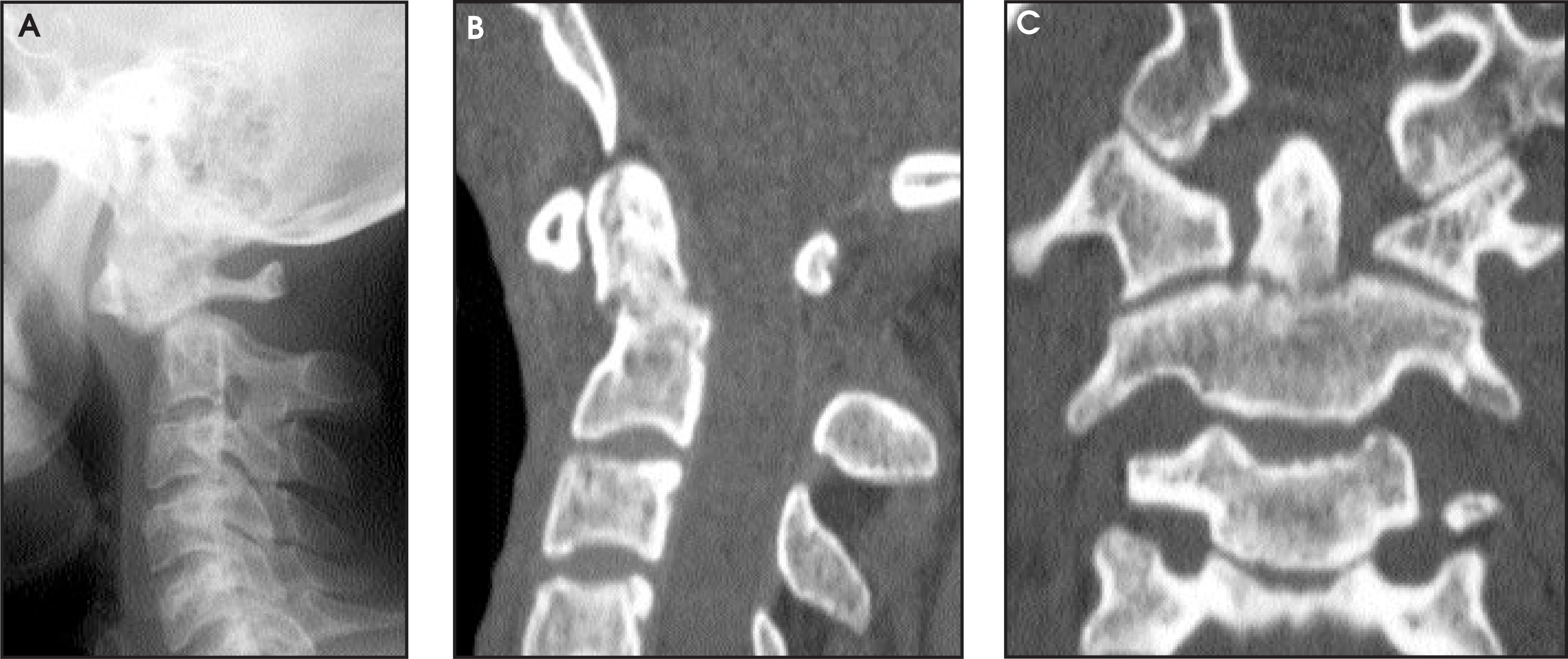
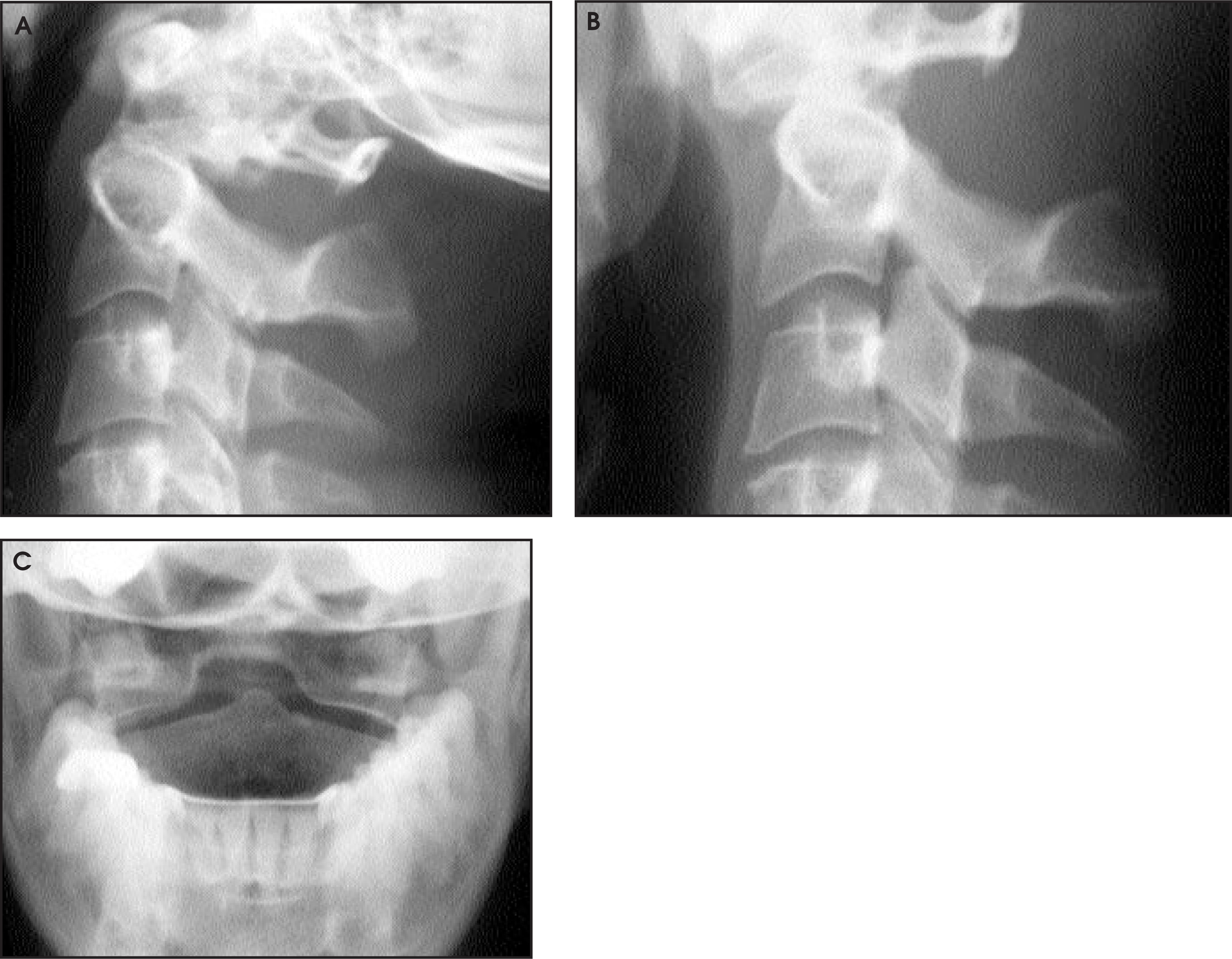
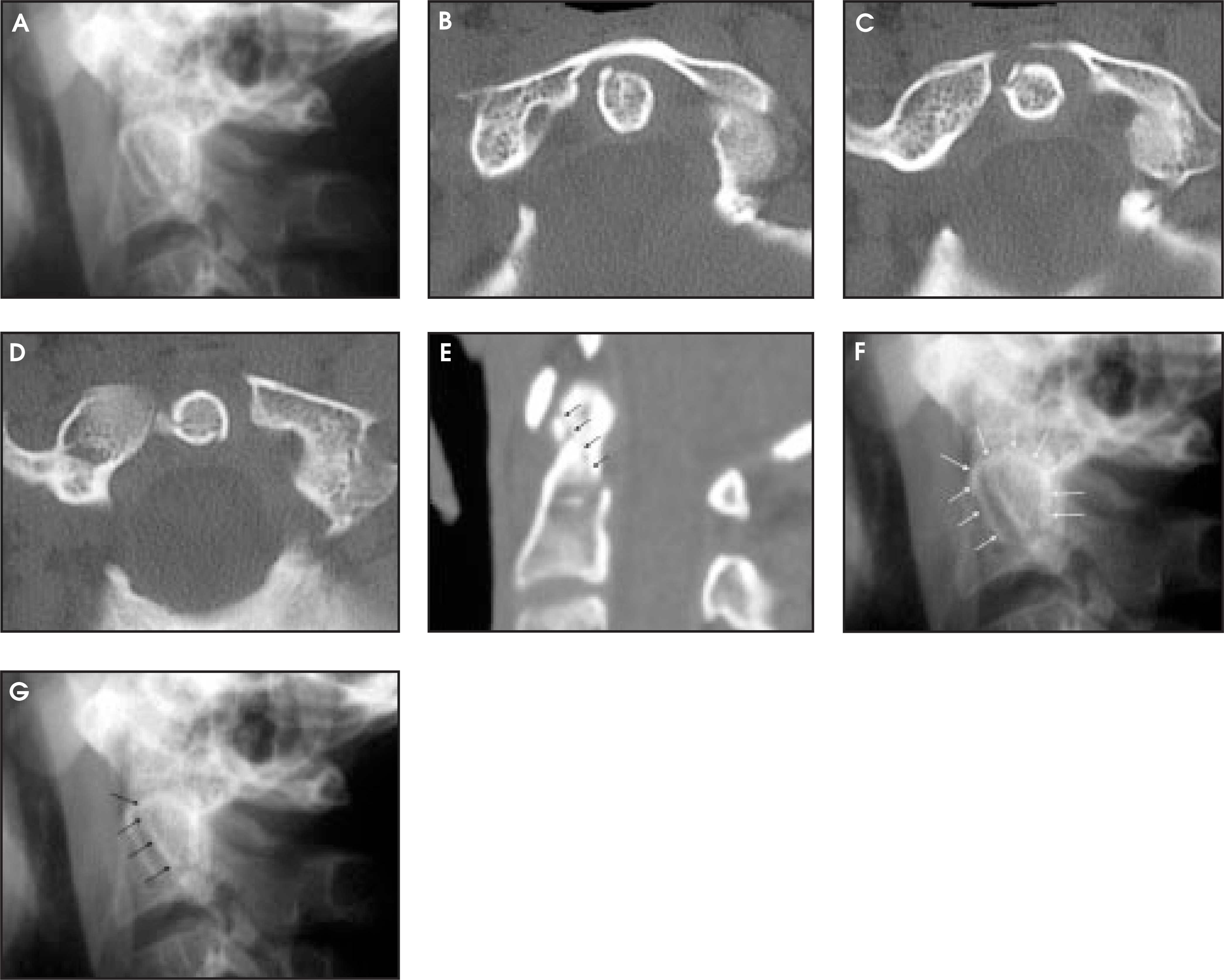
Using thick CT sections (3 to 5 mm), type II fractures may be difficult to detect on axial CT. Our experience using 2.5-mm axial images made with the bone algorithm is that a type II dens fracture is almost always visible on axial CT images as a cortical break at the base of the dens. We routinely make a second set of 1.25-mm axial images with the standard algorithm for high-quality sagittal and coronal reformatted images. Rarely, a minimally displaced horizontal type II dens fracture is aligned perfectly with the plane of axial scanning, making it difficult to detect on axial images. Fortunately, the fracture is very easy to detect on high-quality multiplanar reformatted images (Figure 5). Displaced type II dens fractures are recognized easily on radiographs or CT scans (Figures 6 through 8).
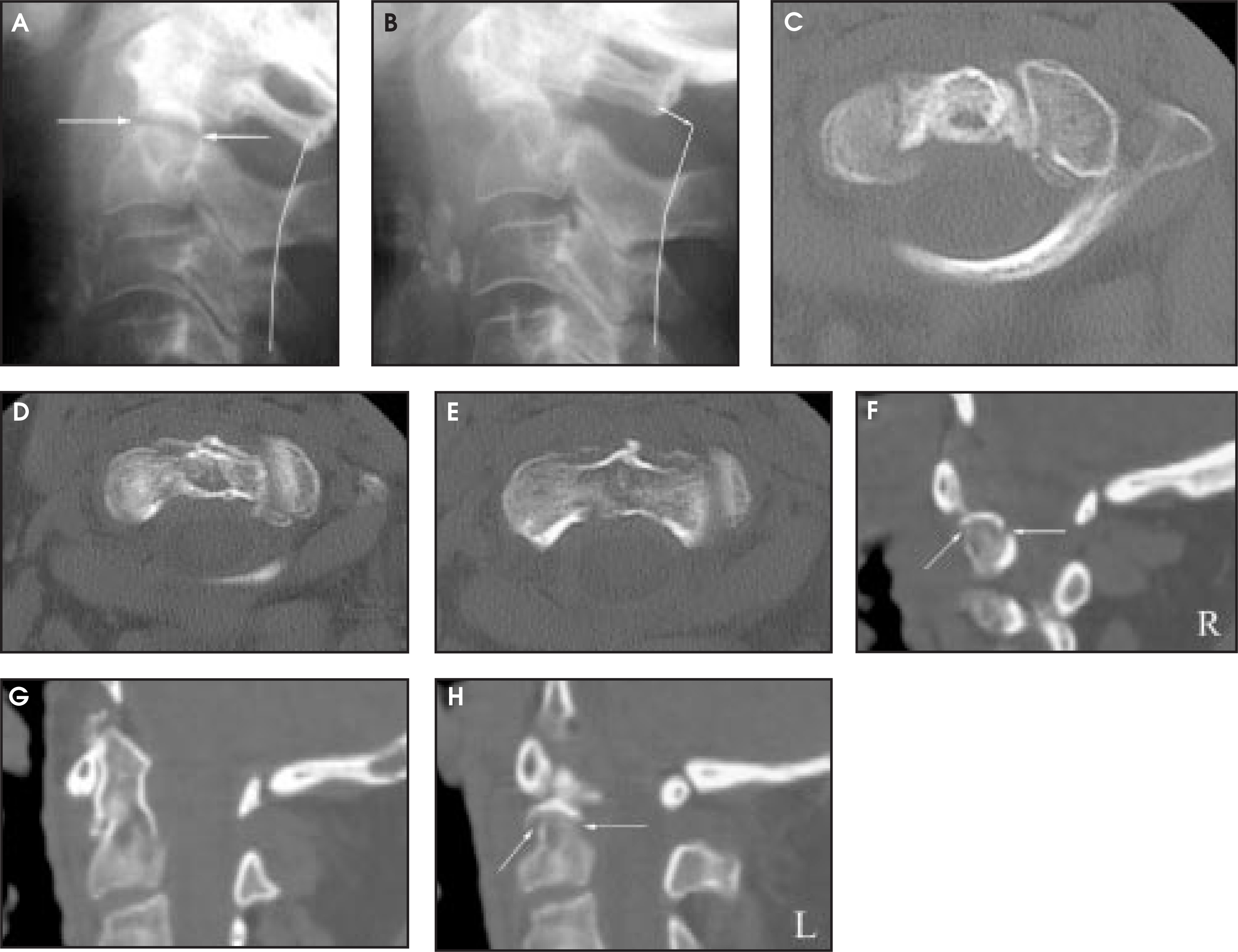
Nonunion of a type II dens fracture occurs with sufficient frequency to present a diagnostic challenge. Well-corticated fracture margins and a history of previous neck injury, or, better still, a history of previous fracture, helps establish the diagnosis. However, the crucial issues are whether or not a quasi-stable fibrous union has formed and whether or not acute re-injury has resulted in instability. The presence of pain, pathologic motion on flexion/ extension lateral radiographs, edema on MRI, and increased radiopharmaceutical uptake on bone scan provides evidence of acute re-injury and instability (Figure 8).
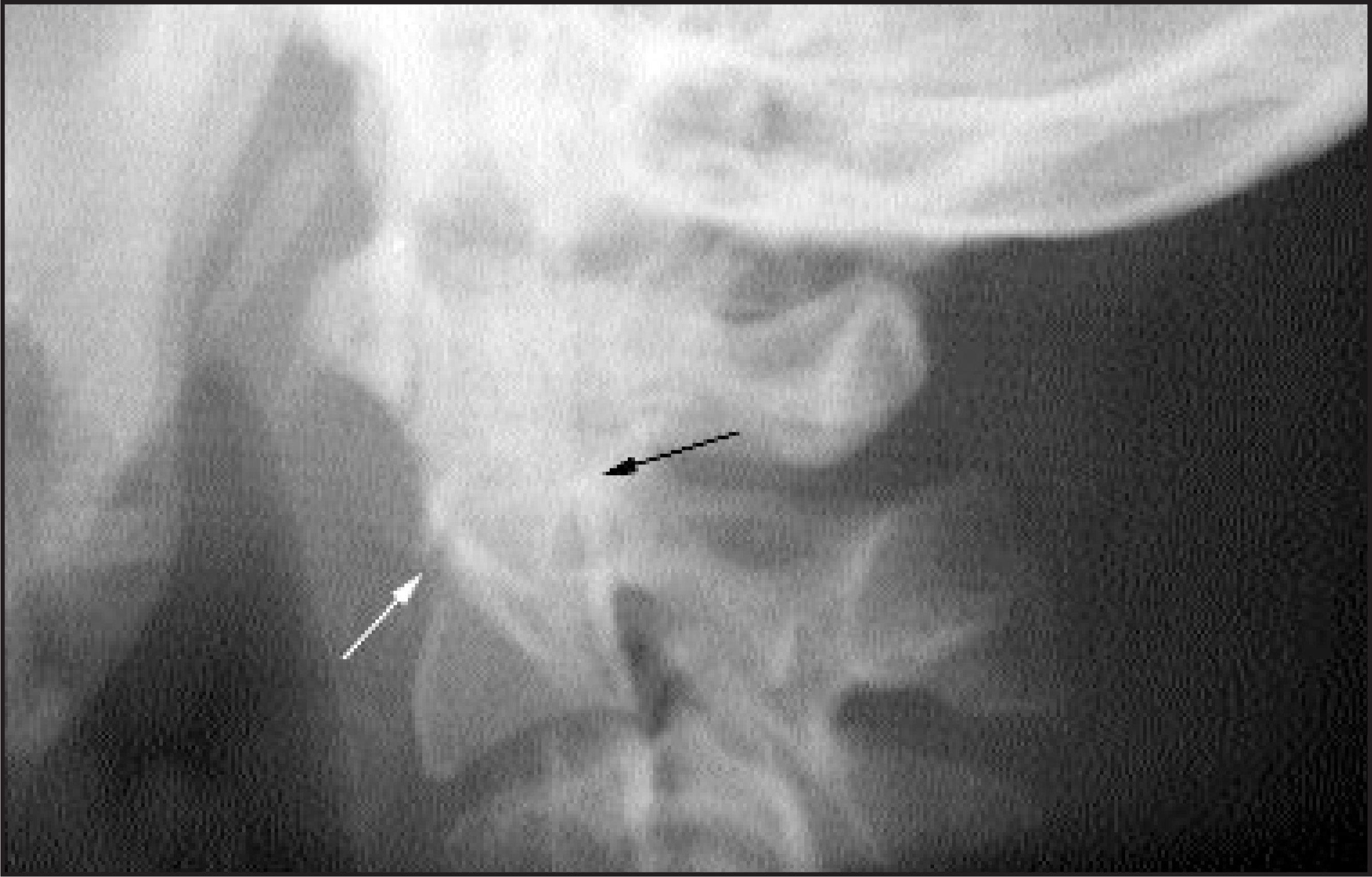
A type III fracture is an oblique fracture at the base of the dens, extending into the cancellous bone in the body of C2. Type III fractures are relatively more stable and less prone to nonunion when compared with type II fractures. Minimally displaced type III fractures may be difficult to detect. Such fractures are usually not apparent in the anteroposterior (AP) projection. Careful analysis of the lateral radiograph with attention to the C2 rings may indicate fracture through the C2 body.5,6 The C2 rings are a pair of composite shadows forming two inverted horseshoes within the C2 body caudal to the base of the dens (Figures 9F and 9G). The C2 rings are superimposed on a perfectly lateral radiograph but are visible as separate structures if the patient is rotated or the X-ray beam is oblique to C2. The anterior margin of this cortical ring is formed by the oblique groove between the body and articular mass. The top is formed by both the notch between the base of the dens and the superior articular facet of C2, and by the superior margin of the superior articular facet itself. The posterior cortex of the C2 body forms the posterior margin. Minimal breaks in the C2 ring are a sensitive means of detecting small fractures (Figures 10 and 11). Displaced type III fractures typically cause interruption of the C2 rings (Figures 12 and 13). Fracture lines often extend into one or both superior articular surfaces.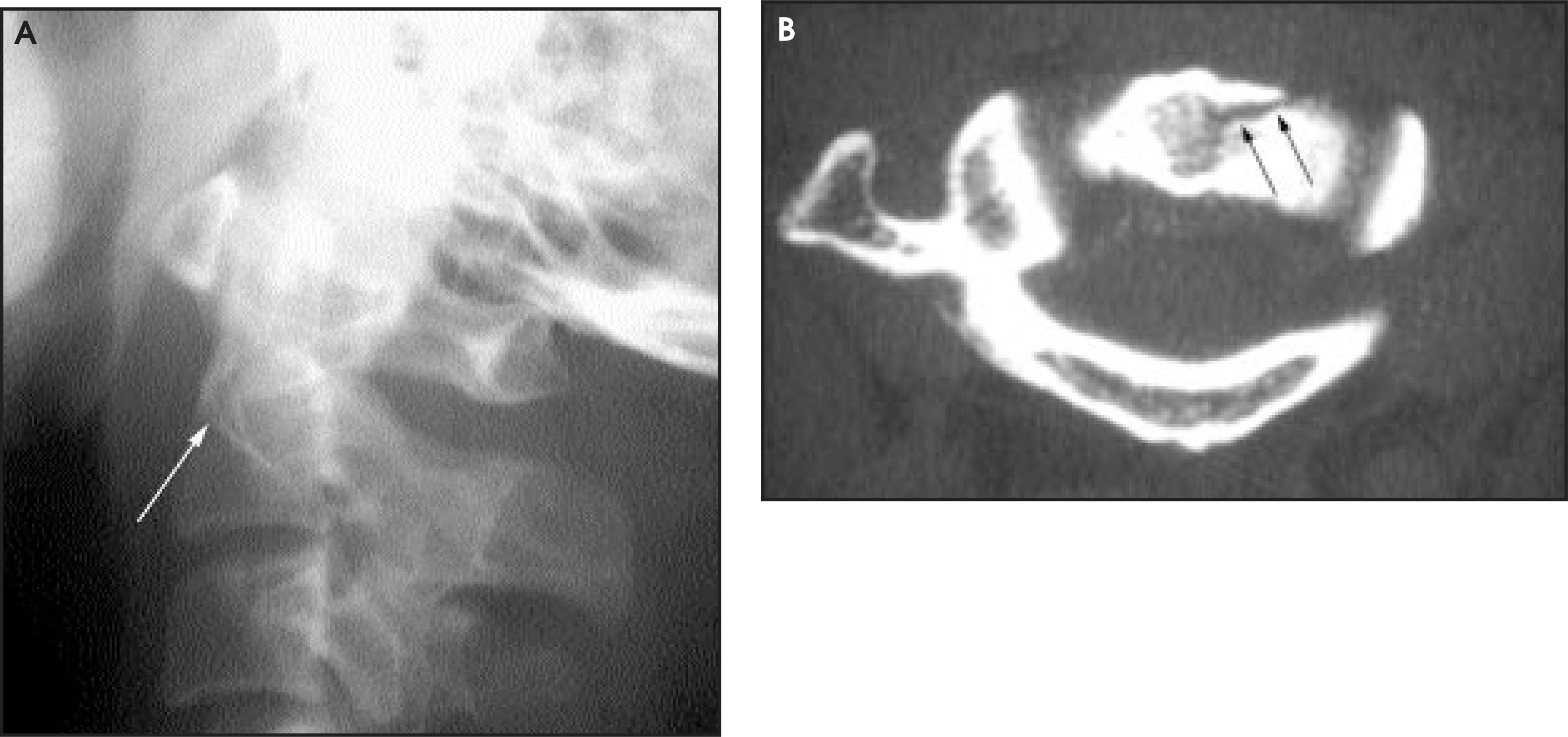
Conclusion
After studying the cases of dens fracture in this pictorial essay, several important facts should be clear. First, fractures of the dens are often, but not always, detectable by radiographs. Second, prevertebral soft-tissue swelling is helpful when present, but the absence of swelling does not exclude a dens fracture. Third, because radiographs are not completely sensitive for the detection of potential unstable injuries in the craniocervical region, screening high-risk trauma patients for injury using CT is suggested.7,8 Fourth, routine use of sagittal and coronal reformatted images is important for detecting minimally displaced type II dens fractures and for characterizing all dens fractures. Fifth, type II dens fractures are commonly encountered in clinical practice—type III fractures are less common; type I fractures are part of more complex craniocervical junction injuries and rarely, if ever, occur as isolated fractures. Finally, the C2 ring sign is a valuable tool in detecting type III dens fractures. AR
References
- Anderson L, D’Alonzo R. Fractures of the odontoid process of the axis.. J Bone Joint Surg Am.. 1974;56:1663-1674.
- Amling M, Hahn M, Wening V. The microarchitecture of the axis as the predisposing factor for fracture of the base of the odontoid process. A histomorphometric analysis of twenty-two autopsy specimens.. J Bone Joint Surg Am.. 1994;76:1840-1846.
- West O, Anbari M, Pilgram T, Wilson A. Acute cervical spine trauma: Diagnostic performance of single-view versus three-view radiographic screening.. Radiology.. 1997;204:819-823.
- Harris J. The cervicocranium: Its radiographic assessment.. Radiology.. 2001;218:337-351.
- Harris J, Burke J, Ray R. Low (type III) odontoid fracture: A new radiographic sign.. Radiology.. 1984;153:353-356.
- Mortelmans L, Geusens E, Sabbe M, Delooz H. Harris or axis ring: An aid in diagnosing low (type 3) odontoid fractures.. Eur J Surg.. 1999;165:1138-1141.
- Link T, Schuierer G, Hufendiek A. Substantial head trauma: Value of routine CT examination of the cervicocranium.. Radiology.. 1995;196:741-745.
- Blackmore C, Mann F, Wilson A. Helical CT in the primary trauma evaluation of the cervical spine: An evidence-based approach.. Skeletal Radiol.. 2000;29:632-639.
Citation
. Imaging of upper cervical spine injuries—Part II: The dens. Applied Radiology. 2003;32(2):30-38. doi:10.37549/AR1165.