Ingested aluminum foreign body
Applied Radiology — Vol. 42 , Issue 1 , pp. 54 -55
DOI: 10.37549/AR1959
Published: January 1, 2013
Categories
Case summary
A 7-year-old child was referred for suspected ingestion of an aluminum ring pull from a can of soda. The patient was awake, alert, cooperative, and displayed no signs of respiratory distress
IMAGING FINDINGS
Anteroposterior and lateral radiographs of the chest and neck, as well as a supine radiograph of the abdomen, revealed clear lungs with no indication of a foreign body and no indirect sign of airway obstruction. The soft tissues of the neck were within normal limits. The abdomen at first look was unremarkable; however, closer examination revealed a barely detectable radiopaque structure in the right iliac fossa intermixed with feces in the cecum (Figure 1). The object corresponded to the suspected aluminum ring pull. Conservative management was undertaken; the child expelled the foreign body one day later with stool.
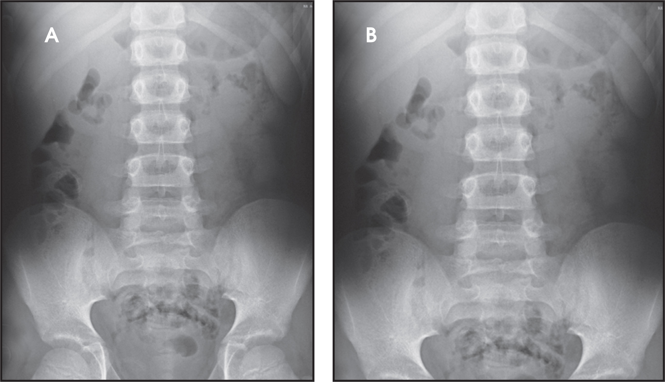
Diagnosis
Ingested aluminum foreign body.
Discussion
Young children often put small objects into their mouth and accidentally swallow them. These foreign bodies may then enter the airway or the gastrointestinal (GI) tract. Those that enter the airway often become lodged distal to the larynx and trachea in the right mainstem bronchus, presenting a potentially life-threatening situation requiring bronchoscopic removal of the object. Most patients present with acute onset of choking, respiratory distress, coughing and wheezing.1 Some other patients may have a silent presentation manifested only by secondary complications.
Most objects that enter the GI tract, meanwhile, pass through without difficulty and can be managed conservatively. However, trapping and potentially severe complications requiring endoscopic or surgical removal may also occur.
The esophagus has 3 areas of narrowing where most foreign bodies become entrapped: the upper esophageal sphincter, the level of the carina and aortic arch, and the lower esophageal sphincter.2 If a foreign body reaches the stomach, depending on the object’s length and diameter, it may fail to pass the pylorus or the duodenal sweep.
If the object reaches the small bowel, the only structural impediment to passage is the ileocecal valve; an object that reaches the cecum is usually expelled with stool.
Foreign bodies retained in the oropharynx and esophagus may damage the mucosa, leading to abscess, perforation, and severe complications like mediastinitis, fistulas, pericarditis or tamponade, or even injuries to the aorta and pulmonary vessels.3 Complications of foreign bodies in the stomach and small bowel consist mainly of perforation and associated infection, including peritonitis.
When a patient is suspected to have inhaled or ingested a foreign body, anteroposterior and lateral chest; lateral neck; and supine abdominal radiographs should be obtained for a complete mouth-to-anus examination. Lateral and AP radiographs are important because some foreign bodies, especially those of discoid shape, may show only in one projection and be undetectable in the other.
The radiological visualization of a foreign body depends on its radiopacity. Radiolucent bodies are undetectable and inhalation may be suspected only when pulmonary obstructive signs are present. In the GI tract, they are invisible unless oral contrast is administered. Gastrografin is preferred over barium because of the risk of associated perforation.
Metal objects like coins are usually easily depicted on radiographs because of their high radiopacity. However, the conspicuity of aluminum foreign bodies is lower than other metals because its atomic number (13) is quite close to that of soft tissue, which is 7.5.4 Although aluminum objects may be detected radiographically in 80% to 90% of cases,5 an accurate review of the images is necessary to identify them. Moreover, aluminum’s low radiodensity (it is 10 times less absorptive than steel)6 is not widely known, so foreign bodies may easily be missed on a superficial reading of the images.
Conclusion
Radiologic detection of ingested metallic foreign bodies is usually a simple task for the radiologist because metal objects are usually strikingly radiopaque and easy to visualize. However, aluminum’s opacity is much lower than that of other metals because of its atomic number. Radiologists must be aware of the low radiodensity of aluminum because superficial assessment of radiographs may result in missed aluminum foreign bodies, and delays in diagnosis may lead to severe complications.
References
- Silva A, Muntz H, Clary R. Utility of conventional radiography in the diagnosis and management of pediatric airway foreign bodies. Ann Otol Rhinol Laryngol. 1998;107:834-838.
- Weller M, Ayshford C. Variable radio-opacity of a metallic foreign body. Emerg Med J. 2004;21:638-639.
- Macpherson R, Hill J, Othersen H. Esophageal foreign bodies in children: Diagnosis, treatment, and complications. AJR Am J Roentgenol. 1996;166:919-924.
- Stewart G, Lakshmi M, Jackson A. Aluminium ring pulls: An invisible foreign body. J Accid Emerg Med. 1994;11:201-203.
- Conners G. Finding aluminum foreign bodies. Pediatr Rev. 2000;21:172.
- Bradburn D, Carr H, Renwick I. Radiographs and aluminium: A pitfall for the unwary. BMJ;308.
Citation
. Ingested aluminum foreign body. Applied Radiology. 2013;42(1):54-55. doi:10.37549/AR1959.