Injected periurethral collagen
Applied Radiology — Vol. 37 , Issue 7 , pp. 25 -27
DOI: 10.37549/AR1625
Published: July 1, 2008
Categories
CASE SUMMARY
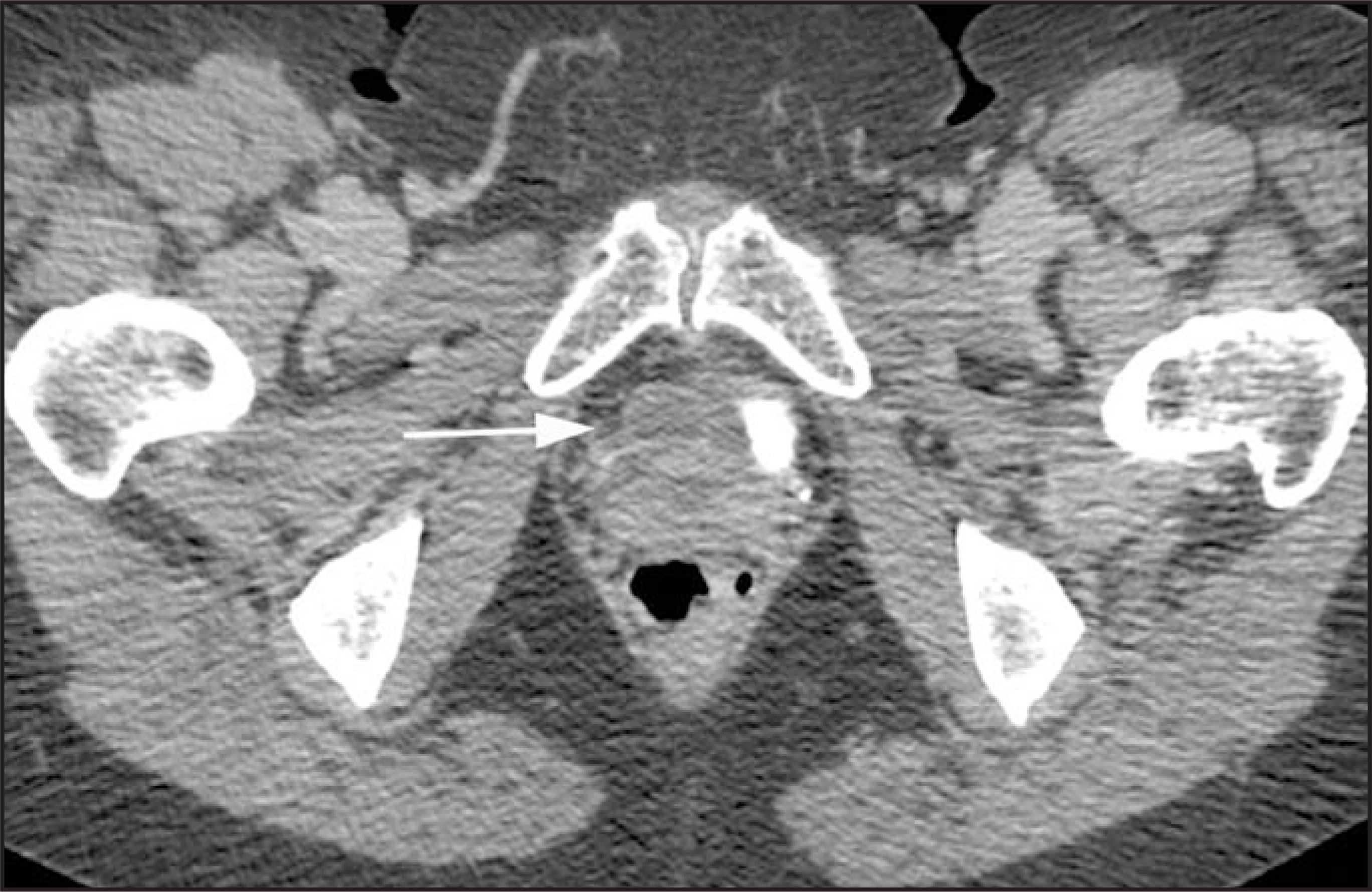
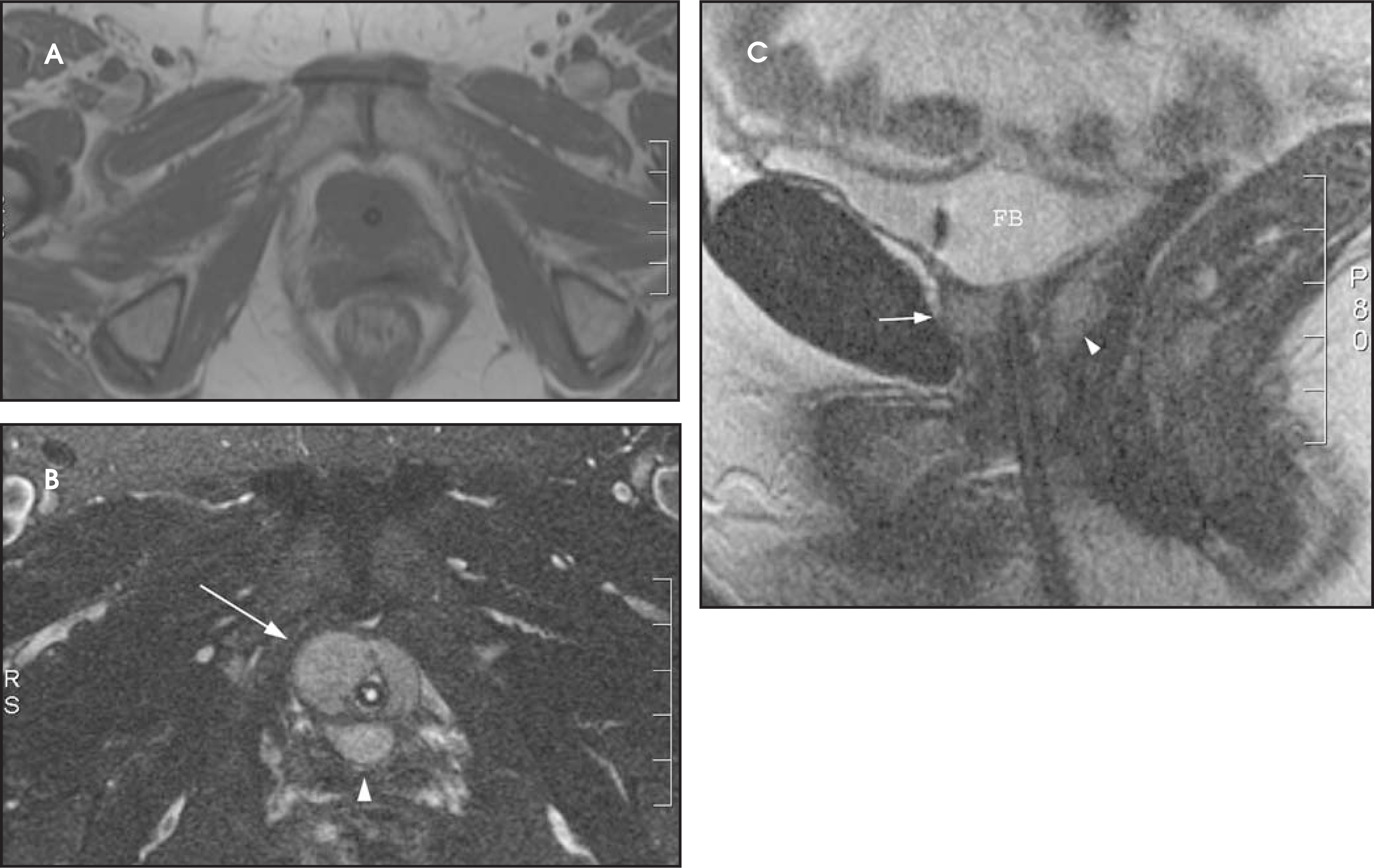
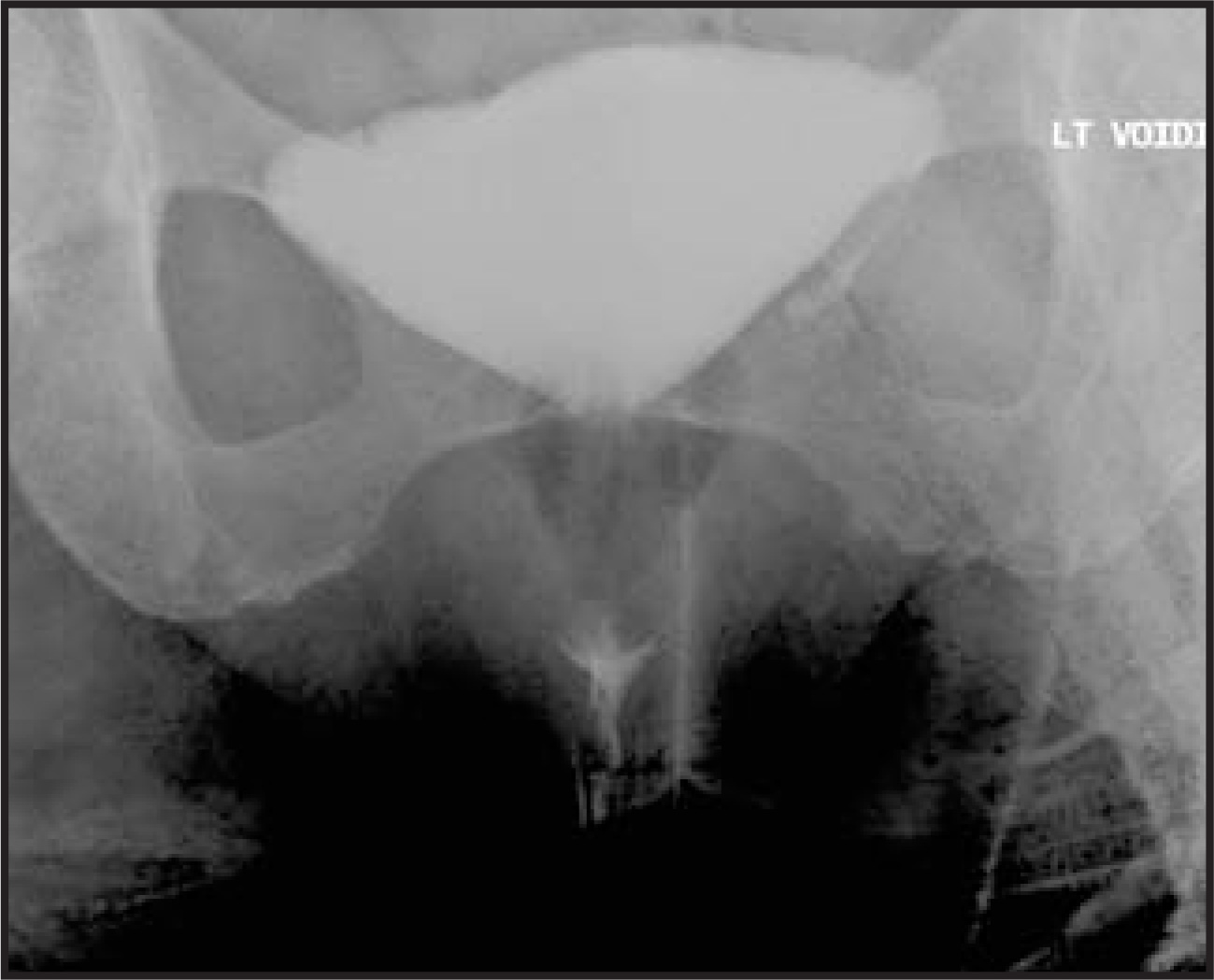
A 68-year-old woman presented with a several-year history of increased urinary frequency and stress urinary incontinence. She reported 6 urinary tract infections within the previous year, all of which had responded well to single-antibiotic therapy. Her pelvic examination and urinalysis were normal. Her medical history was remarkable for an uncomplicated urethral caruncle excision 12 years prior. In addition, she underwent nonsurgical intervention for her urinary incontinence. Her current complaints of urinary tract infections prompted precontrast (not shown) and postcontrast (Figure 1) computed tomography (CT) of the abdomen and pelvis. This was followed by pelvic magnetic resonance imaging (MRI) (Figure 2) and a voiding cystourethrogram (VCUG) (Figure 3).
IMAGING FINDINGS
The CT examination of the abdomen and pelvis showed a well-defined, asymmetric, multiloculated, cystic-appearing, hypodense structurelocated anteriorly and posteriorly to the proximal urethra (Figure 1). The largest cystic component was located to the right and anteriorly withrespect to the urethra and measured approximately 2× 2 cm. Because of this finding, a periurethral diverticulum of the proximal urethra was suspected, and MRI was performed for confirmation (Figure 2). The pelvic MRI showed disruption of the normal, target-like appearance of the urethra and confirmed the multiloculated structure surrounding the proximal urethra, with right-sided eccentricity. The structure exhibited low signal on T1-weighted (T1W) imaging (Figure 2A) and an intermediate signal intensity, less than that of urine, on T2-weighted (T2W) imaging (Figure 2, B and C). The diagnosis of a urethral diverticulum containing complex fluid was considered, although concern was expressed as to the inherent intensity of the lesion and its position with respect to the urethra.
At cystoscopy, no abnormality of the urethra or bladder was identified. A voiding cystourethrogram was subsequently performed to further delineate the anatomy of the urethra (Figure 3). During filling of the bladder, a mild cystocele was identified. While voiding, the urethra was normal in caliber without filling of a urethral diverticulum. There was prominent descent of the bladder base below the level of the inferior pubic rami, suggesting moderate pelvic floor laxity (Figure 3).
DIAGNOSIS
Injected periurethral collagen, which had been administered for the treatment of urinary incontinence
DISCUSSION
An acquired urethral diverticulum is the sequelae of infection in the periurethral glands that progresses to fistulization into the urethra.1 Acquired diverticula occur more frequently in women, who may present with urinary frequency, urgency, and dysuria. More specific symptoms of dribbling, dyspareunia, hematuria, or a tender cystic swelling protruding into the vagina, anteriorly, occur less commonly. In addition, such diverticula can be discovered incidentally on imaging studies.2 Urethral diverticula are usually located within the midurethra at the level of the pubic symphysis. Typically, a diverticulum extends along the posterolateral wall of the urethra, where the paraurethral glands are predominately located. Larger, more complex lesions can assume a horseshoe configuration around the urethra. Complications associated with diverticula include recurrent infections, calculi, and malignancy.2 The differential diagnosis includes submucosal cysts, abscess of Skene’sglands, or injected collagen into the periurethral region for the treatment of urinary incontinence.1
Acquired urethral diverticula can be diagnosed by direct physical examination in combination with radiologic studies including MRI, CT,VCUG, and positive pressure urethrography.3 MRI is a useful modality for the evaluation of pelvic pathology and has been reported to havea higher sensitivity for the depiction of urethral diverticula than VCUG or urethroscopy.3 It allows for detailed evaluation of anatomic relationships between the diverticulum and adjacent structures. This is useful in surgical planning, as treatment requires surgical excision of the diverticulum and reconstruction of the urethra. In addition, MRI can be useful in detecting surrounding inflammation or a tumor within a diverticulum. Although extremely rare, adenocarcinoma, transitional cell carcinoma, and squamous cell carcinoma can arise within urethral diverticula.2
This patient’s imaging studies revealed a well-defined, multiloculated, cystic-appearing structure located near the proximal urethra with a predominantly anterior and lateral location, unlike the typical location for a urethral diverticulum. After VCUG and urethro scopy failed to reveal anorifice to a urethral diverticula, the clinical history of periurethral collagen injection was obtained.
Injected collagen at the bladder base for the treatment of stress urinary incontinence can mimic a periurethral mass; therefore, history of thisprocedure is critical for the correct diagnosis.1,4 Stress urinary incontinence is treated using multiple different approaches, including pelvic floor muscle retraining, periurethral injectables, and surgery.4 Periurethral injection of glutaraldehyde cross-linked collagen became popular because of its widespread use and safety profile in plastic surgery procedures. The cystoscopic procedure is easily performed with a low-pressure injection through a long, small-caliber needle; and, in many cases, multiple treatments are necessary to achieve efficacy. Improvement in the symptoms of stress urinary incontinence occurs in a significant percentage of treated patients.4,5
Carr et al6 described the MR appearance of periurethral collagen as discrete foci that appear brighter than the usual surrounding submucosaon T1W imaging. On T2W imaging, they appear hyperintense, but not to the degree of a urethral diverticulum. Injected collagen is usually seen anterior to the urethra. Collagen can migrate circumferentially around the urethra and can also be seen as ectopic foci located in close proximity to the bladder or within the surrounding soft tissues.6
CONCLUSION
MRI of the female pelvis is a useful, adjunctive diagnostic tool for the diagnosis of female genitourinary pathology because of its excellent inherent soft tissue contrast and multiplanar imaging capabilities. The correct preoperative diagnosis is critical to determine an appropriate therapy. Due to the relatively similar appearance of an acquired urethral diverticulum and periurethral collagen, correlation with the clinical history is necessary to make the correct diagnosis.
References
- Hahn W, Israel G, Lee V. MRI of female urethral and periurethral disorders. AJR Am J Roentgenol J Urol.
- Kim B, Hricak H, Tanagho E. Diagnosis of urethral diverticula in women: Value of MR imaging. AJR Am J Roentgenol. 1993;161:809-815.
- Ryu J, Kim B. MR Imaging of the male and female urethra. RadioGraphics. 2001;21:1169-1185.
- Bridges M, Petrou S, Lightner D. Urethral bulking agents: Imaging review. AJR Am J Roentgenol. 2005;185:257-264.
- Shortliffe L, Freiha F, Kessler R. Treatment of urinary incontinence by the periurethral implantation of glutaraldehyde cross-linked collagen. J Urol. 1989;141:538-541.
- Carr L, Herschorn S, Leonhardt C. Magnetic resonance imaging after intraurethral collagen injected for stress urinary incontinence. J Urol. 1996;155:1253-1255.
Citation
. Injected periurethral collagen. Applied Radiology. 2008;37(7):25-27. doi:10.37549/AR1625.