Intraductal papilloma with focal atypia
Applied Radiology — Vol. 37 , Issue 6 , pp. 37 -38
DOI: 10.37549/AR1620
Published: June 1, 2008
Categories
CASE SUMMARY
A 45-year-old African American woman was referred by her primary care physican for mammography with a chief complaint of bloodydischarge from 1 duct near the areola. No palpable breastabnormality was present.
IMAGING FINDINGS
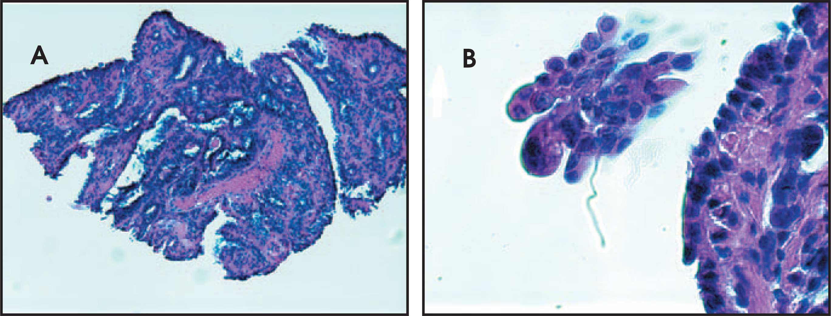
Diagnostic full-field digital craniocaudal and mediolateral oblique projections were obtained and showed no evidence of abnormality(Figure 1). The patient’s concerning history prompted a physical examination, which was nonrevealing, and sonographic evaluation (Figure 2). This single representative transverse image was obtained with a 12-MHz linear transducer and shows a single hypoechoic dilated duct(at the 9 o’clock position approximately 1 cm from the nipple) that contained a single ovoid hyperechoic mass, which was suggestive ofpapilloma.Color Doppler ultrasound was performed (not shown) but did not reveal appreciable flow.An ultrasound-guided core needle biopsy was performed. Histologic examination made the diagnosis of intraductal papilloma with focal atypia (Figure 3).
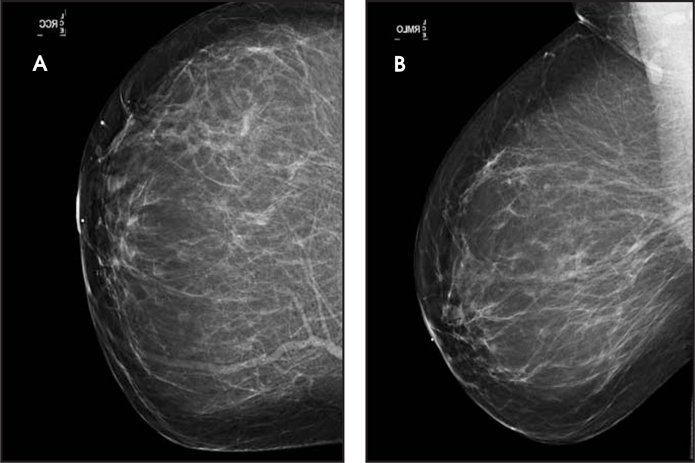
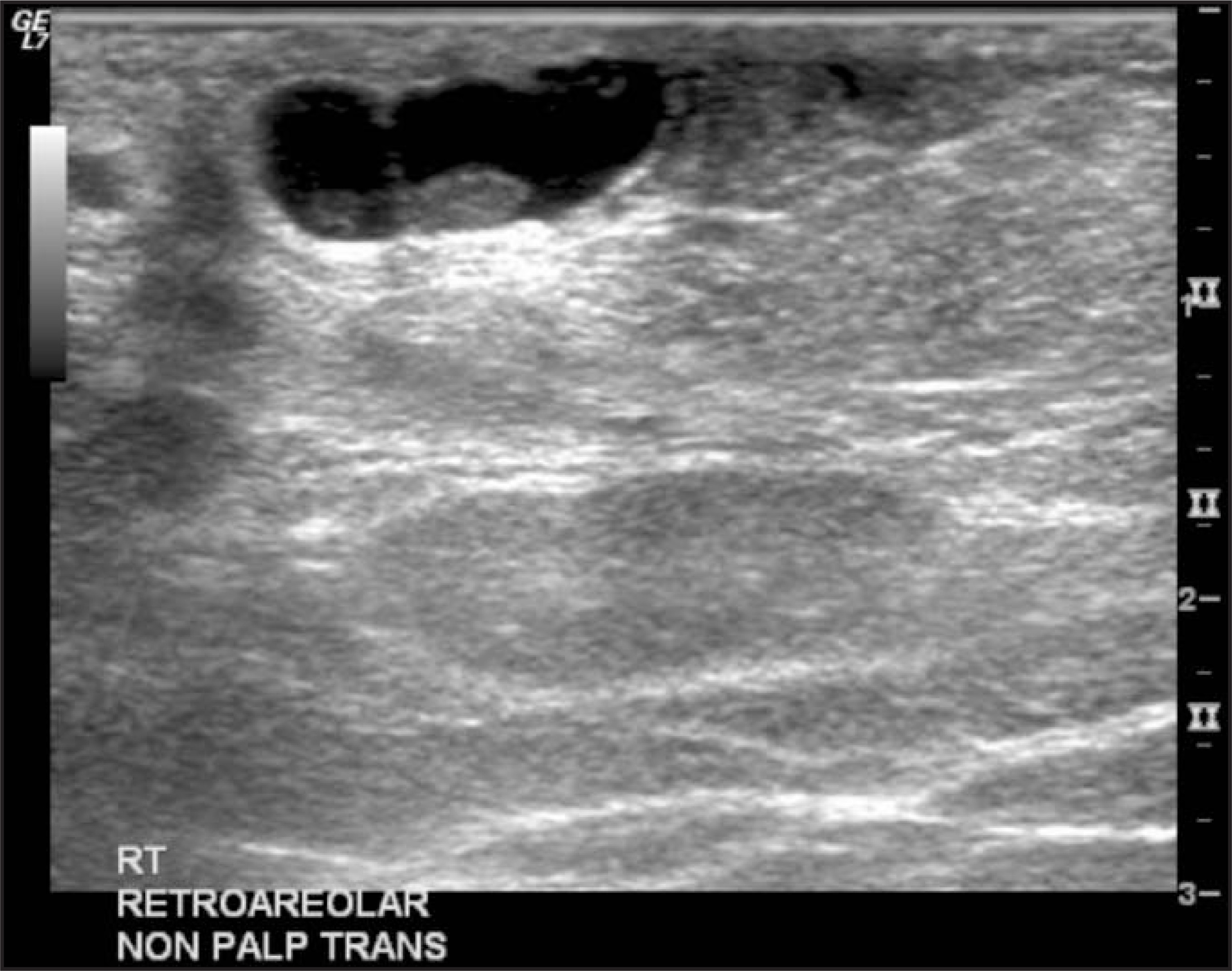
A mass within a dilated duct can result from debris, papilloma, ductal carcinoma in situ (DCIS), invasive ductal carcinoma, or papillary carcinoma. Causes of a bloody or serosanguineous nipple discharge from a single duct include hyperplasia, papilloma, DCIS, invasive ductal carcinoma, and pregnancy. Pathologic differential diagnosis includes papillomatosis (epitheliosis), a term that de scribes microscopic duct hyperplasia.
DIAGNOSIS
Intraductal papilloma with focal atypia
DISCUSSION
Intraductal papillomas are either solitary or multiple and can be benign or can contain atypical cells, as in this case. Papillomas occur inwomen in the fifth through seventh decades, with an average age of presentation of 48 years. It arises most often in the central part of the breastfrom a lactiferous duct but can occur in any quadrant of the breast. Patients typically report a bloody or clear nipple discharge lasting <6months.1
Often physical examination, mammography, and ultrasound are nonrevealing. Rarely, a subareolar mass may be palpable with a centralsolitary papilloma. On mammography, a round, well-circumscribed mass, which may contain calcifications, can be seen in the subareolarregion.2 In 90% of cases, papillomas arise within 1 cm of the nipple.3Ultrasound can reveal a solid oval, round, or microlobulated hypoechoic mass that can contain internal cystic spaces. Nipple discharge occurs in most patients with a central papilloma, as also seen in this case.Ultrasound can show a solid mass in a fluid-filled subareolar duct. Galactography can depict an intraductal or intraluminal filling defect.2
This intraductal papilloma exhibited cytological and architectural atypia on histopathological evaluation—as evidenced by the presence of delicate fibrovascular stalks and foci of atypical ductal hyperplasia (Figure 3A). Most duct epithelial cells were oriented perpendicular to the fibrovascular stalks, with an essentially uniform glandular pattern. The proliferating epithelial cells showed hyperchromatic nuclei and rare mitotic activity. Apocrine metaplasia was absent (Figure 3B). A typical benign intraductal papilloma shows a haphazard arrangement of epithelial cells, normochromic nuclei, apocrine metaplasia, a complex glandular pattern, and prominent fibrovascular cores and florid nonatypical epithelial hyperplasia.The degree of cytological and architectural atypia did not attain the level of carcinoma in this specimen.
Treatment is typically surgical and follow-up is controversial. In patients in whom atypia, nonconcordant imaging findings, or papillarycarcinoma is found, surgical excisional biopsy is universally recommended, as occurred in this case. When benign papilloma is found oncore biopsy, a conservative approach is surgical excision, and the use of follow-up imaging alone is controversial.2 Adherence to screening mammography is strongly encouraged3 because these women have a 1.5- to 2-time relative risk of developing invasive breast carcinoma in their lifetimes.4
CONCLUSION
Intraductal papillomas are fairly common lesions, and a high degree of suspicion is necessary in their evaluation. They may containatypical cells, and in these cases surgical excision is universally recommended. Close follow-up with screening mammography is recommended in those cases where benign histology is noted.
References
- Pisano E, Braeuning M, Burke E. Diagnosis please. Case 8: Solitary intraductal papilloma. Radiology. 1999;210:795-798.
- Ikeda D. Breast Imaging: The Requisites. 2004:113-116.
- Osuch J. Benign lesions of the breast other than fibrocystic change. Obstet Gynecol Clin North Am. 1987;14:703-710.
- Hartmann L, Sellers T, Frost M. Benign breast disease and the risk of breast cancer. N Engl J Med. 2005;353:229-237.
Citation
. Intraductal papilloma with focal atypia. Applied Radiology. 2008;37(6):37-38. doi:10.37549/AR1620.