Islet-cell tumors of the pancreas: Spectrum of MDCT findings. A pictorial essay.
Applied Radiology — Vol. 38 , Issue 11 , pp. 10 -28
DOI: 10.37549/AR1725
Published: November 1, 2009
Categories
Although pancreatic islet-cell tumors account for only 1% to 5% of all pancreatic neoplasms, they represent an important subset ofpancreatic neoplasms due to their substantially improved prognosis compared to pancreatic adenocarcinoma.1,2Pancreatic islet-celltumors, also referred to as neuroendocrine tumors, may present with a broad spectrum of clinical and imaging manifestations, sometimes quite dramatic, depending on whether lesions are solitary or multiple, syndromic or nonsyndromic, inherited or sporadic, or benign or malignant. The role of the radiologist in imaging pancreatic islet-cell tumors is to localize the tumor and evaluate the extent of disease, which in turn helps determine whether the patient is an operative candidate and helps guide therapy.
Ninety-five percent of pancreatic islet-cell tumors are solitary and sporadic. Generally, these do not exhibit hormonal hypersecretion (non-syndromic) and are clinically silent until they grow large enough to cause symptoms.
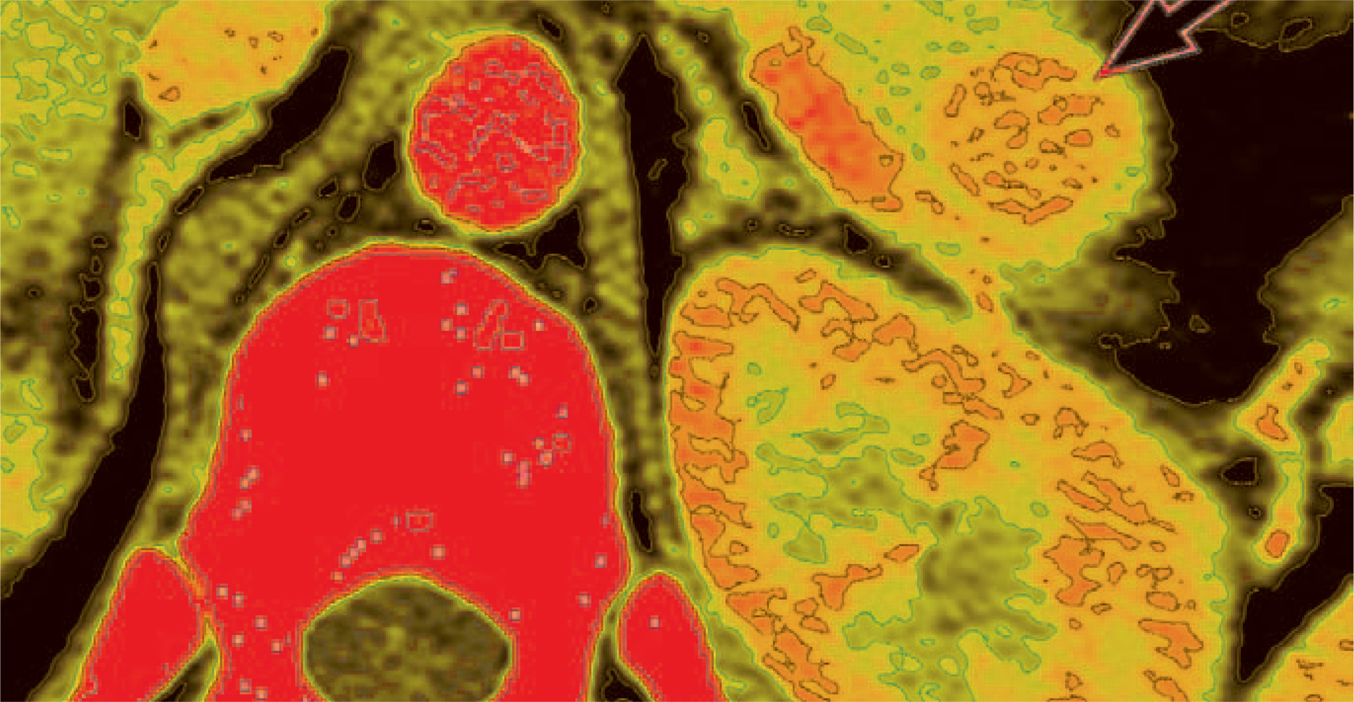
On the other hand, hyperfunctioning islet-cell tumors (referred to as syndromic islet-cell tumors) manifest much earlier, and at a much smaller size due to symptoms caused by hormonal oversecretion. Often these syndromic tumors are barely visible by imaging, in spite of a dramatic clinical presentation. These tumors are classified according to the predominant hormone of oversecretion: insulinoma, gastrinoma, vasoactive intestinal peptide-secreting tumors (VIPoma), glucagonoma, somatostatinoma, and growth-hormone-releasing factor-secreting tumors (GRFomas).
Insulinomas account for 50% of all islet-cell tumors and are the most common islet-cell tumor subtype.6 The majority of insulinomas (80% to 90%) are benign, solitary and are <2 cm at presentation.7These tumors cause fluctuations in serum glucose levels in patients, with associated morbidity related to severe hypoglycemia, occasionally leading to multiple seizures. Surgical resection of insulinomas is thetreatment of choice, with symptoms promptly resolving after resection.
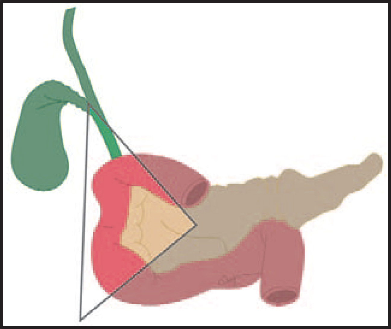
Pancreatic islet-cell tumors other than insulinomas have a higher malignancy rate, approaching 60% to 90%.1 Gastrinomas are the second most common islet-cell tumor, with approximately 25% associated with multiple endocrine neoplasia (MEN-1).4 Resection of gastrinomas is often difficult due to locally invasive features or metastatic disease in more than half of patients.1 When a gastrinoma is suspected, one should carefully inspect the “gastrinoma triangle,” an anatomic triangle bounded by the junction of the cystic duct insertion on the common bile duct, the body of the pancreas, and the junction of the second and third portions of the duodenum (Figure 1). About 85% to 90% of gastrinomas are present within the gastrinoma triangle; the search for this tumor should not be limited solely to the pancreas but should include the boundaries of this anatomic triangle. Similarly, VIPomas, somatostatinomas, and GRFomas may also arise outside of the pancreas.4
Unlike sporadic islet-cell tumors, inherited pancreatic islet-cell tumors are usually multiple in location and most often related to MEN-1. They can also be seen with other syndromes such as Von Hippel-Lindau, tuberous sclerosis and neurofibromatosis type 1.3 Whether pancreatic lesions associated with MEN-1 are more likely benign or malignant is still much debated. Differentiation between inherited and sporadic etiologies is important not only because of their different imaging presentation, but also because they undergo different therapies.4,5
Imaging is helpful in determining extent of disease, with implications on the patient’s overall prognosis. Tumor growth and spread is considered the major determinant of patient survival. Pancreatic islet-cell tumors spread first to regional lymph nodes, then to the liver, bone, and rarely, to distant sites such as the lung or brain. Lymphovascular invasion is an important feature correlating to survival.8 Fifty percent to seventy percent of deaths in patients with metastatic disease are thought to be caused by tumor progression.4
Imaging options
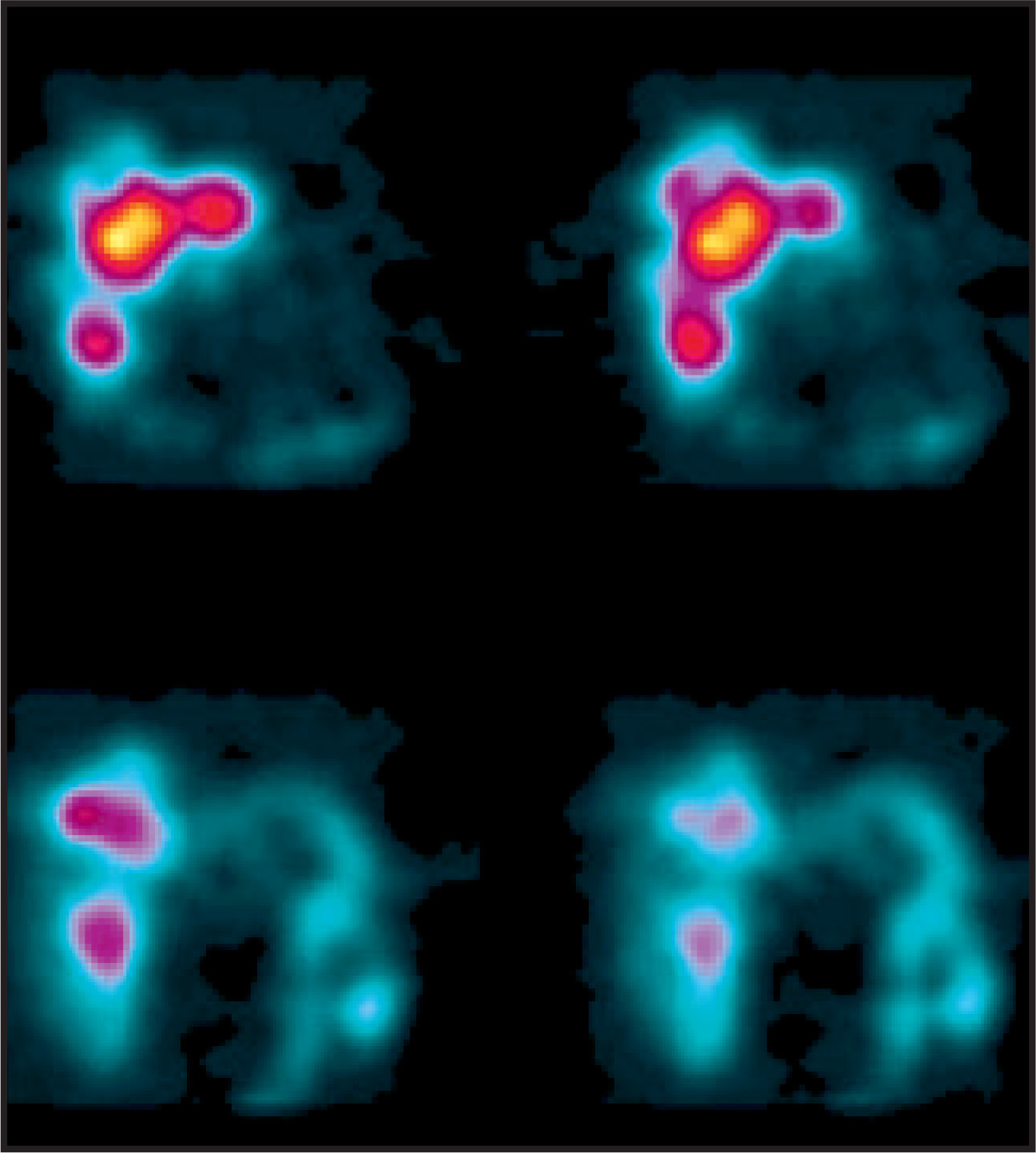
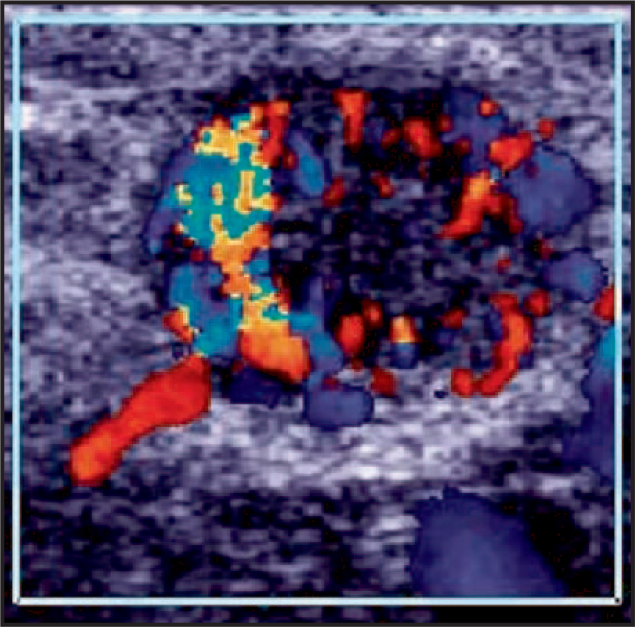
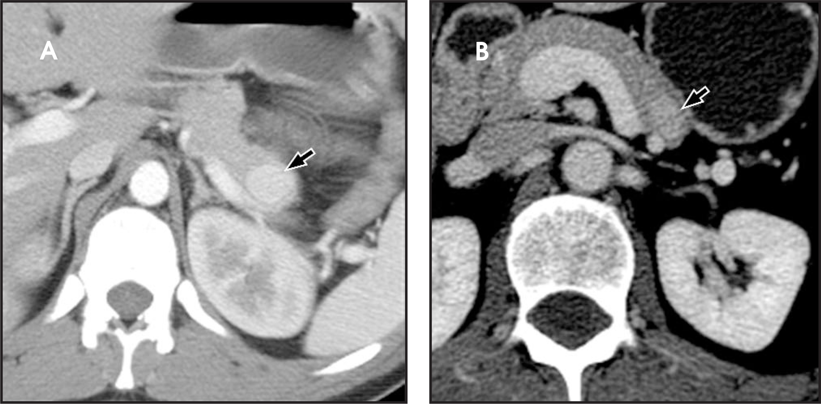
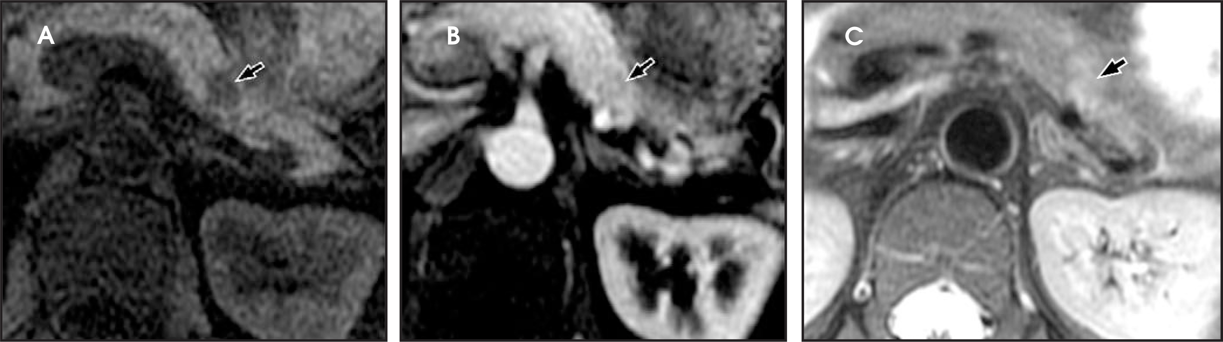
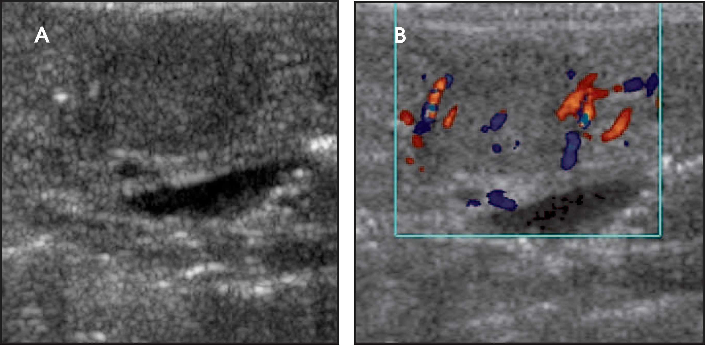
Computed tomography (CT) is the initial imaging modality of choice, but is estimated to detect only 60% to 70% of primary pancreatic insulinomas and gastrinomas.9,10 Magnetic resonance imaging (MRI), intraoperative ultrasound, endoscopic ultrasound, selective angiography and portal venous sampling may serve as adjunctive methods of evaluating disease.11 Recent literature suggests that MRI may be at least as sensitive as multiphasic CT in detecting pancreatic islet-cell tumors.12 In our experience, a multimodality approach is useful for diagnosing and staging pancreatic islet-cell tumors as well as helping to guide surgery (Figures 2-6).
Treatment
Preoperative imaging evaluation is important for surgical planning because resection is the only definitive curative treatment, regardless of benign or malignant pathology. One should determine the location of the tumor within the pancreas, and assess presence of multiple tumors, including tumors outside of the pancreas, and looking for evidence of local invasion or metastatic disease.13 These findings will affect surgical candidacy and help guide the type of surgery to be performed, such as enucleation, partial, or rarely, total pancreatectomy.14
There are several therapeutic options for treating advanced disease from islet-cell tumor. Liver metastases in patients with insulinomas and gastrinomas are estimated to be present in up to 35% to 74% of cases.4 Chemotherapy has been used for metastatic disease with some success in limited trials.15 Interferon and octreotide, as well as hepatic artery embolization or ligation, have also been used.16,17 In select cases liver transplantation has been performed.14 There is some controversy over the efficacy of debulking for treatment of metastatic disease.15
CT technique
At our institution, most patients are initially evaluated with a pancreatic-protocol CT. We use 8- and 16-detector CT scanners in our hospital (Lightspeed, GE Healthcare, Chalfont St. Giles, U.K.) and a 64-detector scanner at our outpatient facility (SOMATOM Sensation 64, Siemens Healthcare, Malvern, PA). Fifteen to thirty minutes prior to the study, patients drink 1350 cc of VoLumen, a neutral oral contrast agent, (Bracco Diagnostics, Princeton, N.J.) and another 450 cc immediately prior to scanning. Oral contrast facilitates luminal distention of the stomach and duodenum. A scout image of the abdomen is initially acquired, followed by limited noncontrast images of the abdomen to localize the pancreas (5 mm thickness). Approximately 140 cc of nonionic contrast (ISOVUE-370, Bracco Diagnostics) is subsequently administered intravenously at an injection rate of 4 cc/sec, with arterial-phase images acquired 40 seconds after injection (1.25 mm thickness, helical acquisition, 0.6 mm interval, 1.35:1 pitch) from the level of the diaphragm to the top of the iliac crests. Portal venous-phase images are obtained at a 60-second delay following injection (2.5 mm thickness, 1.25 mm interval, 1.35:1 pitch). Delayed images are routinely acquired through the kidneys 3 minutes after contrast administration (5 mm thickness).
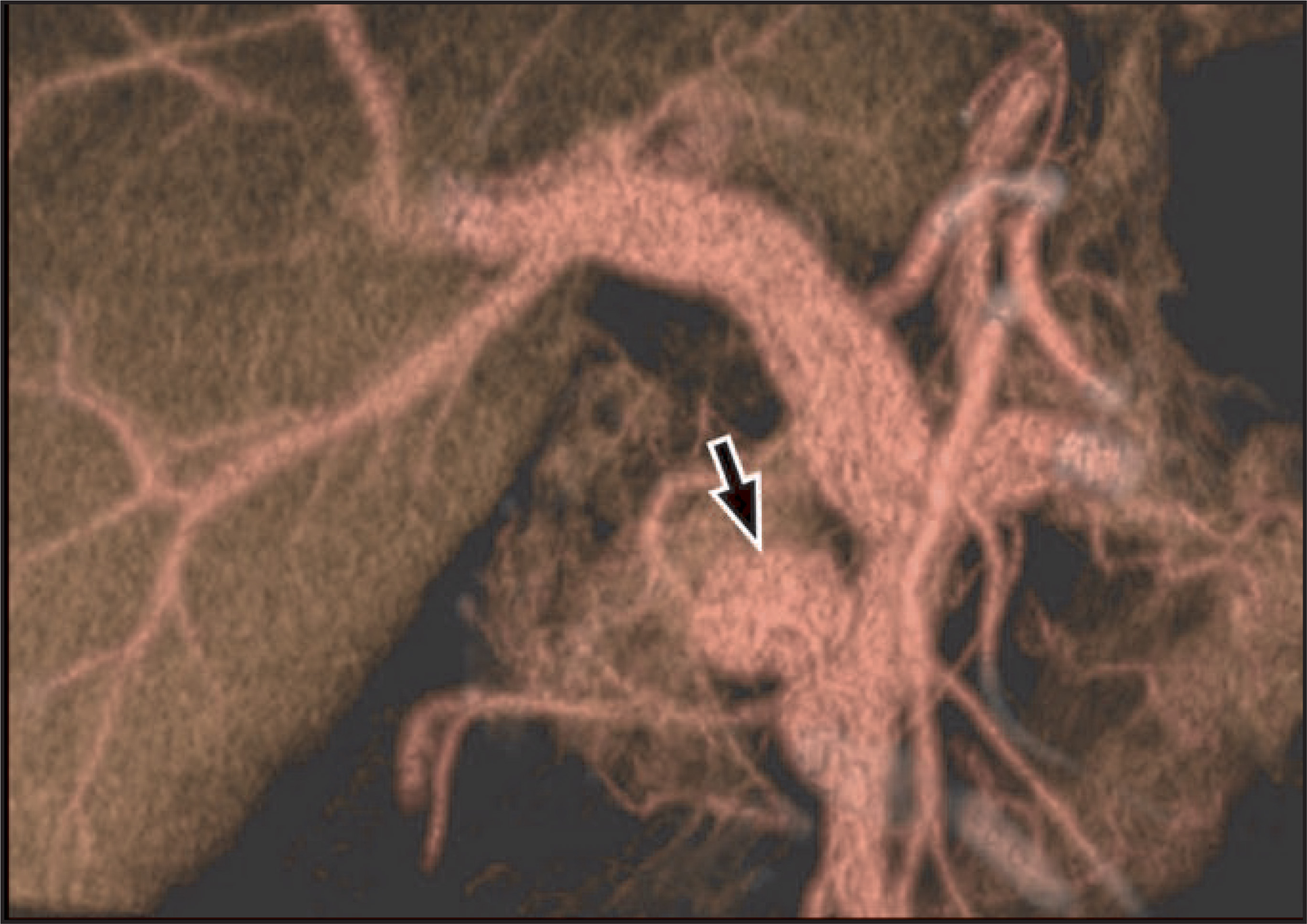
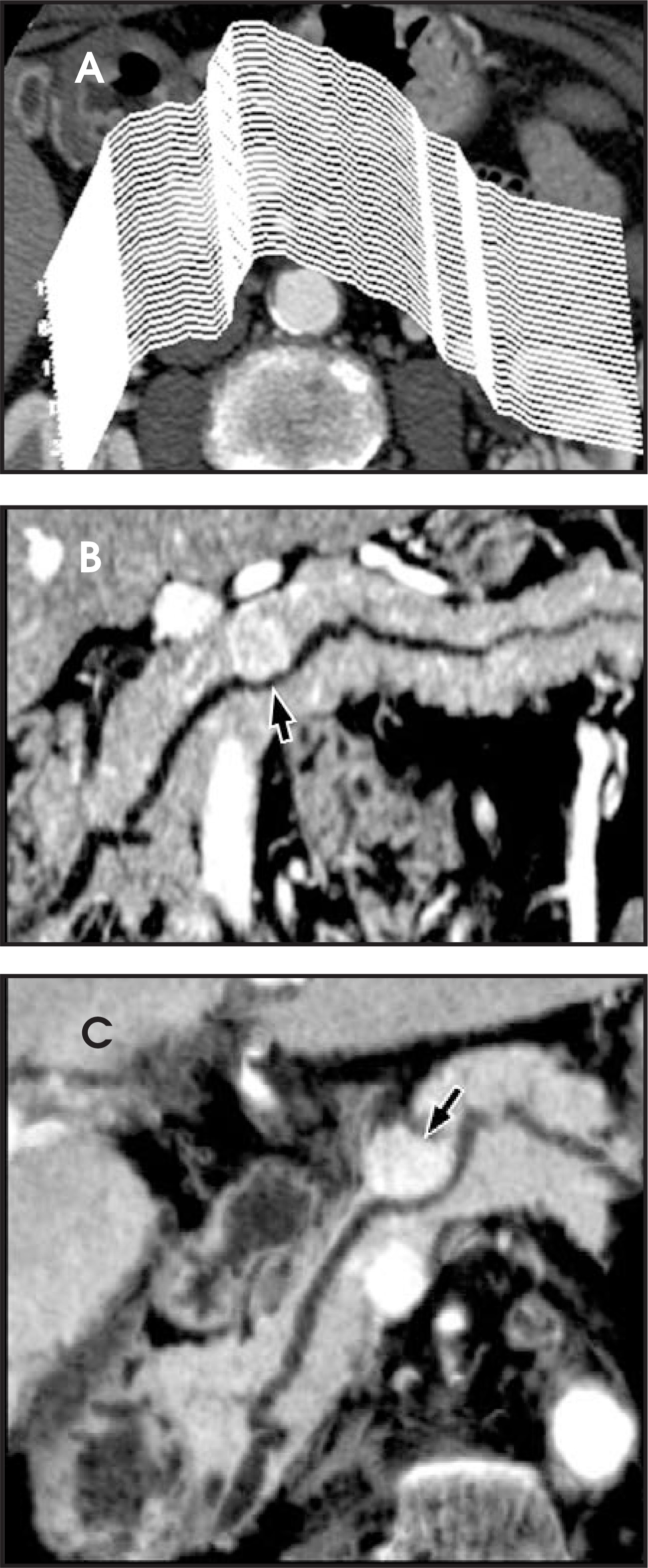
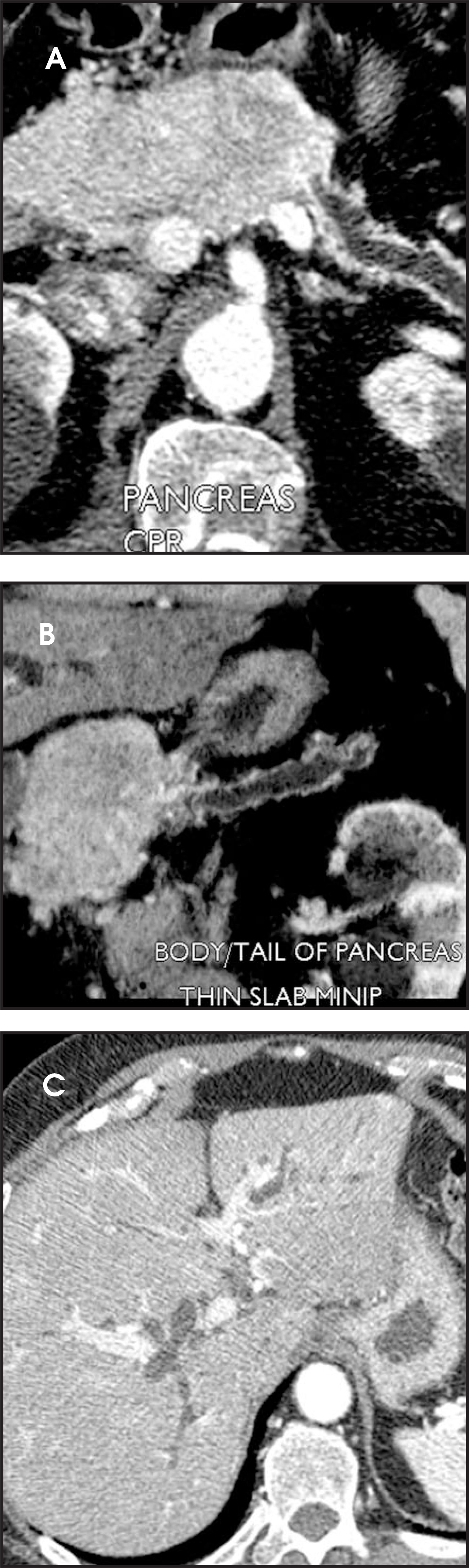
The helically acquired volumetric data is postprocessed by specially trained technologists in our dedicated 3-dimensional laboratory to generate multiplanar reconstruction images including maximum intensity projections (MIPs), minimum intensity projections (MINIPs) and curved planar reformations (CPRs). These reconstructions provide better anatomic definition of keystructures commonly affected by locally invasive pancreatic neoplasms (Figure 7), including thin-slab MIPs of the celiac origin, portal and splenic veins; CPRs of the superior mesenteric vein and artery, hepatic artery, gastroduodenal artery, splenic artery, distal common bile duct and pancreatic ducts; thin-slab MINIPs of the pancreatic ducts; and, coronal MPRs in the plane of the main portal vein through the entirety of the pancreas.
Typical imaging findings
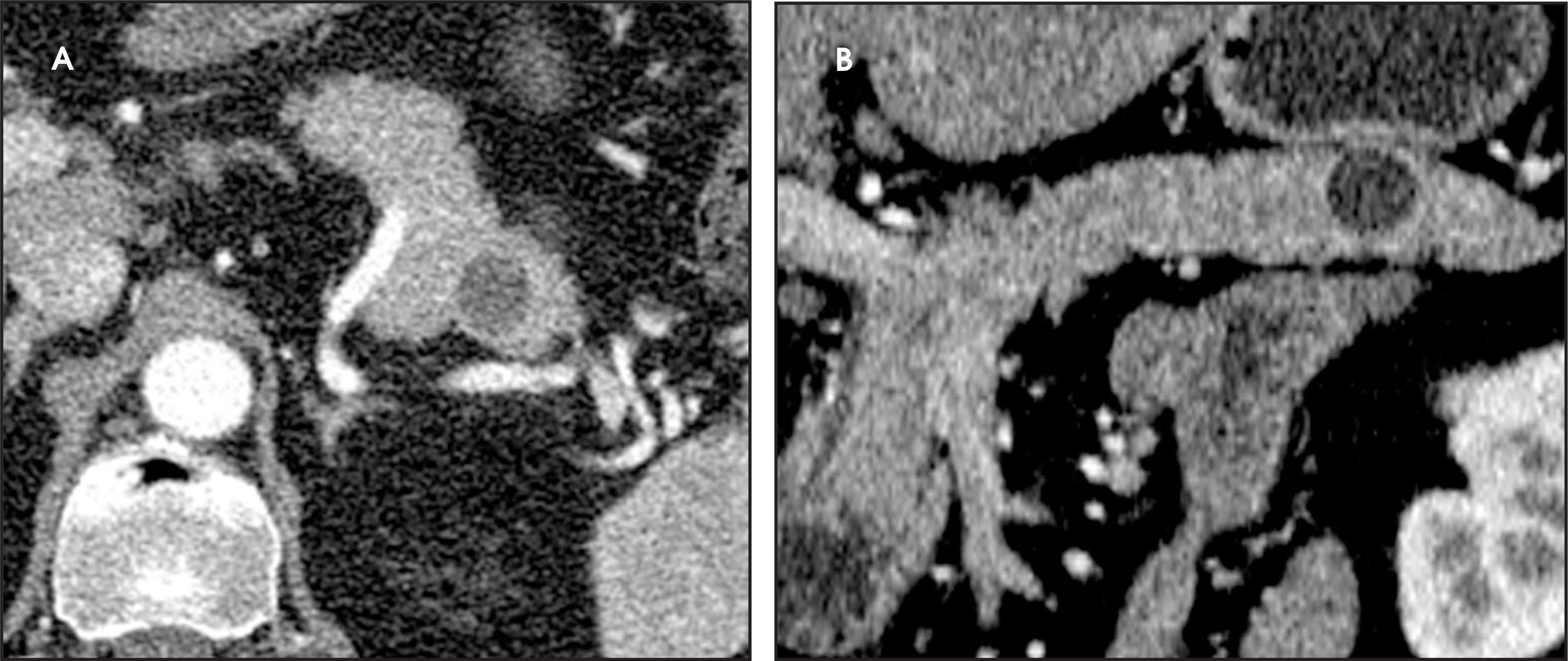
The typical appearance of a pancreatic islet-cell tumor is a well-circumscribed, solitary, hypervascular mass (Figure 4). Because islet-cell tumors are generally soft, and lack associated desmoplastic reaction, they do not usually cause pancreatic duct obstruction or dilation. Mild distortion of the duct may be seen due to adjacent mass effect or if the tumor is located near the main pancreatic duct (Figure 8).
Typically, pancreatic islet-cell tumors and metastases are best seen during the late arterial phase (Figures 4, 9). On portal-venous phase, tumors will characteristically demonstrate late retention of contrast (Figures 4, 10). While arterial enhancement patterns are accepted as the most common imaging appearance, atypical enhancement patterns do occur, and varying opinions on optimal phase ofenhancement have been reported.9,12,18,19,20 Given the variability in peak enhancement and enhancement patterns, multiphasic imaging isgenerally accepted as the best method to ensure optimal detection of pancreatic islet-cell tumors.
On ultrasound, pancreatic islet-cell tumors are generally hypoechoic and well circumscribed. Color Doppler imaging will reveal prominent vascularity of the tumor. Often the tumor is difficult to palpate surgically due to its soft texture. Intraoperative ultrasound is helpful in localizing small tumors and delineating their relationship to the main pancreatic duct and adjacent vessels (Figure 6).
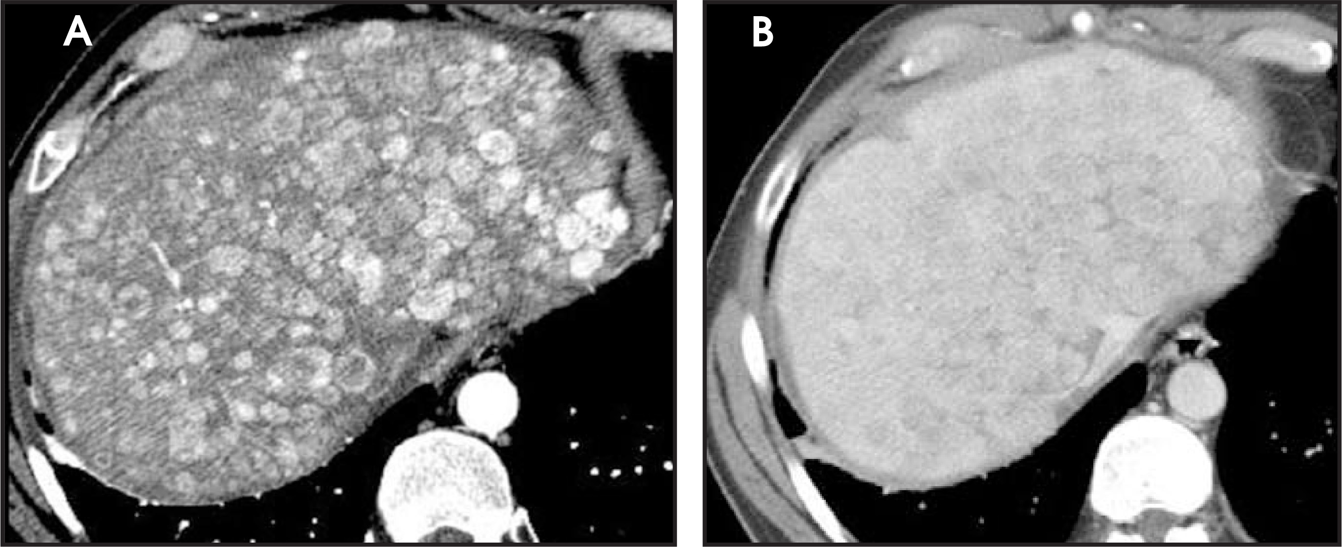
In patients with inherited islet-cell tumors, multiple hypervascular pancreatic masses are often present (Figures 11 and 12). Additionally, other pancreatic lesions are commonly seen, such as multiple pancreatic cysts in patients with Von Hippel-Lindau syndrome (Figures 12 and 13).
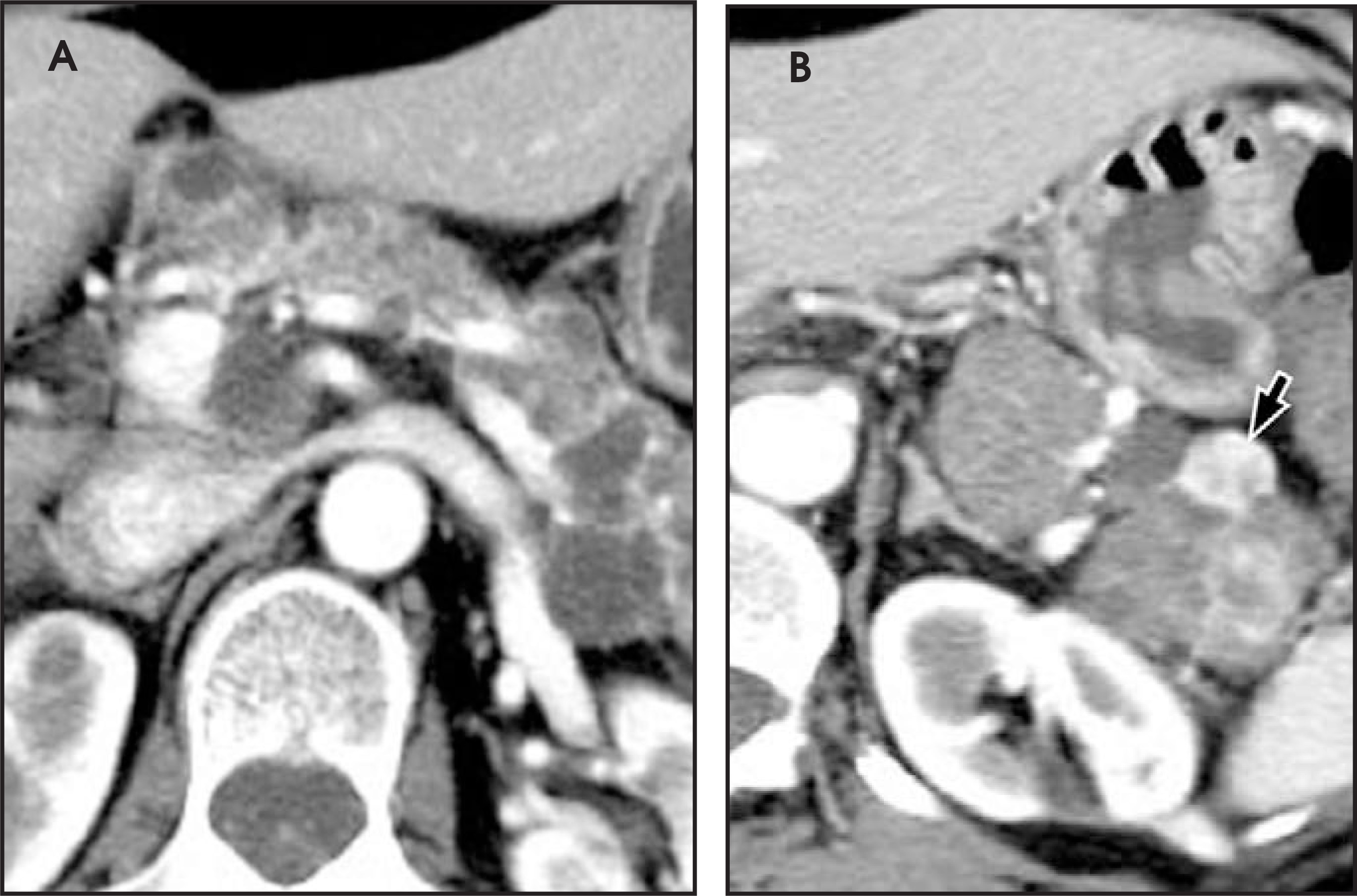
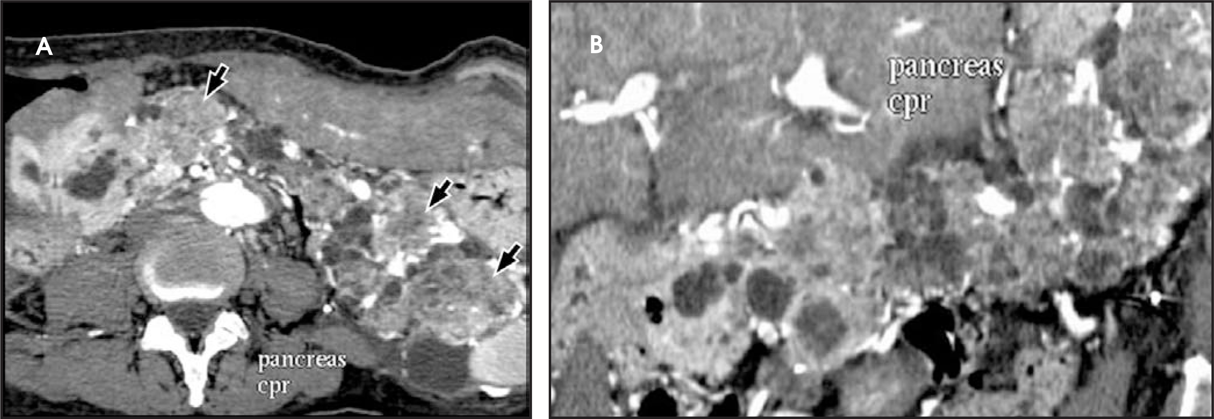
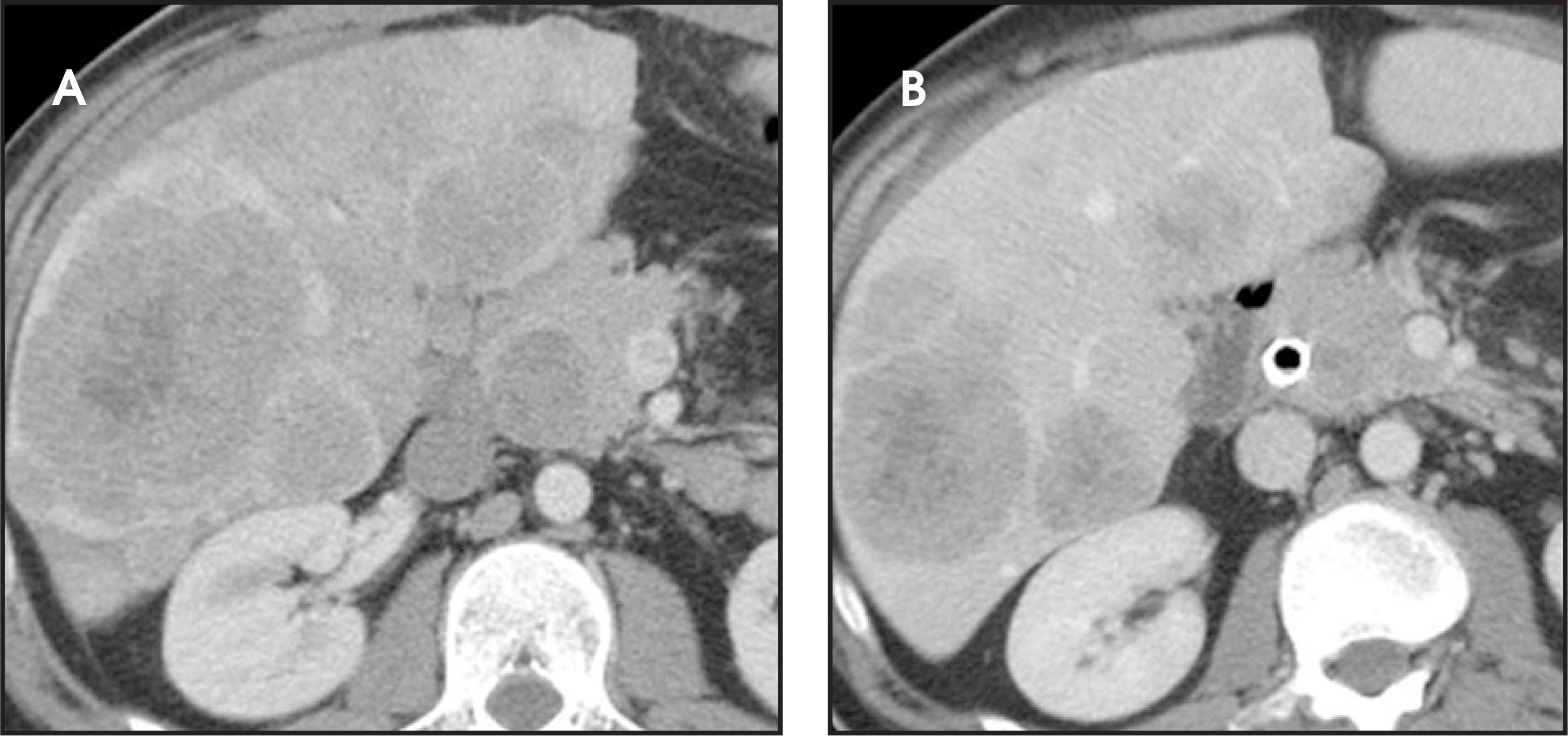
Metastatic lesions occurring in the liver are generally hypervascular in appearance on late arterial-phase scans, with similar enhancement characteristics as the primary pancreatic neoplasm (Figure 14). Rapid washout from hepatic metastases may render them isodense on venous-phase acquisitions. It is therefore essential to perform arterial-phase imaging to detect liver metastases. As with the primary tumor, liver metastases can become centrally necrotic as they increase in size (Figure 15). Regional adenopathy, liver, bone and pulmonary metastases are additional imaging features of advanced disease (Figures 16 and 17).
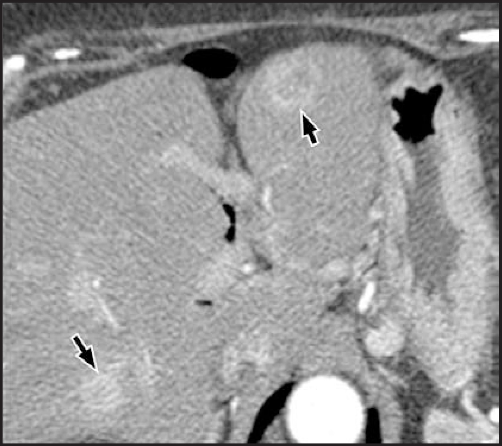
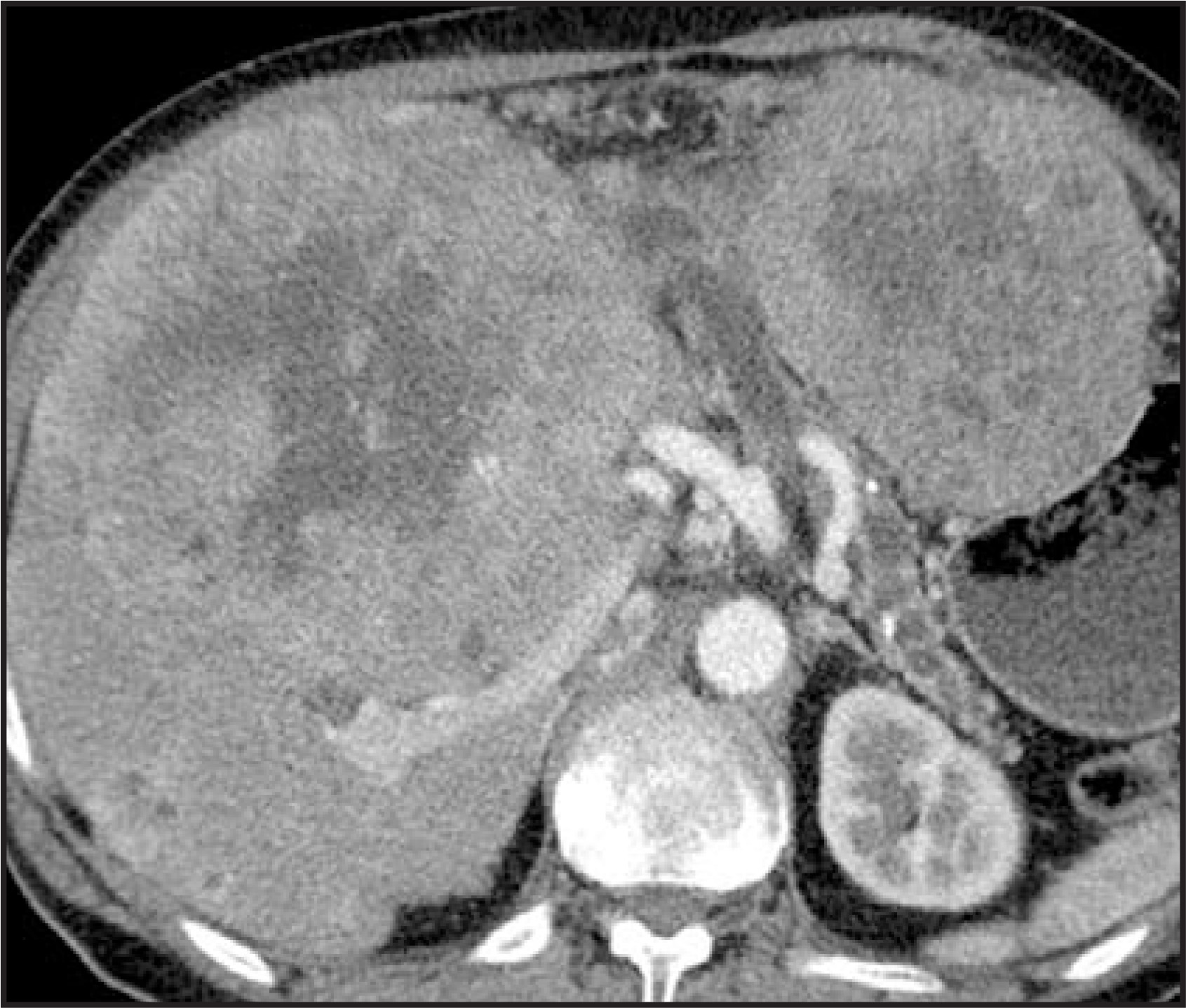
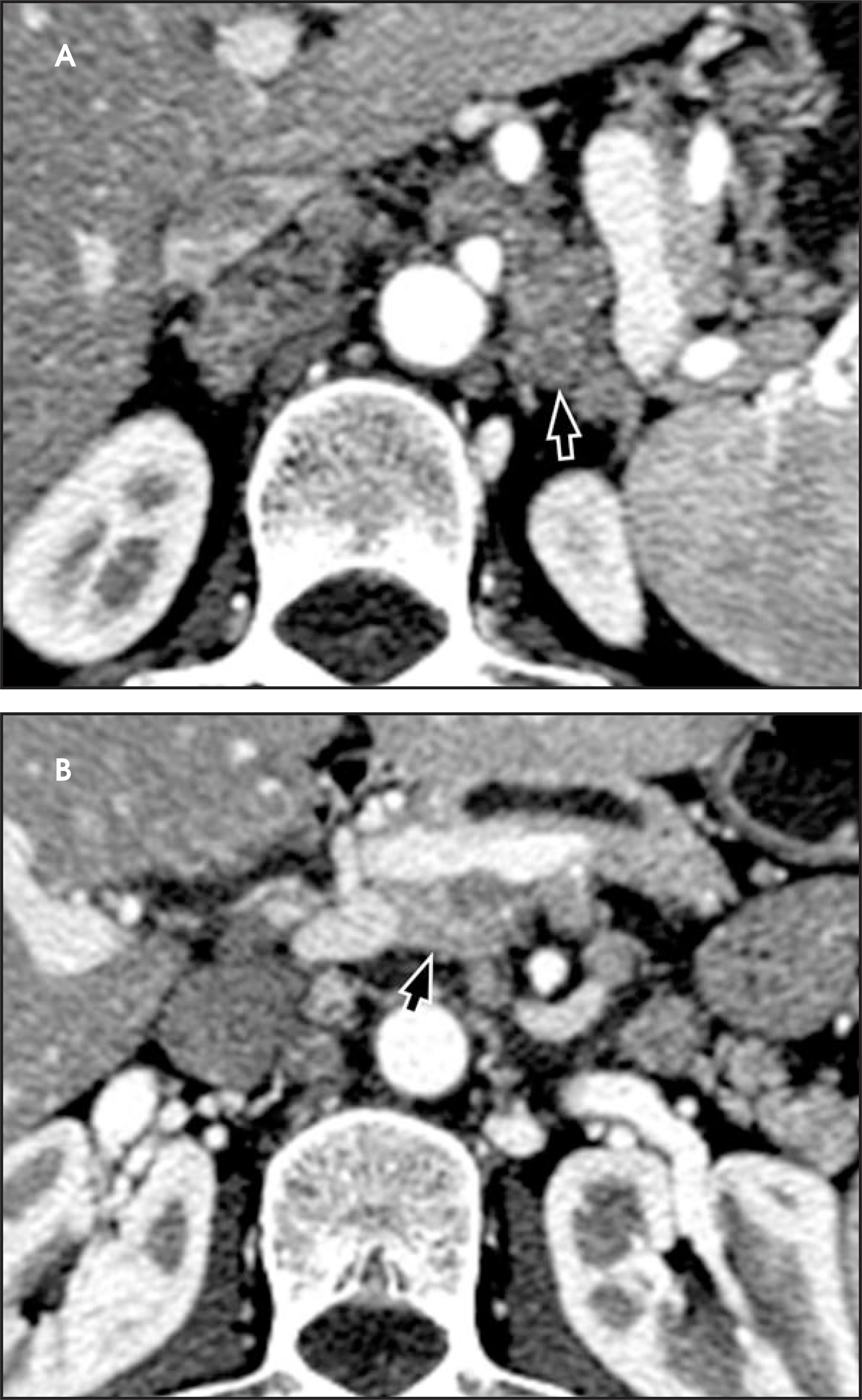
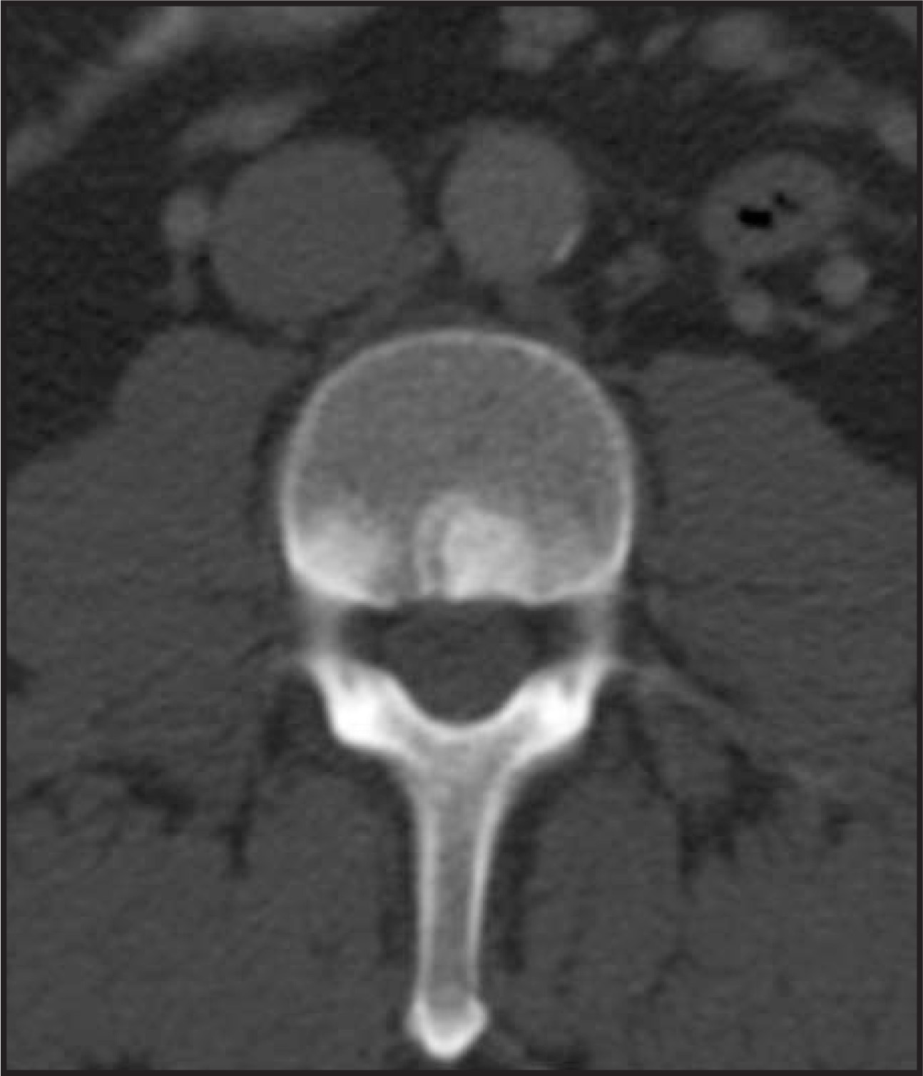
A commonly cited reason for difficulty in visualizing pancreatic islet-cell tumors is the presence of adjacent enhancing vessels. Because pancreatic islet-cell tumors are often intensely vascular, their appearance in the axial cross-section can either mimic, or be obscured by, adjacent vessels of similar caliber.9 Multiplanar reformations as well as multiphasic imaging can be helpful to avoid this potential pitfall. Occasionally, hypervascular metastases to the pancreas can occur, most commonly from renal-cell carcinoma. While rare, these can be mistaken for a pancreatic islet-cell tumor due to similar enhancement characteristics.21
Atypical imaging findings
Atypical imaging appearances of pancreatic islet-cell tumors are not uncommon. While most islet-cell tumors are well-visualized on CT due to their hypervascular nature, some may be isoattenuating to the rest of the pancreas and challenging to visualize. In cases where the lesion is not immediately apparent, subtle pancreatic duct indentation or subtle changes in pancreatic duct caliber may be helpful clues in identifying the location of the lesion (Figure 18). Indeed, small isoattenuating lesions can be particularly difficult to delineate by CT and in some cases may be better seen on endoscopic ultrasound or intraoperative ultrasound.
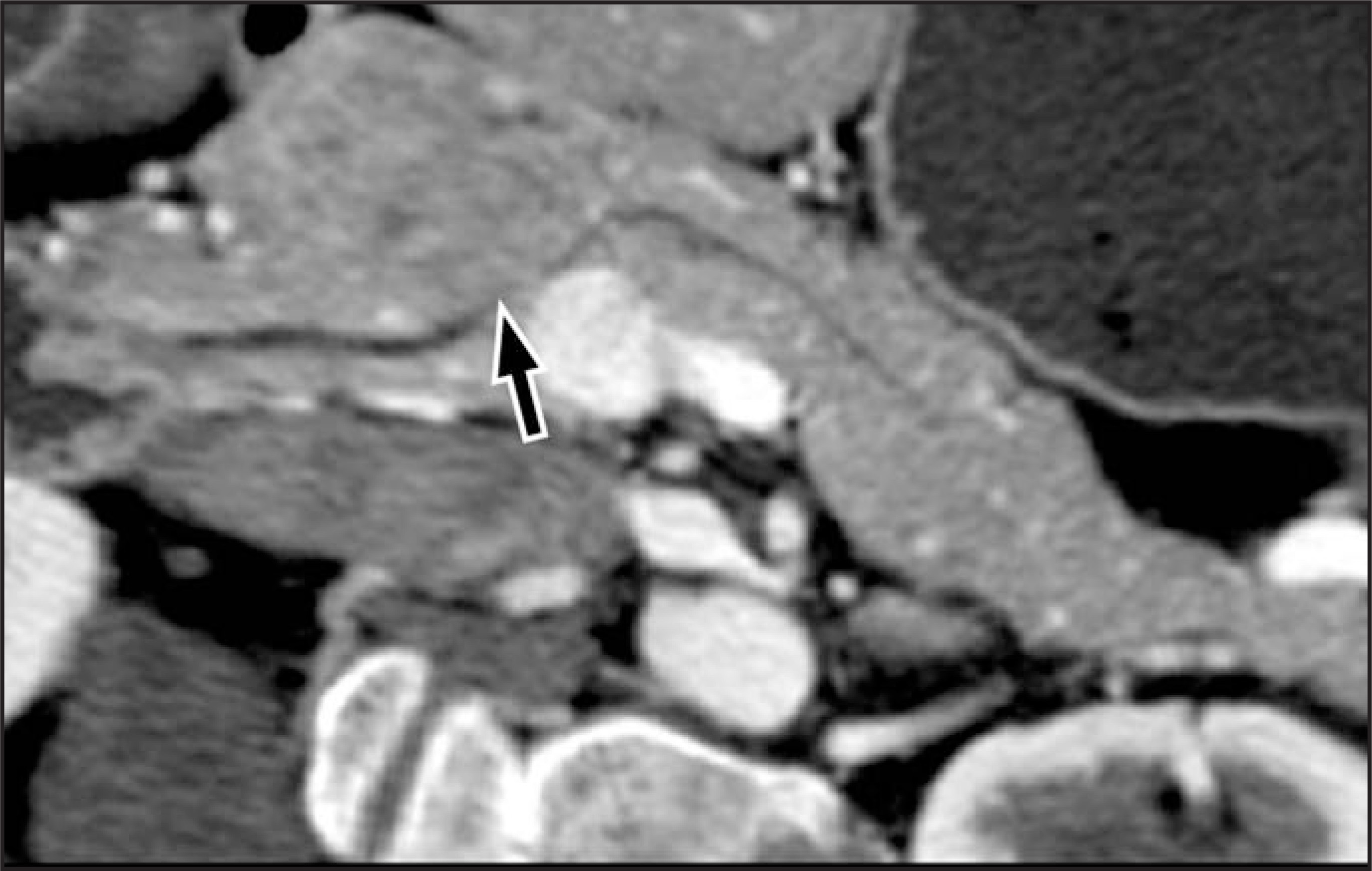
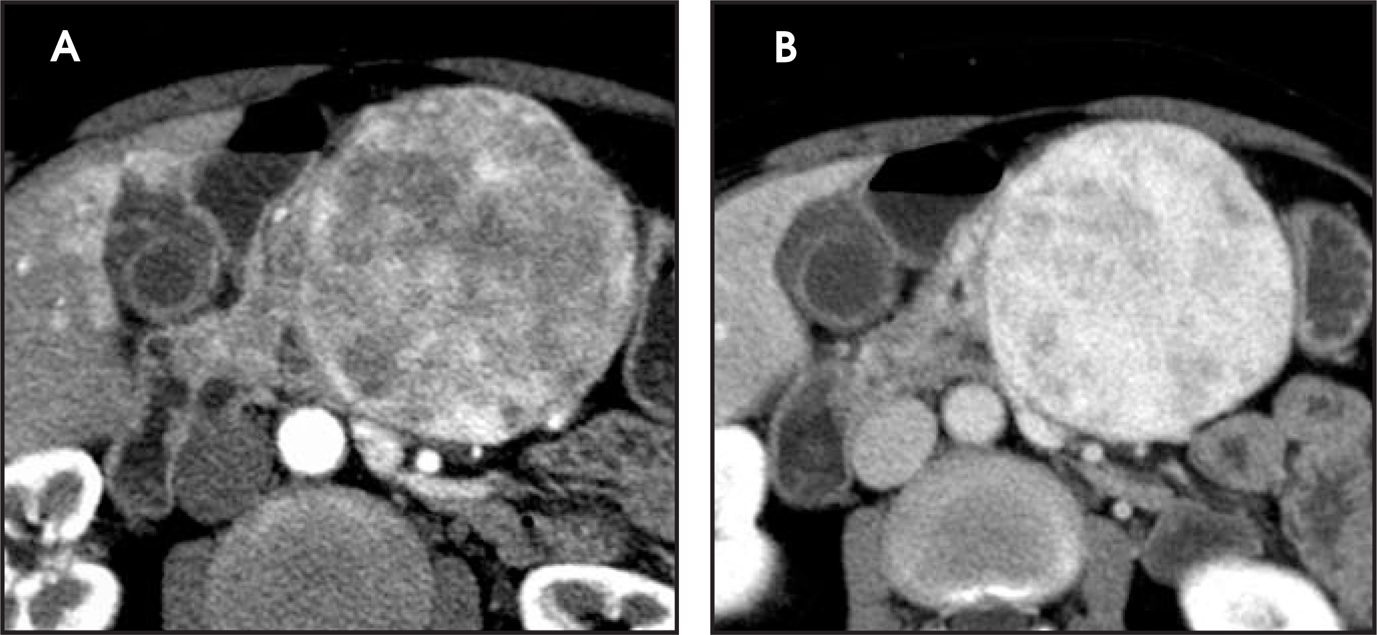
Another atypical appearance of islet-cell tumors is that of cystic or necrotic change. Central necrosis is more commonly seen in larger nonsyndromic pancreatic islet-cell tumors that outgrow their blood supply. Often the lesion periphery will remain hypervascular while the center does not enhance (Figures 19 and 20).22 In our experience in one very rare instance, a pancreatic islet-cell tumor presented with a purely cystic appearance (Figure 21).
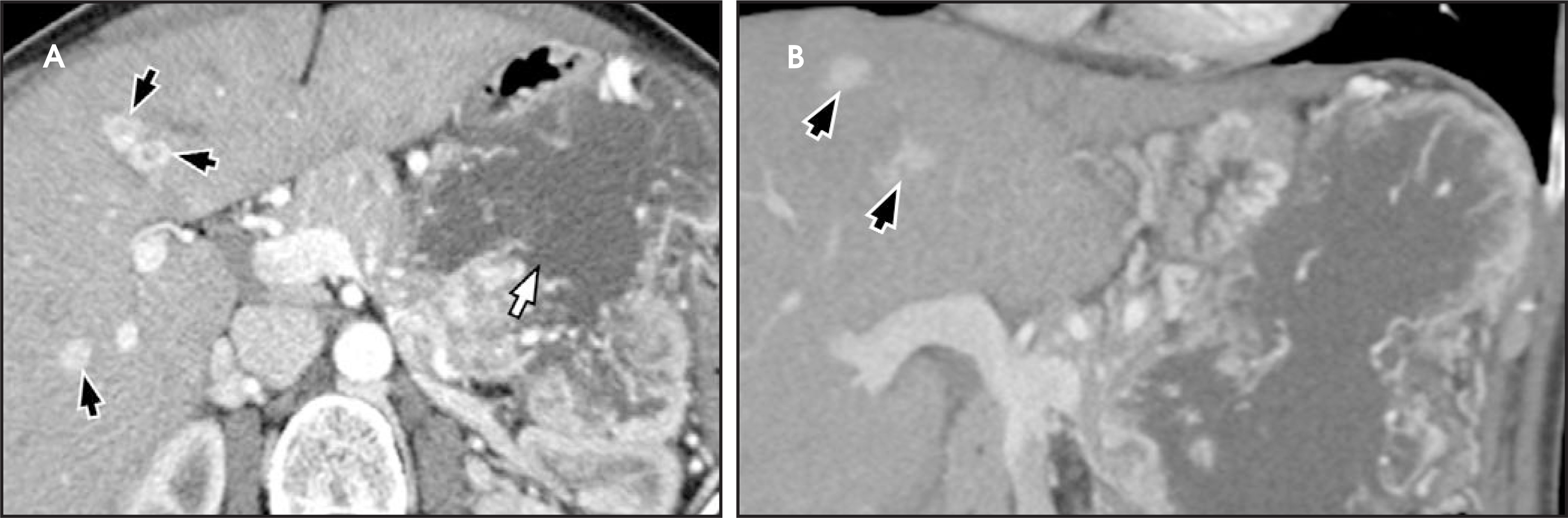
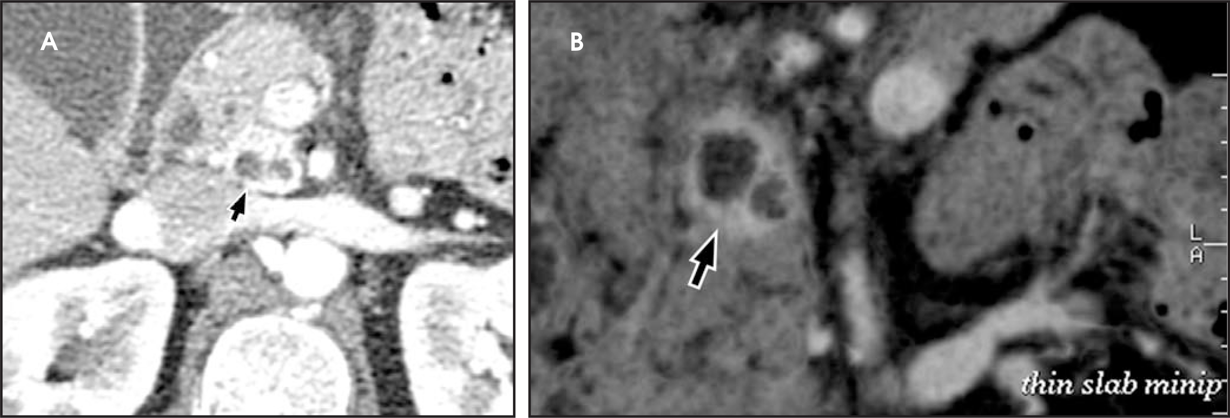
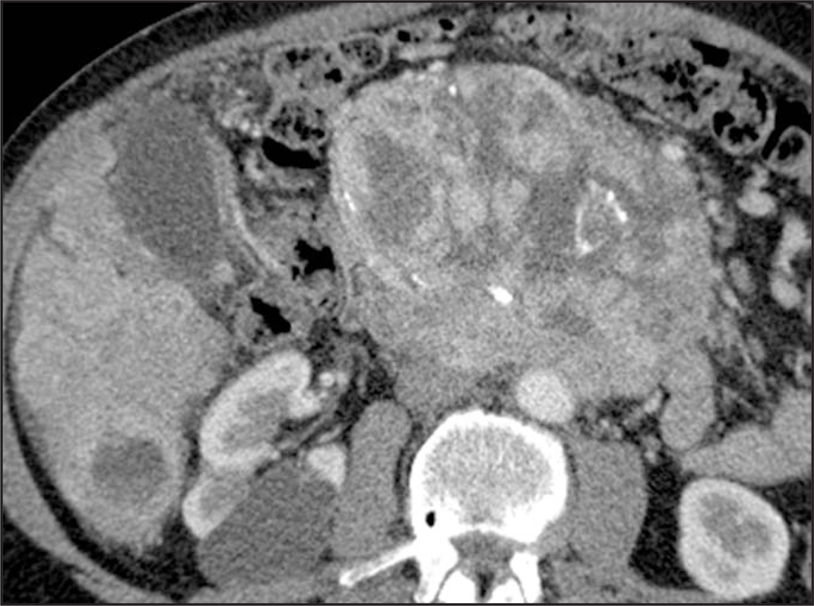
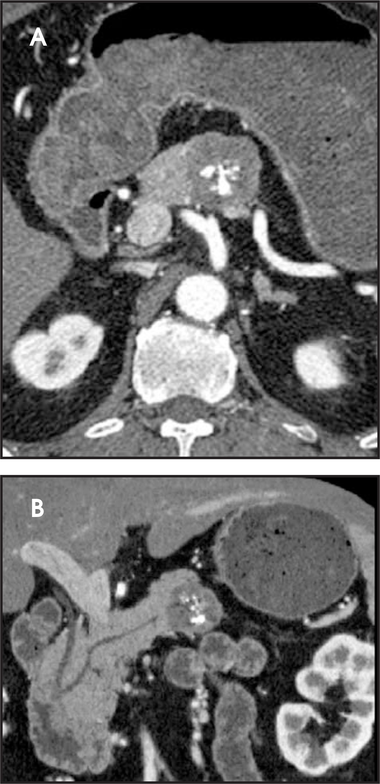
Calcifications are not a typical imaging feature of pancreatic islet-cell tumors but can be seen in up to 20%, best delineated on CT (Figures 22 and 23). The presence of calcifications is more suggestive of a pancreatic islet-cell tumor and can be helpful in differentiating a lesion from adenocarcinoma of the pancreas.6
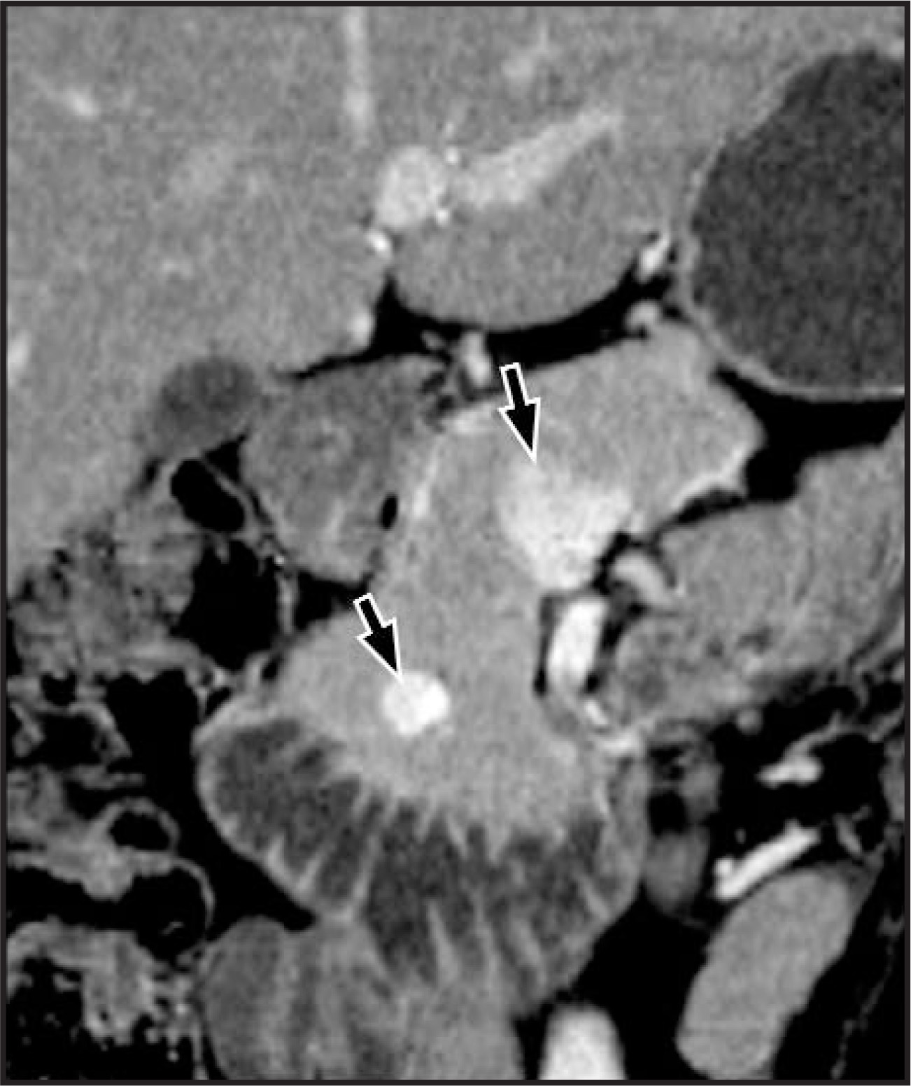
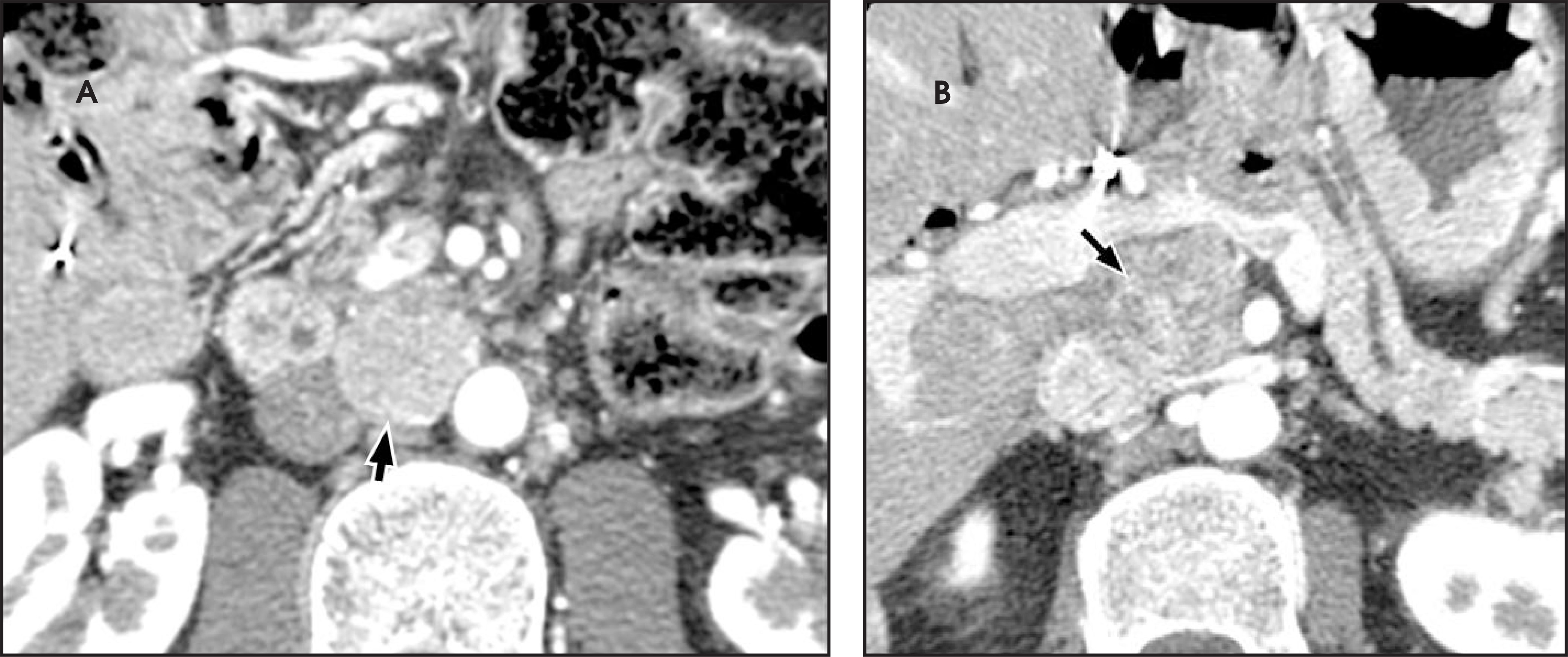
Vascular encasement, narrowing, and/or frank vascular invasion are aggressive findings commonly seen in pancreatic adenocarcinoma. Occasionally, these findings may also be seen with pancreatic islet-cell tumors. In these atypical islet-cell tumors, the degree of vascular encasement or occlusion is often disproportionally less than that found in a similarly sized pancreatic adenocarcinoma (Figures 24–28). Beyond encasement, direct invasion of the portal vein with resulting tumor thrombus can rarely occur in islet-cell tumors, againmimicking an adenocarcinoma of the pancreas (Figure 29).
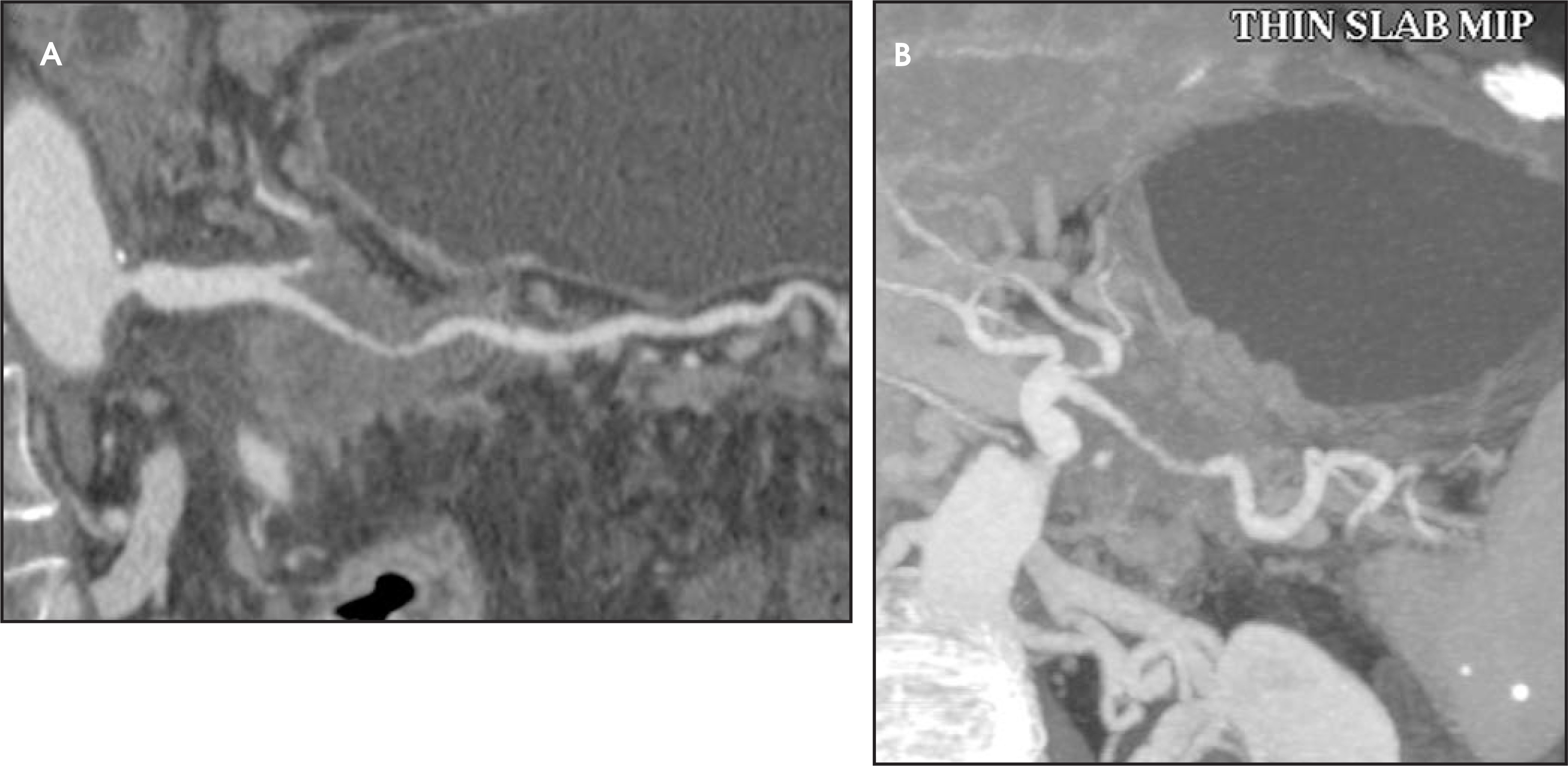
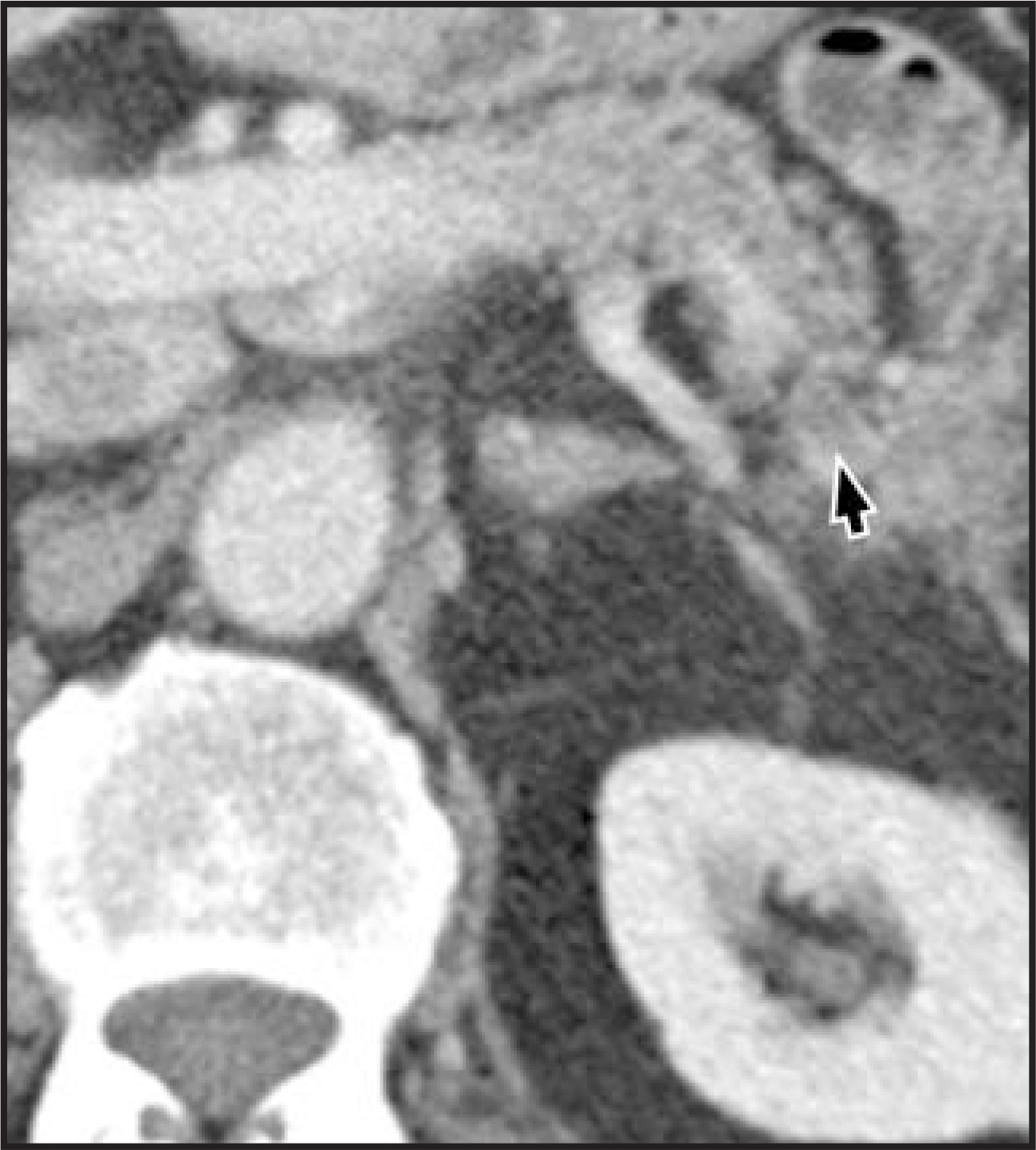
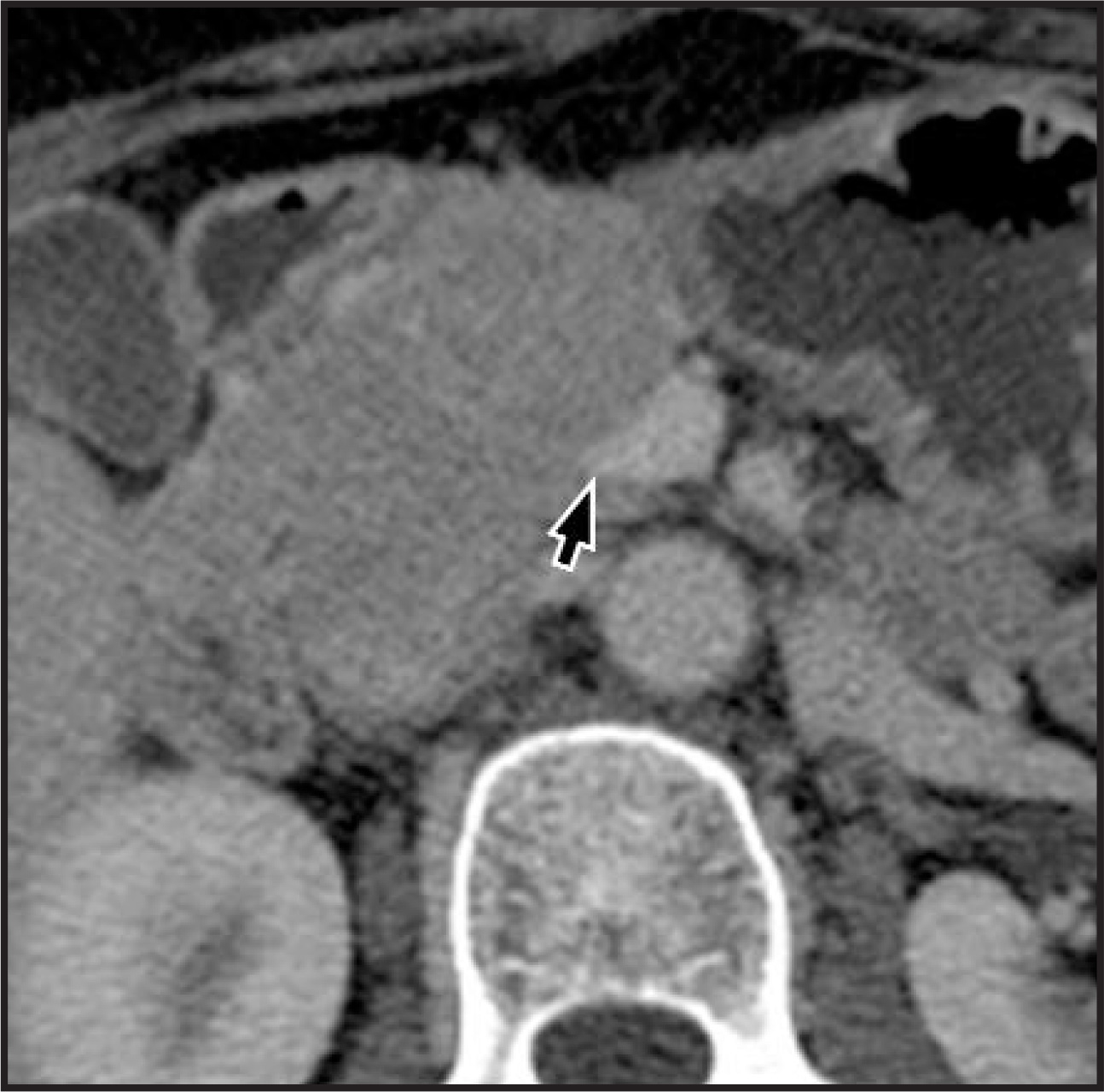
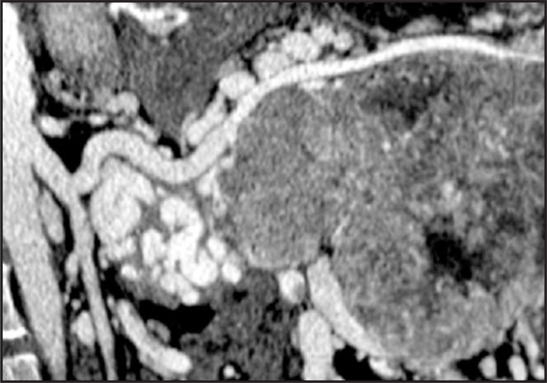
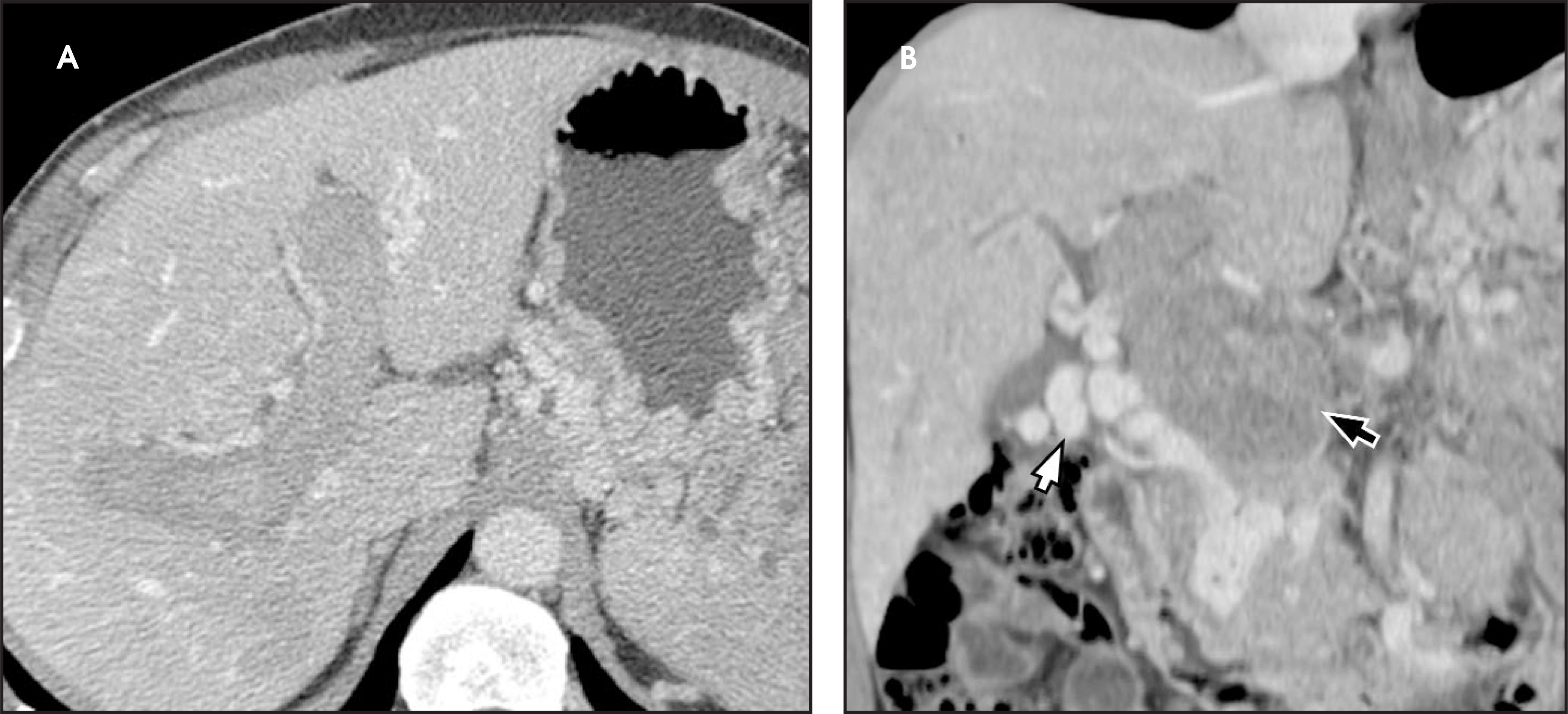
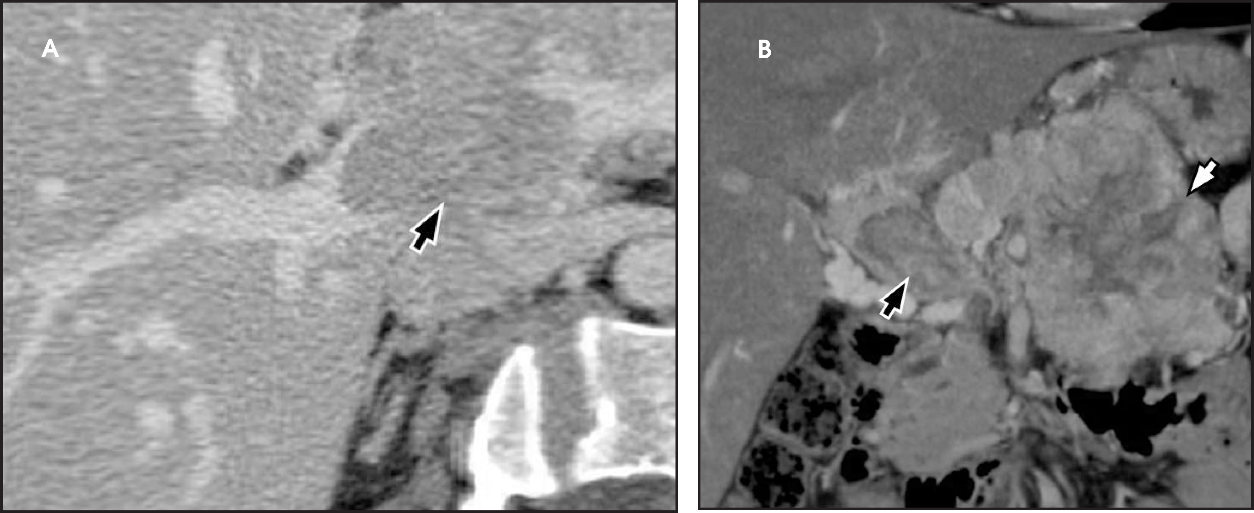
Another uncommon appearance of islet-cell tumors is pancreatic duct involvement. While mild mass effect upon the pancreatic duct is commonly seen, occasionally significant narrowing or pancreatic duct obstruction and subsequent dilatation can be present (Figure 30). Obstruction may be so severe that intrahepatic biliary ductal dilatation may also be present (Figure 31). In these cases, the degree of obstruction is again usually less than would be expected with adenocarcinoma.
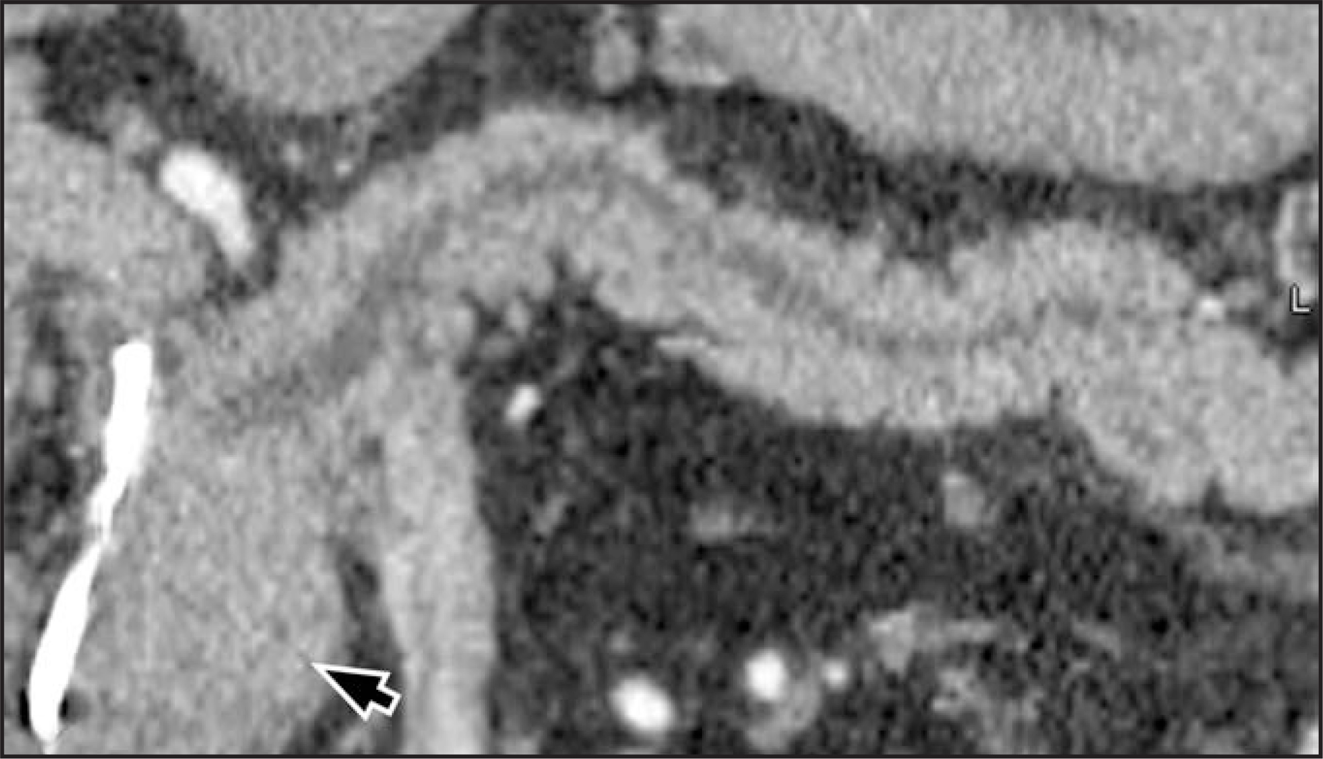
Once the diagnosis and staging of a pancreatic islet-cell tumor is performed, imaging is helpful in determining the success of surgical treatment and monitoring for signs of recurrence. Recurrence may occur locally in the postoperative site, present as regional lymphadenopathy, or appear as distant metastatic disease in the liver, lungs or bone (Figure 32). Patients who are not surgical candidates are also imaged to assess for therapy response (Figure 33).
Conclusion
The radiologist should be familiar with both typical and atypical imaging appearances of pancreatic islet-cell tumors and the appearance of metastatic disease to help determine whether a patient is a surgical candidate. The classic appearance of pancreatic islet-celltumors is a solitary, well-circumscribed hypervascular lesion that does not disrupt the pancreatic duct. Advanced disease may present as regional adenopathy or distant metastases to liver, bone and/or lungs. Atypical appearances include isodense lesions, cystic changeand internal calcifications. Rarely, islet-cell tumors may mimic a pancreatic adenocarcinoma by narrowing or obstructing the pancreatic duct or causing vascular encasement, occlusion or invasion.
Acknowledgments
The authors wish to thank Jeslyn A. Rumbold for editorial assistance.
STATEMENT OF OWNERSHIP MANAGEMENT AND CIRCULATION (REQUIRED BY 39 U.S.C. 3685)
Title of Publication: APPLIED RADIOLOGY
Publication Number: 0160-9963
Filing Date: 11/9/09
Frequency: Monthly except combined
July/August and January/February
Number of Issues Published Annually: 10
Annual Subscription Price: $115
Mailing Address and Business Headquarters:
APPLIED RADIOLOGY
Anderson Publishing, Ltd.
180 Glenside Avenue
Scotch Plains, NJ 07076
Publisher: O. Oliver Anderson
Editor: Stuart E. Mirvis, MD, FACR
Managing Editor: Mark Palacio
Owners: O. Oliver Anderson
Brenda M. Anderson
| Average # copies for 12-month period | Actual # copies for September 2009 issue | |
|---|---|---|
| Total # copies (Net Press Run) | 24,121 | 23,693 |
| Paid and/or Requested Circulation through | ||
| Dealers | – | – |
| Mail subscriptions | 12,482 | 12,197 |
| Total Paid and/or Requested | 12,482 | 12,197 |
| Outside County Nonrequested Distribution | 10,922 | 10,907 |
| Free Distribution Outside of Mail | 295 | 200 |
| Total Nonrequested Distribution | 11,217 | 11,107 |
| Total Distribution | 23,699 | 23,304 |
| Copies Not Distributed | 422 | 389 |
| Return from News Agents | – | – |
| TOTAL | 24,121 | 23,693 |
| Percent Paid and/or Requested Circulation | 52.7% | 52.3% |
| I certify these statements are correct and complete. | ||
| O. Oliver Anderson, Publisher | ||
| November 9, 2009 | ||
| PS Form 3526-R |
References
- Kumar V, Fausto N. Robbins Basic Pathology. 2007.
- Bartsch D, Schilling T, Ramaswamy A. Management of nonfunctioning islet cell carcinomas. World J Surg. 2000;24:1418-1424.
- Jensen R. Pancreatic endocrine tumors: Recent advances. Ann Oncol. 1999;10(Suppl 4):170-176.
- Haubrich W, Berk J. Endocrine Tumors of the Pancreas, Bockus Gastroenterology. 1995:3002-3034.
- Grant C. Insulinoma. Best Pract Res Clin Gastroenterol. 2005;19:783-798.
- Noone T, Hosey J, Firat Z, Semelka R. Imaging and localization of islet-cell tumours of the pancreas on CT and MRI. Best Pract Res Clin Endocrinol Metab. 2005;19:195-211.
- Power N. Imaging pancreatic islet cell tumours. Imaging. 2002;14:147-159.
- Kazanjian K, Reber H, Hines O. Resection of pancreatic neuroendocrine tumors: Results of 70 cases. Arch Surg. 2006;141.
- Fidler J, Fletcher J, Reading C. Preoperative detection of pancreatic insulinomas on multiphasic helical CT. AJR Am J Roentgenol. 2003;181:775-780.
- Gouya H, Vignaux O, Augui J. CT, endoscopic sonography, and a combined protocol for preoperative evaluation of pancreatic insulinomas. AJR Am J Roentgenol. 2003;181:987-992.
- McAuley G, Delaney H, Colville J. Multimodality preoperative imaging of pancreatic insulinomas. Clin Radiol. 2005;60:1039-1050.
- Ichikawa T, Peterson M, Federle M. Islet cell tumor of the pancreas: Biphasic CT versus MR imaging in tumor detection. Radiology. 2000;216:163-171.
- Bilimoria K, Bentrem D, Merkow R. Application of the pancreatic adenocarcinoma staging system to pancreatic neuroendocrine tumors. J Am Coll Surg. 2007;205:558-563.
- Nakakura E, Bergsland E. Islet cell carcinoma: Neuroendocrine tumors of the pancreas and periampullary region. Hematol Oncol Clin North Am. 2007;21.
- Kouvaraki M, Solorzano C, Shapiro S. Surgical treatment of non-functioning pancreatic islet cell tumors. J Surg Oncol. 2005;89:170-185.
- Strosberg J, Hoffe S, Gardner N. Effective treatment of locally advanced endocrine tumors of the pancreas with chemoradiotherapy. Neuroendocrinology. 2007;85:216-220.
- Ho A, Picus J, Darcy M. Long-term outcome after chemoembolization and embolization of hepatic metastatic lesions from neuroendocrine tumors. AJR Am J Roentgenol. 2007;188:1201-1207.
- Horton K, Hruban R, Yeo C, Fishman E. Multi-detector row CT of pancreatic islet cell tumors. Radiographics. 2006;26:453-464.
- King A, Ko G, Yeung V. Dual phase spiral CT in the detection of small insulinomas of the pancreas. Br J Radiol. 1998;71:20-23.
- Van Hoe L, Gryspeerdt S, Marchal G. Helical CT for the preoperative localization of islet cell tumors of the pancreas: Value of arterial and parenchymal phase images. AJR Am J Roentgenol. 1995;165:1437-1439.
- Ascenti G, Visalli C, Genitori A. Multiple hypervascular pancreatic metastases from renal cell carcinoma: Dynamic MR and spiral CT in three cases. Clin Imaging. 2004;28:349-352.
- Buetow P, Parrino T, Buck J. Islet cell tumors of the pancreas: Pathologic-imaging correlation among size, necrosis and cysts, calcification, malignant behavior, andfunctional status. AJR Am J Roentgenol. 1995;165:1175-1179.
Citation
. Islet-cell tumors of the pancreas: Spectrum of MDCT findings. A pictorial essay.. Applied Radiology. 2009;38(11):10-28. doi:10.37549/AR1725.