Magnetic resonance imaging of injury patterns in the athlete’s knee
Applied Radiology — Vol. 32 , Issue 2 , pp. 21 -27
DOI: 10.37549/AR1162
Published: February 1, 2003
Categories
Magnetic resonance imaging (MRI) has become the imaging modality of choice for evaluation of posttraumatic internal derangement of the knee. Although athletes may sustain an “isolated” injury to a specific ligament or meniscus, “combined” injuries to more than one ligament, with or without associated meniscal and/or osseous injuries, are commonly encountered by the orthopedic surgeon and by the radiologist interpreting the patient’s MRI examination. This article reviews specific patterns of combined injuries seen on MRI studies of the knee in athletes, with reference to specific mechanisms of sports-related trauma.
Functional anatomy
The anterior cruciate ligament (ACL) (Figure 1) attaches proximally to the posteromedial aspect of the lateral femoral condyle and distally onto the tibial plateau as a broad expansion in front of the intercondylar eminence. The primary function of the ACL is to resist anterior tibial translation. A secondary function is to resist tibial rotation.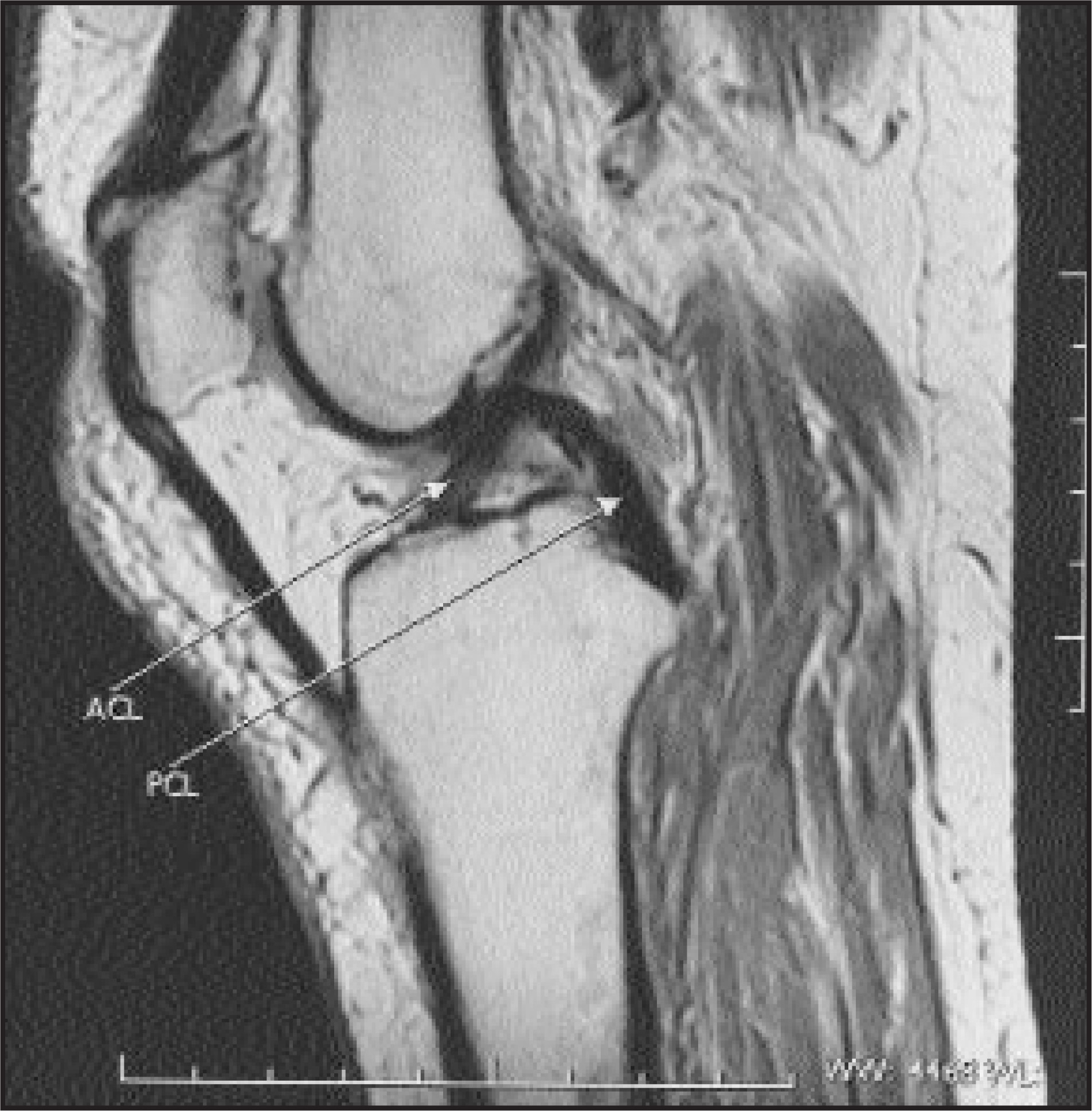
The posterior cruciate ligament (PCL) (Figure 1) attaches proximally to the lateral aspect of the medial femoral condyle and distally to the posterior aspect of the tibia approximately 1 cm below the joint line. The primary function of the ligament is to resist posterior tibial translation, especially when the knee is flexed. A secondary function of the PCL, like the ACL, is to resist tibial rotation.
The medial collateral ligament (MCL) (Figure 2A) extends from the medial femoral condyle at the adductor tubercle to the medial aspect of the tibia several centimeters below the joint line. The ligament has a superficial and a deep layer. When intact, the superficial layer is always visualized on MR images. The deep layer, which attaches to the medial meniscus and the posteromedial capsule, is visualized less consistently. The MCL is the primary stabilizer of the medial aspect of the knee, serving to resist valgus forces as well as rotation forces of the tibia. A secondary function is to resist anterior tibial translation.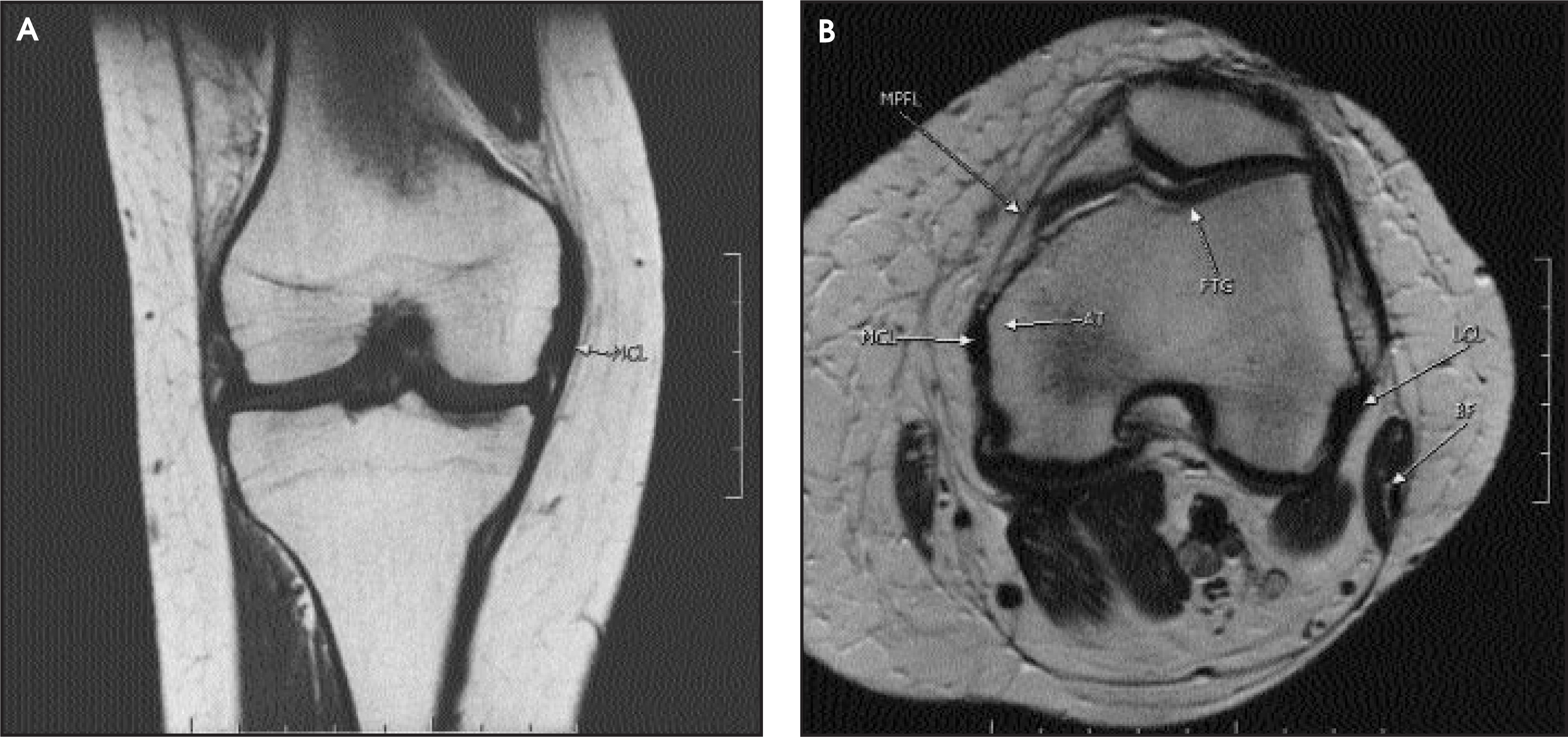
The adductor tubercle (Figure 2B) also serves as a point of attachment for the medial patellofemoral ligament (MPFL), which extends anteriorly to the medial patella. The MPFL is the most important static restraint of lateral patellar displacement.1
The lateral collateral ligament (LCL) proper (also called the fibular collateral ligament or femorofibular ligament) extends from the lateral femoral epicondyle to the posterior aspect of the fibular head (Figure 3A). The primary function of this ligament is to resist varus forces applied to the knee. While a detailed description of the anatomy of the lateral knee is beyond the scope of this article, the reader should be aware that the LCL is only one of several structures, including the iliotibial band (Figure 3B), the lateral patellar retinaculum, the arcuate ligament, the fabellofibular ligament, and the popliteus muscle and tendon, that contribute to the support of the lateral aspect of the knee. These lateral structures also function to limit external rotation of the tibia. Many radiologists refer to the iliotibial band, fibular collateral ligament, and the tendon of the biceps femoris muscle (Figure 3C) as the lateral collateral ligament complex, which is reasonable considering that these three structures are visualized consistently and easily on MR images of the knee. Orthopedic surgeons view the posterolateral region (or posterolateral “corner”) of the knee as a functional tendoligamentous unit, which is termed the arcuate ligament complex. This complex includes the LCL and biceps femoris tendon, as well as the popliteus muscle and tendon, the lateral gastrocnemius muscle, and several capsular ligaments that are often difficult to isolate on MRI. Static stabilizers of the lateral knee include the LCL and the lateral joint capsule. Dynamic stabilizers of the knee include the iliotibial tract, the popiteus muscle/ tendon, and the biceps femoris muscle/tendon.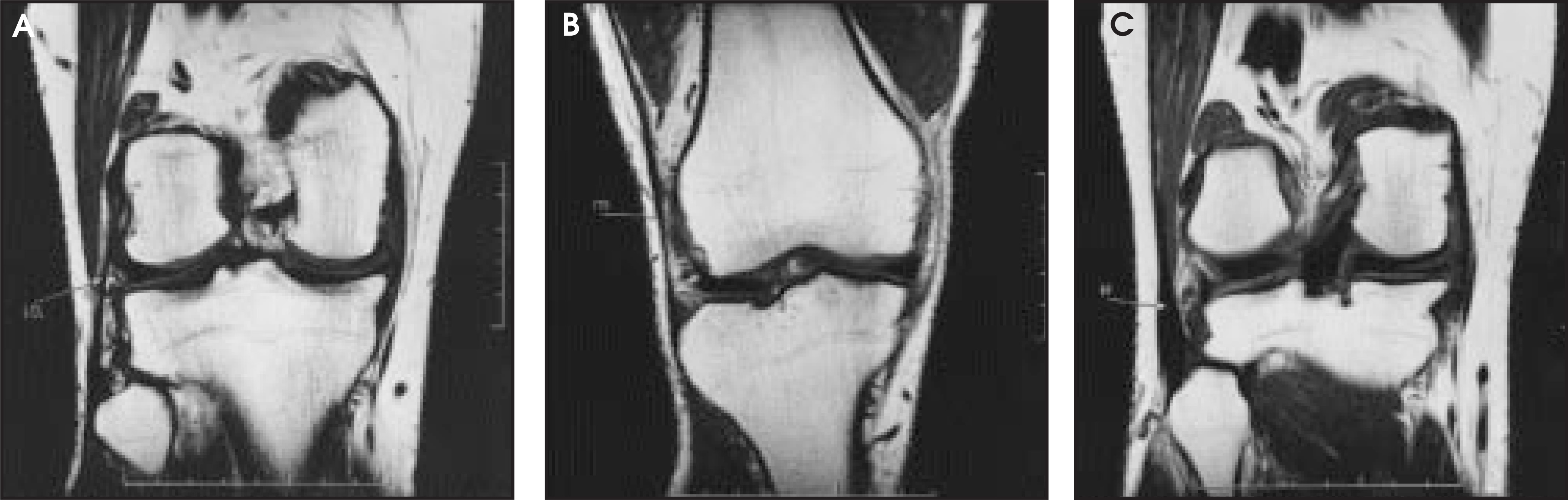
Patterns associated with ACL tears
The association of bone contusions with rupture of the ACL is perhaps the best-known pattern of injury seen on MRI examinations of the knee. Mink and Deutsch2 described this association in 1989. In 1993, Graf et al3 emphasized that the posterior third of the lateral tibial plateau and the middle third of the lateral femoral condyle were particularly susceptible to contusions following an acute ACL tear. These two regions strike one another when the tibia subluxes anteriorly with respect to the femur following ACL rupture, leading to poorly circumscribed regions of edema-type signal in the subchondral marrow (Figure 4). The abnormal signal is of low intensity (relative to marrow) on T1-weighted images (Figure 4B), and increased signal intensity on T2-weighted images (Figure 4D). The signal is often slightly hypointense to marrow on non–fat-suppressed, proton-density–weighted images (Figure 4C). Adding fat suppression increases conspicuity of the high-signal edema on T2-weighted images (Figure 4A). The precise location of the femoral contusion may vary slightly depending upon the degree of flexion of the knee at the time of injury.4 Impaction is rarely severe enough to cause a fracture through the posterior aspect of the lateral tibial plateau. More commonly, a depression along the articular surface of the lateral femoral condyle occurs, which is the MRI correlate of the “lateral notch sign” seen on lateral radiographs of the knee (Figure 4D). Contusions of the lateral tibial plateau and lateral femoral condyle may also be caused by valgus forces that accompany the injury. An additional contusion of the posteromedial tibial plateau is seen occasionally; it is thought to be the result of a “contrecoup” mechanism.4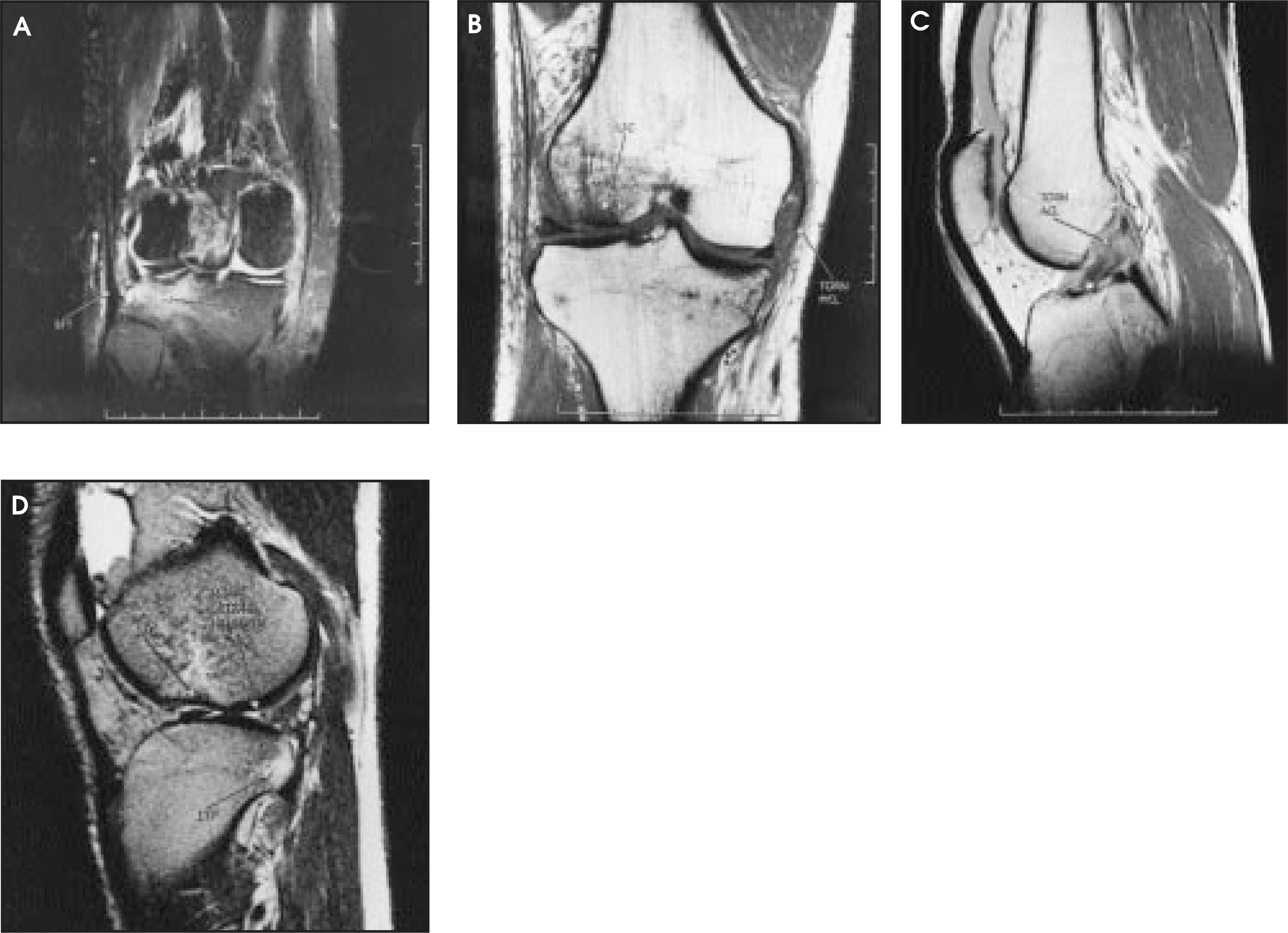
The mechanism of injury of isolated complete ruptures of the ACL is usually deceleration and change of direction, as opposed to contact with another player.5 More commonly, however, ACL tears are associated with injuries to the MCL and/or menisci. Concomitant injuries of the ACL and MCL represent the most common combined lesion occurring in sports, frequent in football, soccer, and basketball players, as well as in skiers.6 Both contact and noncontact injuries are responsible. A blow to the lateral aspect of a fixed, weight-bearing limb results in a valgus force, often with a component of external tibial rotation as well. This mechanism of combined ACL-MCL injury is observed most often in football and soccer players. Noncontact injuries leading to combined ACL-MCL injuries occur when the athlete plants his or her foot on the ground and cuts in the opposite direction, pivoting on the weight-bearing foot. A valgus-external tibial rotation mechanism is common in skiers when the ski tip gets caught in the snow. Motorcyclists may also injure their ACL and MCL when using the right foot as a fulcrum to rapidly turn their vehicle.
On MRI studies, an abnormal-appearing MCL can be classified as 1) sprained, 2) partially torn, or 3) completely torn. These classifications correspond approximately to the three grades of MCL injury described clinically based on physical examination. On T2-weighted images, grade 1 injuries (sprains) show increased signal in the soft tissues around the superficial fibers of the MCL. Grade 2 injuries (partial tears) show increased signal both within and surrounding the ligament. Grade 3 injuries (complete tears) show disruption of the MCL (Figure 4B). An isolated MCL injury does not typically require operative management, as an intact ACL usually provides enough support to allow sufficient healing. In patients with grade 2 or 3 MCL injuries, however, there is a high incidence of ACL tears. Since the ACL acts as a secondary restraint to valgus laxity, lack of structural support from the ACL will adversely affect healing of the MCL. Similarly, since the MCL acts as a secondary stabilizer to anterior tibial translation, a lax MCL can hinder healing of an ACL graft and lead to graft failure.6 In general, if the ACL is torn, and a grade 1 or 2 MCL injury is present, the patient is usually treated with an ACL reconstruction and a valgus support brace with early aggressive rehabilitation. Treatment of combined ACL and grade 3 MCL tears is more controversial. Often the MCL and the ACL will be repaired, particularly if the MCL tear extends into the posteromedial capsule.7
When confronted with a torn MCL and an equivocally torn ACL, the radiologist may use several clues to help ascertain ACL integrity. The presence, or absence, of effusion is important (Figure 4C). Frequently, isolated MCL injury is not associated with significant effusion. If the ACL is also torn, significant effusion is usually present (although with complete MCL tears, effusion may be smaller than expected owing to loss of capsular integrity).8 The status of the menisci is also important. The presence of an associated medial meniscal tear is rare with an isolated MCL injury but meniscal injuries are common with combined ACL-MCL injury.
O’Donoghue9 described a triad of injury involving the ACL, MCL, and the medial meniscus. More recent studies have revealed, however, that the lateral meniscus is torn more frequently than is the medial meniscus in patients with combined ACL-MCL injury (Figure 4D).10 Interestingly, patients with combined ACL and grade 3 MCL tears often do not show meniscal injury. Finally, lateral compartment “kissing” bone contusions may be seen in patients with an isolated MCL injury due to a pure valgus injury (usually due to direct contact), but bruising of the posterior aspect of the lateral tibial plateau (Figure 4A) should raise the suspicion of an ACL tear.
While a valgus-external tibial rotation force is a common mechanism for an ACL tear, a varus-internal tibial rotation force can also cause rupture of the ACL, albeit much less commonly. As expected, the varus component of the force may cause injury to the lateral supporting structures of the knee as opposed to the medial structures. The majority of injuries to the lateral structures occur in conjunction with an ACL rupture. When the ACL is stressed in younger patients, however, the ligament may avulse a small fragment of bone from the tibial plateau rather than be torn directly (Figure 5). Isolated fibular collateral ligament injuries are rare, most commonly occurring in wrestlers after straight varus stress. Noncontact injuries of the ACL and LCL, which may occur in sports such as downhill skiing or basketball, result from sharp deceleration with the knee in internal rotation.6 The forward motion of the body forces the tibia forward and in adduction, resulting in an ACL injury. The ACL tear directly leads to impactions of the posterolateral tibia and lateral femoral condyle, mimicking the familiar bone bruises of the combined ACL-MCL injury previously discussed (Figure 5B). The adductors and extensor mechanism, however, place considerable stress on the lateral supporting structures of the knee, which may rupture. Additional areas of bone edema may result from avulsion fractures of the fibular head or the lateral tibial condyle (Figure 5A); the latter is usually a capsular avulsion (Segond) fracture that has a high association with ACL rupture. The posterolateral corner structures, as well as both menisci, are also at risk for injury.11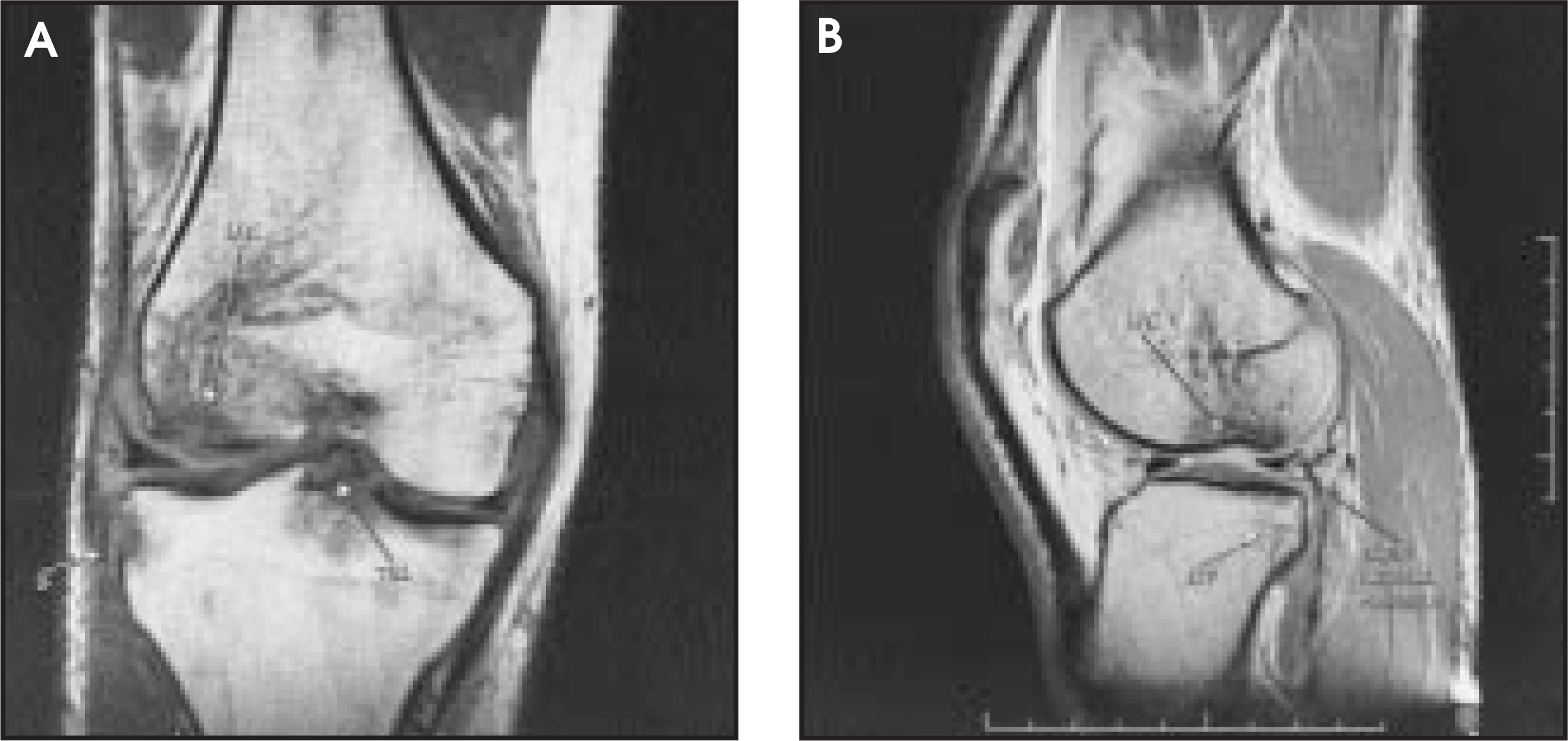
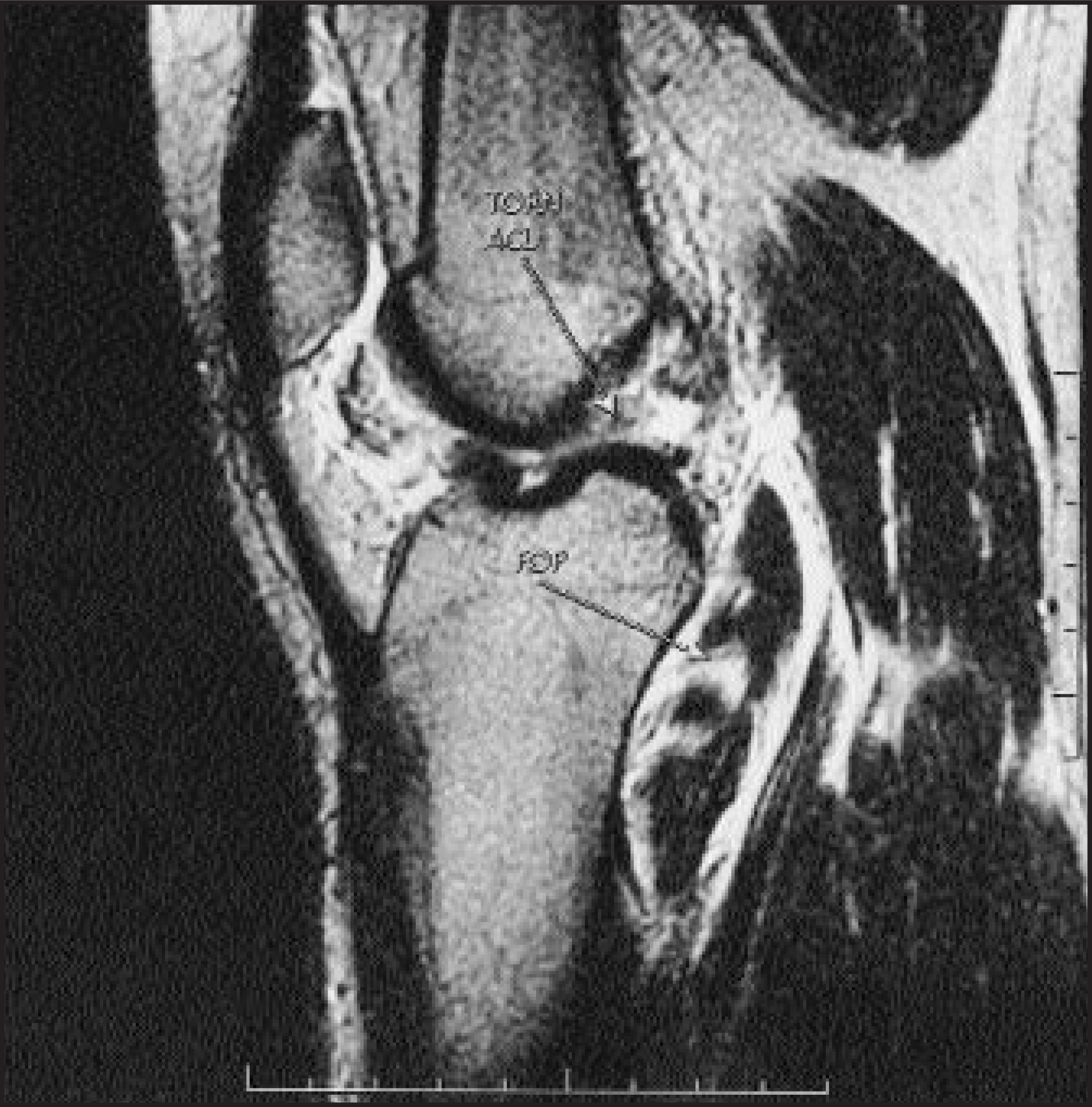
Usually, the typical bone contusions associated with an ACL tear involve the posterolateral tibial plateau and the lateral femoral condyle. However, if the ACL is torn and contusions are identified in the anteromedial tibial plateau and femoral condyle, then a varus-hyperextension injury should be considered (Figure 6). These injuries are often contact injuries that resulted from a blow to the anteromedial aspect of the leg.6 An acute combined ACL-LCL injury can cause immediate and severe disability, with the patient requiring assistance to stand up and leave the playing field. As would be expected with a blow to the anteromedial leg, the structures of the posterolateral corner are at risk of injury. Avulsions of the biceps femoris tendon from the fibular head and injuries to the popliteus muscle (usually at the myotendinous junction) should not be overlooked (Figures 6C and 7). If injuries to the posterolateral corner associated with ACL injury are not recognized, reconstruction of the ACL alone will not always correct the patient’s symptoms of instability. Furthermore, many orthopedic surgeons attempt to treat patients surgically with posterolateral corner injuries acutely to avoid poor results.12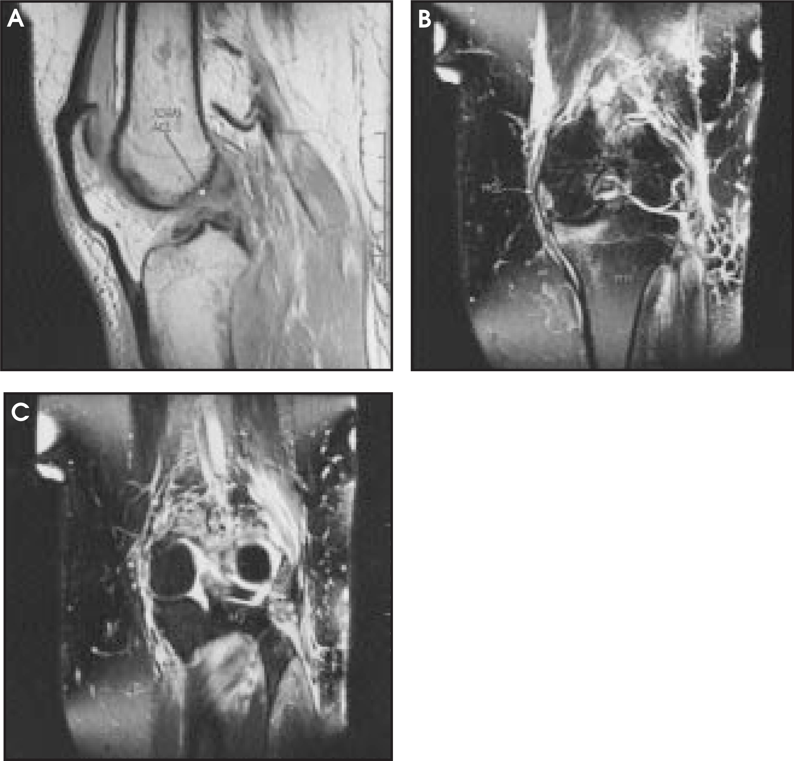
Patterns associated with PCL tears
Injury to the PCL is much less common than injury to the ACL. In the minority of cases, the PCL is injured in isolation (ie, without concomitant ligamentous injury).7 In sports, this usually occurs when the athlete falls forward on a flexed knee with the ipsilateral foot in plantar flexion, applying force to the tibial tuberosity and tearing the PCL.6 A similar posteriorly directed force on the anterior aspect of a flexed knee occurs when the dashboard strikes the knee in a motor vehicle accident. A bone contusion may or may not be seen at the anterior aspect of the proximal tibia (Figure 8). The isolated PCL tear without capsular or other ligament involvement seldom includes a meniscal tear.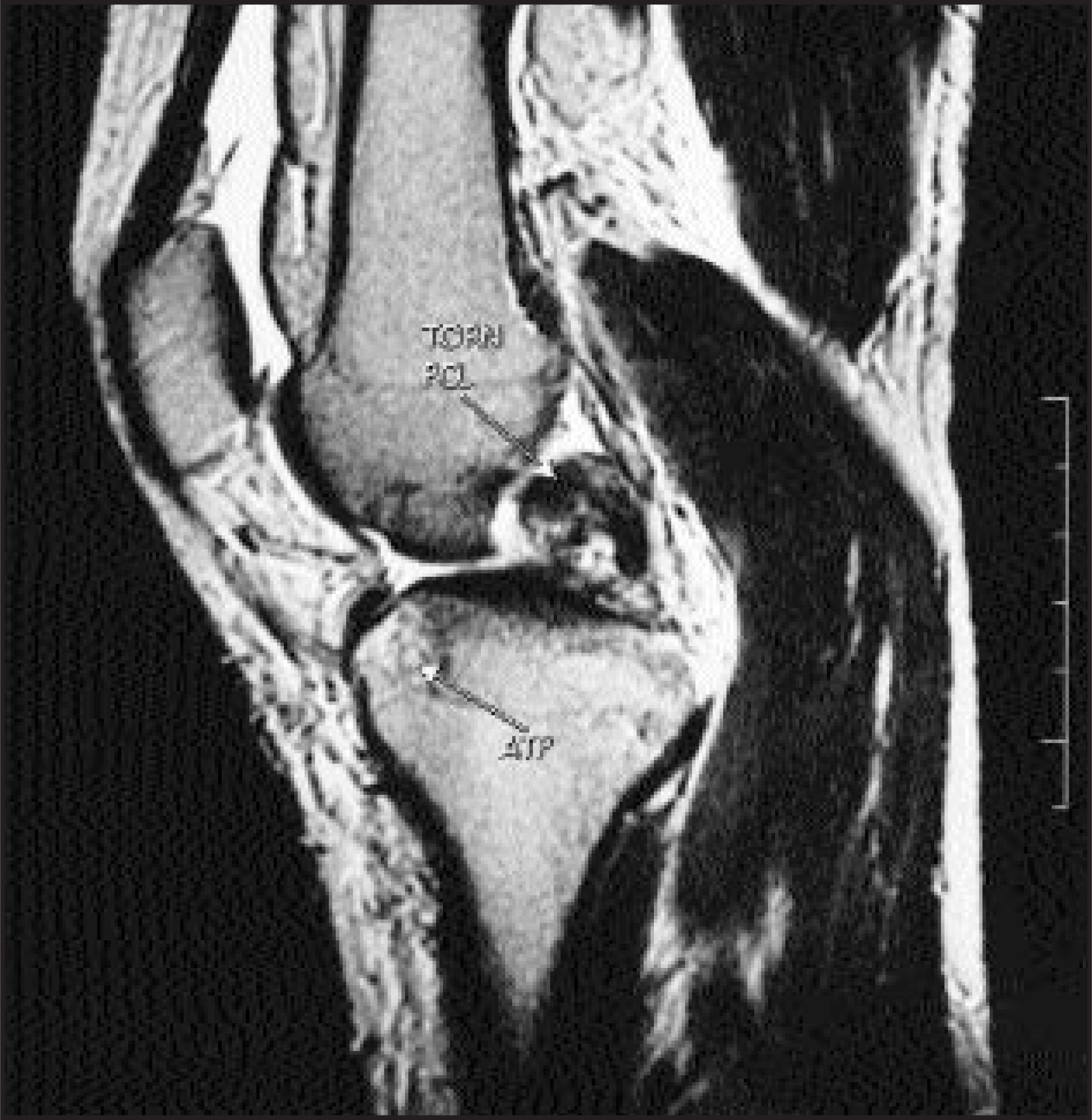
In the majority of cases of PCL disruption, additional structures are injured. In sports, such combined injury occurs when the athlete plants his or her foot on the ground and subsequently falls forward or is struck anteriorly on the knee, resulting in extension of the joint. Although this mechanism may tear the ACL, the PCL, the posterior capsule, and, ultimately, the neurovascular bundle may also be injured. Meniscal tears are seen in 60% of these injuries, with equal involvement of the medial and lateral menisci. Disruptions of the capsule are rare but imply possible injury to the posterior neurovascular bundle; an arteriogram should be considered in such cases. Central anterior tibial and femoral bone contusions may be present with pure hyperextension injuries. Adding a valgus component to the mechanism of injury may result in MCL and/or posteromedial corner injury, and would lateralize the anterior bone bruises (Figure 9). The posterolateral corner should also be scrutinized for injury.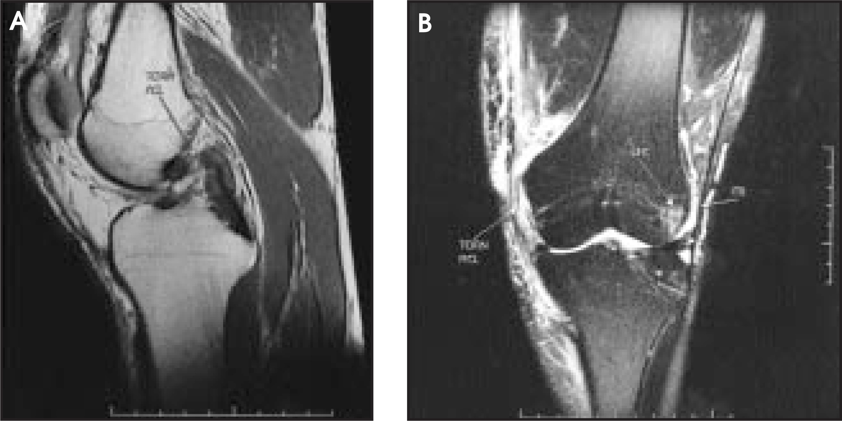
Because of the significant forces associated with PCL injuries, the possibility of a spontaneously reduced knee dislocation should be considered. Knee dislocations may result from tears of the ACL, PCL, and at least one of the collateral ligaments.6 Dislocation represents one of the most serious athletic injuries that can occur to the lower extremity, as it is a major threat to the viability of the lower limb. Reparative ligamentous surgery should be deferred until the arterial status has been determined by observation and/or arteriography.13
Transient lateral patellar dislocation
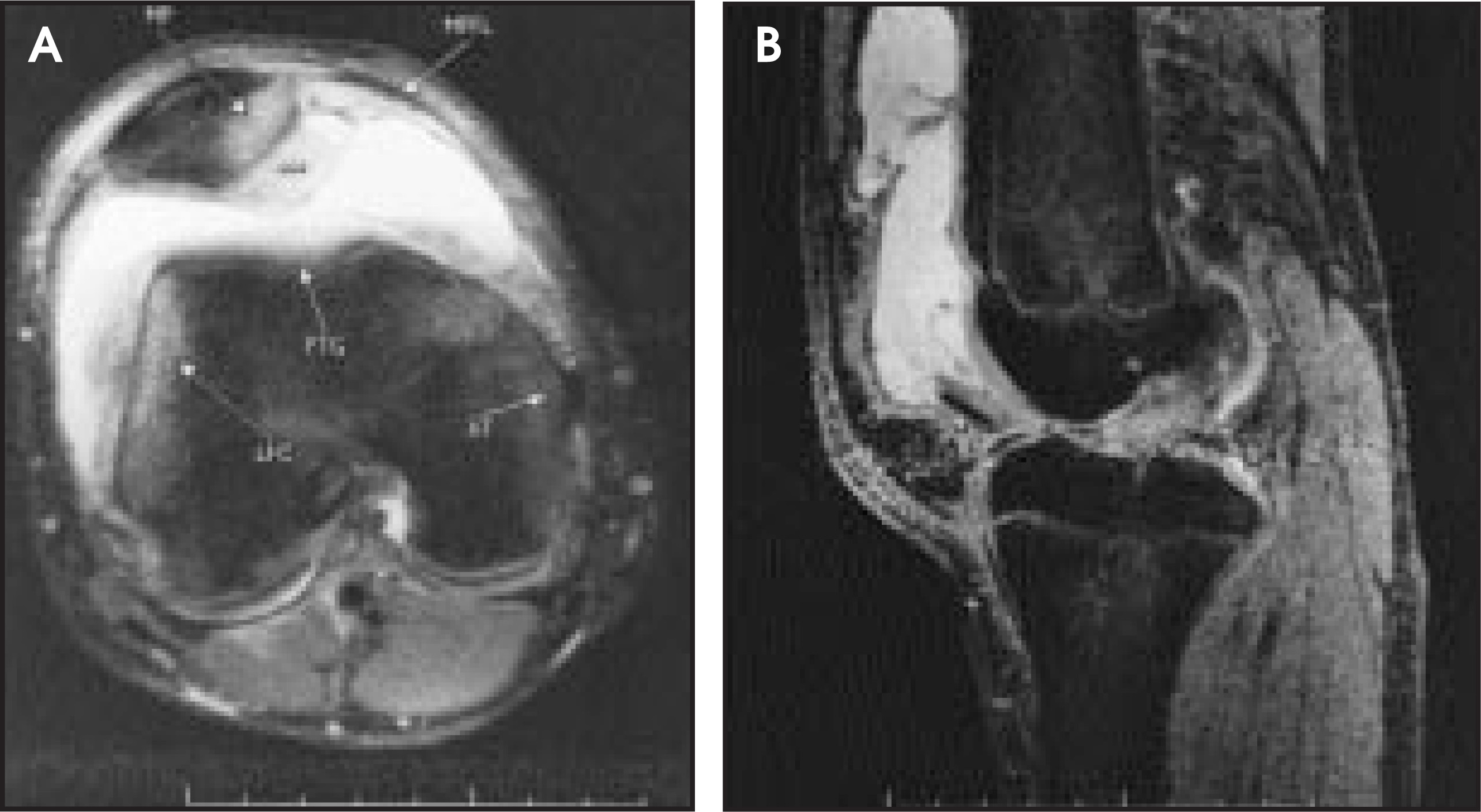
If a contusion of the lateral (or anterolateral) femoral condyle is seen, but a tibial bruise is absent, the radiologist should consider the possibility of a transient lateral patellar dislocation. Acute dislocation occurs as a result of a direct blow to the anteromedial aspect of the patella or as a result of internal rotation of the femur on the fixed tibia during a sudden twisting movement (eg, swinging a bat while the back leg is planted). After dislocation, the patella almost always relocates spontaneously with extension of the knee; actual patellar malalignment may or may not be apparent on MRI. Such dislocations, however, often result in characteristic contusions of the anterolateral femoral condyle and the inferomedial aspect of the patella (Figure 10), which should alert the radiologist to the mechanism of injury. Frank osteochondral injuries of the inferomedial patella should be noted, and displaced intra-articular fragments should be sought (Figure 11). The MPFL, extending from the adductor tubercle to the superior patella may be injured, as can additional retinacular structures, the vastus medialis obliquus, or even the MCL. If transient patellar subluxation is suspected, a search for findings of femoral trochlear dysplasia should be attempted. A shallow trochlear groove (which predisposes one to patellar dislocation) can be diagnosed on MRI if the depth of the groove is <3 mm at a level 3 cm above the femorotibial joint line (Figures 10 and 11).14 In severe cases, the “groove” may actually be convex.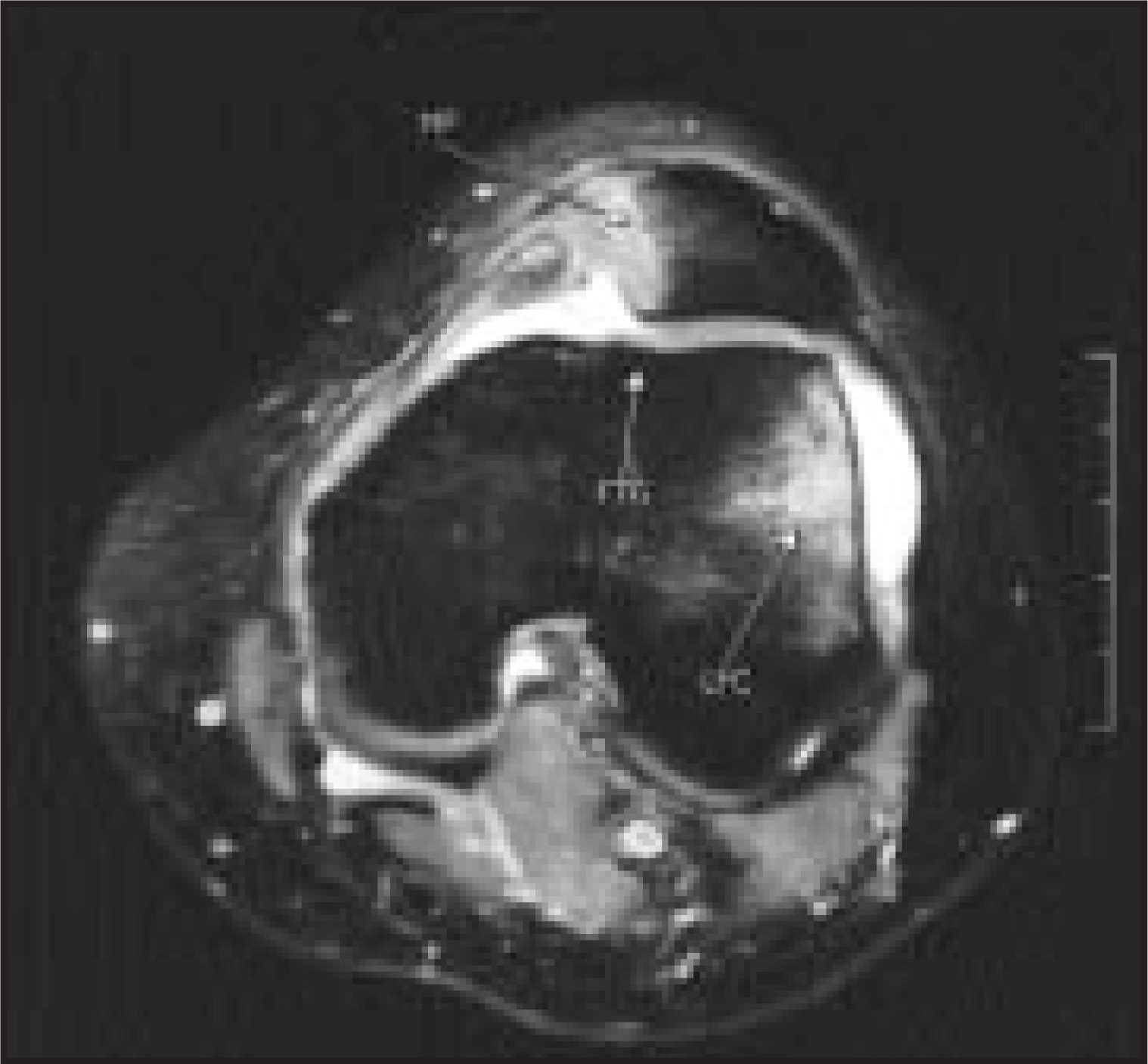
Conclusion
Injury to multiple supporting structures of the knee is common following sports-related trauma. Patterns of injury specific to the mechanism of trauma can often be recognized on MRI of the knee. The locations of bone contusions, for example, often provide a clue to the mechanism of injury, and hence should prompt the radiologist to evaluate specific stabilizing structures. Noting the integrity (or lack thereof) of such structures will greatly assist the referring orthopedic surgeon.AR
References
- Starok M, Lenchik L, Trudell D. Normal patellar retinaculum: MR and sonographic imaging with cadaveric correlation.. AJR Am J Roentgenol.. 1997;168:1493-1499.
- Mink J, Deutsch A. Occult cartilage and bone injuries of the knee: Detection, classification, and assessment with MR imaging.. Radiology.. 1989;170:823-829.
- Graf B, Cook D, DeSmet A. “Bone bruises” on magnetic resonance imaging evaluation of anterior cruciate ligament injuries.. Am J Sports Med.. 1993;21:220-223.
- Sanders T, Medynski M, Feller J. Bone contusion patterns of the knee at MR imaging: Footprint of the mechanism of injury.. RadioGraphics.. 2000;20:S135-S152.
- Feagin J, Curl W. Isolated tear of the anterior cruciate ligament: 5-year follow-up study.. Am J Sports Med.. 1976;4:95-100.
- Aglietti P, Zachcherotti G, DeBiase P, Tria A. Ligaments of the Knee.. 1995:207-259.
- Burger R, Larson R, Larson R, Grana W. The Knee: Form, Function, Pathology, and Treatment.. 1993:513-598.
- Rettig A, Rubinstein R, Scott W. The Knee.. 1994:703-722.
- O’Donoghue D. Surgical treatment of fresh injuries to major ligaments of the knee.. J Bone Joint Surg.. 1950;32A:721-738.
- Shelbourne K, Nitz P. The O’Donoghue triad revisited.. Am J Sports Med.. 1992;19:474-477.
- Hayes C, Brigido M, Jamadar D. Mechanism-based pattern approach to classification of complex injuries of the knee depicted at MR imaging.. RadioGraphics.. 2000;20:S121-S134.
- Veltri D, Warren R. Operative treatment of posterolateral instability of the knee.. Clin Sports Med.. 1994;13:615-627.
- Feagin J. The Crucial Ligaments: Diagnosis and Treatment of Ligamentous Injuries About the Knee.. 1994:93-101.
- Pfirrmann C, Zanetti M, Romero J. Femoral trochlear dysplasia: MR findings.. Radiology.. 2000;216:858-864.
Citation
. Magnetic resonance imaging of injury patterns in the athlete’s knee. Applied Radiology. 2003;32(2):21-27. doi:10.37549/AR1162.