Management of thyroid cancer
Applied Radiology — Vol. 33 , Issue 11 , pp. 34 -45
DOI: 10.37549/AR1296
Published: November 1, 2004
Categories
The incidence of differentiated thyroid cancer (papillary and follicular cancer and their variants) has gradually increased in the last few decades. It is estimated that in 2004 there will be 23,600 new cases of thyroid cancer and 1460 cancer-related deaths in the United States.1 In iodine-sufficient regions, as in the United States, the relative proportions of papillary and follicular cancer are approximately 85% and 15%, respectively. Higher proportions of follicular cancer are seen in areas of iodine deficiency and endemic goiter.
Exposure to radiation is the only known environmental factor associated with thyroid cancer. Accidental ingestion of radioactive iodine, as after the Chernobyl reactor incident, and external neck radiation in childhood have resulted in increased incidence of thyroid cancer, primarily of the papillary type.2,3 The risk of radiation-induced thyroid cancer is greatest in children, presumably due to increased susceptibility of rapidly proliferating cells to mutation and to increased thyroidal trapping of iodine resulting from increased expression of sodium iodide symporter (NIS) mRNA.4
Both papillary and follicular cancer may metastasize to regional or distant sites; regional lymph node metastases occurring more frequently in papillary cancer, and distant metastases, in follicular cancer.5,6 Distant metastases are present in approximately 5% to 10% of patients at initial presentation and 10% to 25% of patients at follow-up. Lung and bone metastases are the most common, although other regions, including the brain, skin, and liver, are not infrequently involved.
Surgery and iodine-131 (I-131) treatment are the mainstays of thyroid cancer management, and they are complementary. The extent of thyroidectomy may infiuence the outcome of subsequent ablative I-131 treatment and long-term prognosis. Additionally, surgical resection of cervical nodal recurrences and of distant metastases, where feasible, are generally associated with a more favorable response to I-131 treatment. The roles of surgery, diagnostic studies, and I-131 treatment are discussed below.
Surgery
Thyroid gland
The extent of thyroidectomy may infiuence the subsequent course of thyroid cancer; total or at least near-total thyroidectomy that leaves only a small portion of one lobe is desirable. Since thyroid cancer is frequently multicentric, total thyroidectomy ensures removal of most, if not all, tumor tissue.7 Moreover, a small remnant is easier to ablate with I-131. Studies of recurrence rates and survival have confirmed the superiority of total over partial thyroidectomy.8,9 Other disadvantages of inadequate surgery include the potential for dedifferentiation of residual tumor, and a “star” artifact at I-131 scintigraphy that may hinder the detection of cervical, mediastinal, and pulmonary metastases.
Gross invasion of extrathyroidal structures by the primary tumor remains a surgical challenge.10,11 Occasionally, the cancer may extend into the major airways, blood vessels, and nerves. Aggressive local disease has a high mortality rate, and frequently may require meticulous shaving of thyroid tissue from vital structures.
Metastases
The neck and mediastinum are frequent sites of lymph node metastases and recurrence, particularly for papillary tumors (Figure 1). Cancer appears to spread first to nodes in the central compartment and lateral compartment on the side of the tumor, and subsequently to nodes in the opposite lateral compartment and mediastinum. Involvement of bilateral cervical and mediastinal lymph nodes is associated with higher risk.6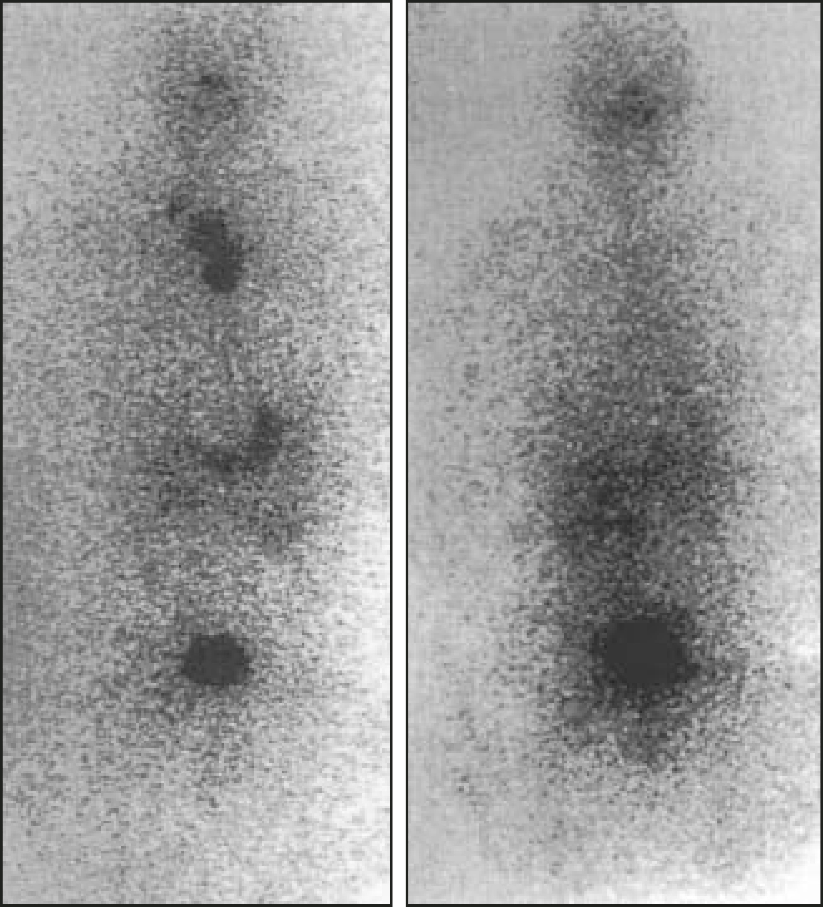
The surgical approach for cervical lymph nodes has varied from “berry-picking” to modified radical neck dissection. Although the impact of these approaches on survival remains controversial, more extensive nodal resections generally result in fewer recurrences and decreased morbidity, and may well be worthwhile, particularly in older patients, who are at higher risk.5,12-14
Distant metastases also may benefit from surgical resection where feasible, particularly if they are large.15 It is difficult, if not impossible, to ablate bulky metastases with I-131, even if they are iodine-avid.
Risk assessment
The clinical course of differentiated thyroid cancer is highly variable, ranging from nearly benign to lethal. Consequently, staging systems that predict the clinical risk play an important role in patient management. Staging is done after the initial surgery for thyroid cancer and includes such prognostic characteristics as age, tumor size, tumor invasion of neck tissues, and distant metastases.6,16-18 The TNM classification (T = primary tumor, N = lymph node metastases, M = distant metastases), developed by the American Joint Committee on Cancer, is used most frequently (Table 1). It takes into account the age, primary tumor size, regional nodal involvement, extrathyroidal invasion, and distant metastases.
Other factors are associated with increased risk and also deserve consideration.5-6,19-20 These include: extensive vascular invasion, brain metastases (compared with other metastases), tumor invasion through the lymph node capsule, certain histological subtypes (eg, Hurthle cell, tall cell, columnar cell, diffuse sclerosing), delay in treatment of ≥1 year after diagnosis, and decreased radioactive iodine uptake or increased fiuorodeoxyglucose (FDG) uptake in metastases (see “Diagnostic evaluation”).
The initial risk assessment as described provides a basis for subsequent diagnostic evaluation and management. The overall prognosis, however, depends not only on the stage of the cancer but also on the treatment. Staging, therefore, should be supplemented by findings at follow-up clinical examinations and diagnostic testing.
Diagnostic evaluation
Radioiodine scintigraphy
Selection of radiotracer—Whole-body imaging with radioiodine generally precedes ablative I-131 therapy, and permits the evaluation of thyroid remnant size and presence of metastases.21 Both I-131, the traditional tracer, and iodine-123 (I-123) have been used for pretherapy imaging, although the relative merits of each continue to be debated. With the use of I-131 imaging, it was found that subsequent uptake of the therapy dose in the thyroid remnant was decreased. This effect was attributed to “stunning” or temporary dysfunction of thyroid tissue, and it was believed to have the potential to decrease the therapeutic efficacy of I-131. More recent studies, however, have improved our understanding of this phenomenon and suggest otherwise.
In addition to stunning, other mechanisms appear to be responsible for the decrease in therapeutic I-131 uptake.22-24 One such mechanism is the destruction of part of the remnant tissue by the diagnostic I-131 dose; another is auto stunning, early damage in the first few hours from the therapy itself, with decrease in I-131 accumulation measured at later intervals. It is also conceivable that these two mechanisms are the predominant reasons for decreased uptake of the therapy dose. Consistent with this premise are recent studies that fail to show an adverse impact of pretherapy I-131 imaging on the outcome of subsequent ablative treatment.22,23
There is another important concern: is I-123 as effective as I-131 in the detection of thyroid tissue? Data obtained from the comparison of sequentially performed I-123 and (pretherapy) I-131 scans suggest that this may not be the case. Using radiotracer amounts in the 2 to 5 mCi range, I-123 appears to be adequate for imaging of neck remnants but is not as sensitive as I-131 for the detection of metastases.25-27 The difference in diagnostic sensitivity is probably related to the longer physical half-life of I-131, which permits imaging at later intervals when target-to-background ratios are higher.25 The efficacy of I-123 may improve if a larger radiotracer amount, (eg, 10 mCi) were used, albeit at a much higher cost, since I-123 is approximately five-fold more expensive than I-131.
Preparation for scintigraphy—Optimal patient preparation is critical for the success of whole-body scintigraphy and ablative I-131 therapy. Preparation is directed primarily at increasing thyroid-stimulating hormone (TSH) levels to stimulate function of tumor foci and enhance the uptake of radioiodine. This can be accomplished by withholding or withdrawing of thyroid hormone, or by the administration of recombinant human TSH (RTSH) (Table 2).
After thyroidectomy, the simplest means of increasing endogenous TSH is withholding of thyroid hormone for 3 to 4 weeks. A level of at least 30 to 50 mU/mL is considered adequate. Generally, higher TSH levels are obtained in a shorter period of time after thyroidectomy than after cessation of chronic suppressive thyroid hormone treatment. Alternatively, triiodothyronine (T3) may be administered postoperatively for 4 weeks, and imaging may be done at approximately 6 weeks. Tri-iodothyronine helps maintain a near-euthyroid state while the circulating thyroxine is eliminated, and it is cleared rapidly from the body when stopped.
For patients on long-term thyroxine (T4) treatment, a number of approaches are possible. First, T4 may be stopped for a period of 5 to 6 weeks. Second, T4 may be replaced by T3, which is stopped after 4 weeks, and imaging is performed at approximately 6 to 7 weeks. Third, RTSH may be administered without stopping T4 treatment. The use of RTSH is discussed later.
A low-iodine diet helps to increase uptake of I-131 in thyroid tissue.28,29 It may be started 1 to 2 weeks prior to radiotracer administration and continued until imaging and treatment are completed. Simplified, rather than stringent, low-iodine diets are more widely used, since they are easier to follow.
Imaging technique—Approximately 4 mCi of I-131 (or I-123) may be used. Imaging is performed at 48 to 96 hours with a high-energy collimator for I-131, and at 24 hours with a low-energy collimator for I-123. Generally, a composite whole-body image is obtained at a scanning speed not exceeding 5 cm/ minute, and it is combined with spot 10-minute images, particularly of the neck and chest.
Posttherapy imaging—The absolute activity in thyroid tissue after I-131 therapy is greater than at diagnostic (pretherapy) imaging. Posttherapy imaging, therefore, is superior for detecting metastases and permits a more complete assessment of the extent of disease both at the initial workup and at follow-up30 (Figure 2). Sarkar and co-workers31 found that posttherapy imaging was three-fold more sensitive for metastases than was diagnostic imaging in patients receiving their first I-131 treatment. The higher count rates from the therapy dose also allow single-photon emission computed tomography (SPECT), so that lesion localization is enhanced.32 Imaging is usually not done until 7 to 10 days after I-131 treatment to ensure optimal target to background ratios.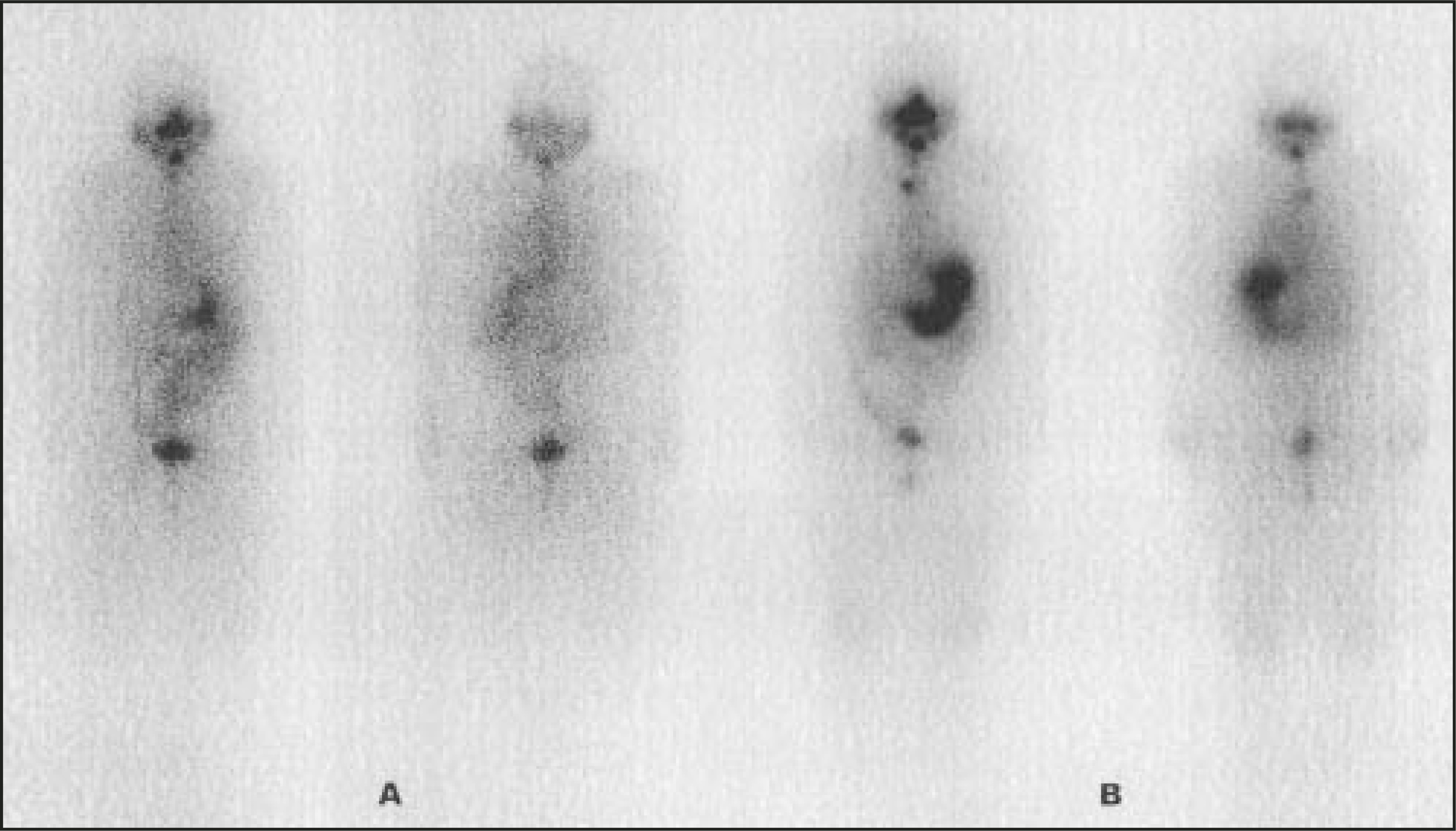
“False-positive” images—Interpretive errors are largely related to the normal physiological distribution of radioiodine.33 Tracer may accumulate in a number of extrathyroidal sites, including the salivary glands, stomach, thymus, and lactating breasts. Activity may be seen in the esophagus related to swallowed saliva, or to such conditions as diverticulum, stricture, Barrett’s esophagus, and gastroesophageal refiux. Activity on the skin may be related to contamination by saliva or urine, secretion by sweat glands (seen characteristically under the hair), and various skin lesions. Other entities associated with radioiodine uptake include infiammatory/ infectious processes, certain tumors, and renal cysts.
Radioiodine uptake and prognosis—Uptake of radioiodine in metastases, together with lesion size, has prognostic significance. For instance, survival rates for patients with small radioiodine-avid pulmonary lesions are significantly better than for those with large tumors that are not visualized at scintigraphy.34 Tumor uptake of radioiodine also appears to be lower in older individuals, consistent with decreased NIS expression and a more aggressive disease course with increasing age.35,36
Tc-99m-methoxyisobutylisonitrile
The traditional myocardial perfusion imaging agents, Tc-99m-methoxyiso-butylisonitrile (MIBI) and Tc-99m-tetrofosmin, also accumulate in thyroid tissue. They may be used to detect lesions not visualized at I-131 scintigraphy.37 The Tc-99m tracers have the advantage that they can be administered in larger amounts (approximately 25 mCi), permitting SPECT and better localization of tumors. Despite the merits of these radiotracers, however, their popularity has waned with the greater availability of positron emission tomography (PET) using FDG, discussed below.
FDG-PET
The use of FDG-PET recently has been extended to the evaluation of thyroid cancer metastases and recurrences.38-40 Occasionally, lesions may accumulate FDG but are not visualized on radioiodine images. This behavior is particularly true of less differentiated and more aggressive tumors. Therefore, FDG-PET complements I-131 for evaluating the extent of disease and prognosis, and is particularly suitable for high-risk patients, who are more likely to have recurrent disease (Figure 3).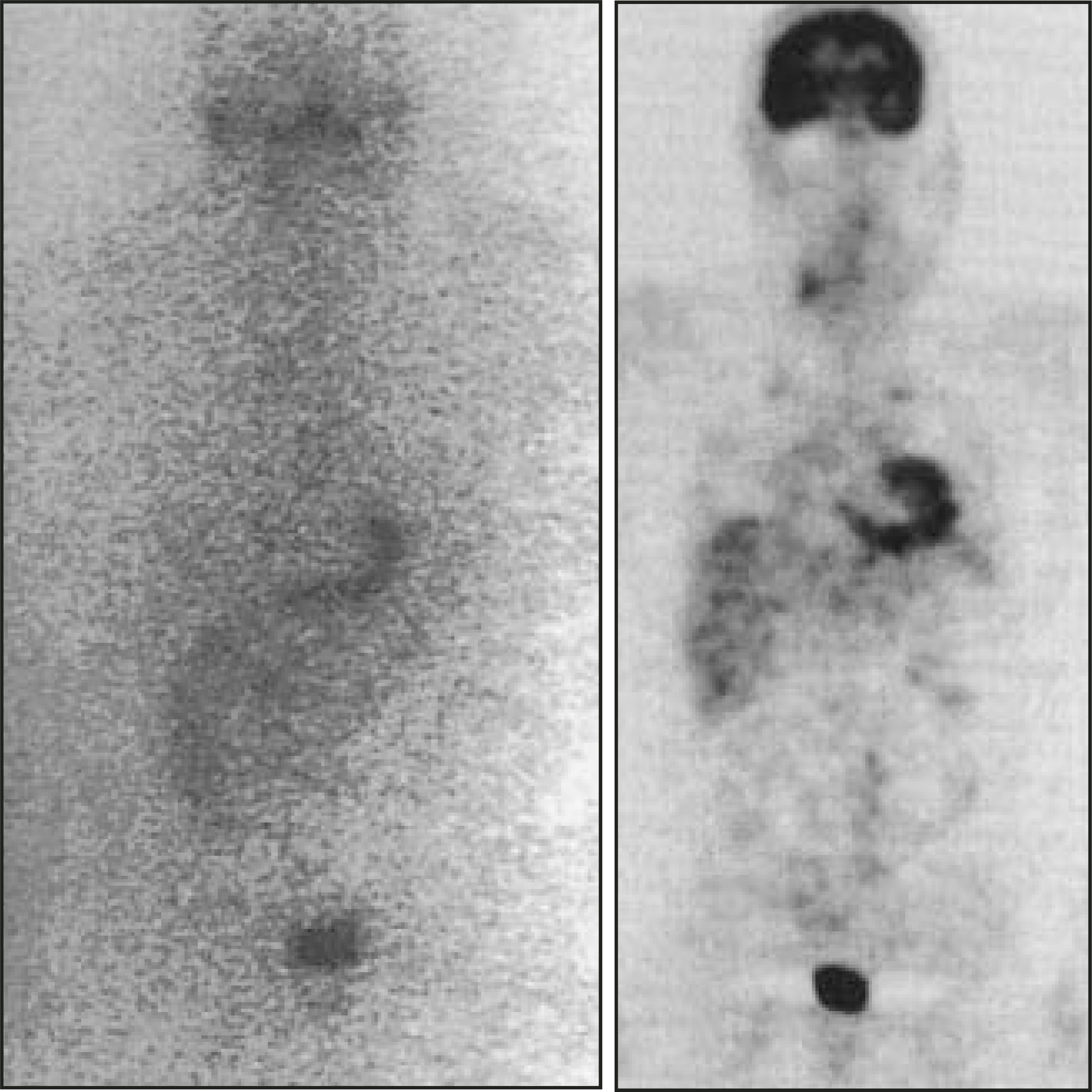
Traditionally, FDG-PET imaging has been performed without stopping thyroid hormone supplements. However, recent studies indicate that high TSH levels significantly improve the diagnostic efficacy,41-43 so that the preparation for FDG-PET should be similar to that for I-131 imaging. The FDG and I-131 studies may be done sequentially after thyroid hormone withdrawal or RTSH administration.
CT, MRI, and ultrasound
In patients at high risk, radioiodine scintigraphy may be combined not only with FDG-PET as noted above, but also with CT and MRI as needed. Gross tumor invasion of the neck should be evaluated with CT/MRI to assess involvement of the larynx, trachea, esophagus, and large vessels.10,44 Imaging with CT also complements FDG-PET for evaluating lung metastases in high-risk cases.38 Vertebral lesions (with the potential for spinal cord compression) and suspected brain metastases require evaluation with MRI imaging because of the potential for tumor swelling and central nervous system complications after I-131 treatment.45 Tumor swelling is also a concern if RTSH is used (see later). CT or MRI may be done at the initial presentation and at follow-up as needed.
Ultrasound of the neck is another imaging option, used primarily at follow-up.46 It offers a relatively inexpensive means of monitoring patients at low risk, in whom recurrences are frequently limited to the lymph nodes. The role of the various imaging modalities is also addressed in the section on “Monitoring and treatment.”
Thyroglobulin
Serum thyroglobulin measurements and imaging play a key role in the follow-up of thyroid cancer.47-49 Thyroglobulin, a glycoprotein stored in the thyroid follicle, is released into the circulation in small quantities from both normal and malignant thyroid tissue. Following thyroidectomy and ablative I-131 treatment, circulating thyroglobulin becomes gradually undetectable, and a subsequent rise signals a recurrence.
The serum thyroglobulin level is most sensitive when TSH is high, and it may be falsely negative if TSH is low, as in patients receiving replacement/suppressive thyroid hormone supplements. In the early period after thyroidectomy and initial I-131 ablation, serum thyroglobulin levels may be positive, despite the absence of persistent disease. They become undetectable over time without further treatment.50 Thus, increasing rather than stable or decreasing thyroglobulin levels following I-131 ablation are a better predictor of persistent disease.
The presence of antithyroglobulin antibody occasionally can be a confounding factor, as it may erroneously increase or decrease the measured thyroglobulin level depending on the assay used.51 Therefore, assays for thyroglobulin levels routinely should be combined with those for antithyroglobulin antibody. These measurements may serve another purpose. The antibody itself may be used as a marker of thyroid tissue, since it refiects the presence of the antigen—thyroglobulin. Antithyroglobulin antibodies disappear from circulation by 2 years after thyroidectomy and successful I-131 ablation, and their reappearance signals a recurrence.51
Recombinant TSH
Adequate TSH stimulation is necessary for I-131 imaging and treatment. While endogenous TSH may be increased by withdrawal of thyroid hormone, the associated hypothyroidism may result in a diminished quality of life and absence from work. Also, patients with serious coexisting medical conditions may not tolerate a prolonged hypothyroid state. The use of RTSH, therefore, offers an attractive alternative to hormone with-drawal.52-54 At the present time, RTSH is approved for diagnostic use only—ie, for the detection of recurrent/persistent disease with stimulated thyroglobulin measurement and scintigraphy—although the compassionate use of RTSH-aided I-131 therapy is permitted in selected patients unable to tolerate a hypothyroid state.55
Since RTSH is approved only for diagnostic evaluation, it is usually limited to patients who are unlikely to harbor active disease, ie, unlikely to require I-131 treatment. Those at low risk by initial staging and with undetectable serum thyroglobulin at low TSH (on levothyroxine treatment) are suitable. Recombinant TSH testing also may be used in patients with moderate- to high-risk stages if at least one prior withdrawal scan is negative and thyroglobulin is undetectable at low TSH.
While RTSH has an established role in the monitoring of differentiated thyroid cancer, a number of limitations are worth noting. First, it is not a perfect substitute for hormone withdrawal. Stimulation of thyroid tissue after RTSH administration is not as intense as after withdrawal, with the result that serum triglyceride (Tg) levels and radioiodine uptake may be lower and lesions occasionally may be missed at scintigraphy.56,57 Why is thyroid stimulation less despite higher serum TSH levels achieved with RTSH? Presumably, the duration of TSH stimulation is also important. With two RTSH injections 24 hours apart, TSH levels remain significantly elevated for only 4 days.53 Notwithstanding this limitation, the value of RTSH has been proven, particularly in low-risk individuals,52-54 and it is likely to play an increasing role in thyroid cancer monitoring.
Serious side effects of RTSH are infrequent. Central nervous system deficits, bone pain, and respiratory difficulty related to rapid swelling of thyroid tissue have been reported.58-60 Needless to say, such complications are largely avoided by careful screening of patients and by limiting the use of RTSH to patients at low risk of metastases/recurrences.
The protocol for the RTSH study is outlined in Table 3. An injection of 0.9 mg RTSH on each of 2 consecutive days is followed 24 hours later by the administration of I-131. Imaging and thyroglobulin measurement are done 2 days after tracer dose. While the standard protocol calls for imaging at 2 days, later imaging is feasible and should be performed if clarification is needed.54
Monitoring and treatment
Serum thyroglobulin is a sensitive marker of thyroid tissue, and an elevated level is frequently the first indicator for recurrent thyroid cancer. Thyroglobulin (with antithyroglobulin antibody) may be measured at 6 and 12 months after the initial I-131 ablation, and yearly thereafter in the absence of active disease. Thyroglobulin levels may be combined with imaging studies for tumor localization. Radioiodine scintigraphy is usually performed every year until the results are negative, and it may be combined with other imaging tests as needed. Examples of management strategies based on thyroglobulin levels and imaging studies are discussed below.
Thyroglobulin elevated at low TSH
If the serum thyroglobulin is increased when the TSH is low (ie, when the patient is on thyroid hormone therapy), a recurrence is likely. In this setting, thyroid hormone is usually withdrawn for tumor localization studies and possible I-131 treatment.
Thyroglobulin undetectable at low TSH
If the serum thyroglobulin is negative during thyroid hormone treatment, a recurrence is not necessarily excluded because thyroglobulin measurements are not sensitive at low TSH.47-49 Whether thyroglobulin should then be measured at high TSH levels depends on the assessed risk. If the risk at initial staging is low and a recurrence is considered unlikely on the basis of prior treatment and diagnostic studies, patients may be followed clinically, or the thyroglobulin level may be obtained after stimulation with RTSH. If the risk and likelihood of recurrence are high, testing after hormone withdrawal is preferable because I-131 treatment may be needed.
Thyroglobulin positive, I-131 scan positive
The combination of these findings confirms the presence and location of cancer, and it is generally followed by I-131 treatment. Prior to treatment, patients may undergo additional imaging with ultrasound, CT, or MRI as needed, and surgical resection of metastases may be performed, where feasible.
Thyroglobulin positive, I-131 scan negative
This is a frequent occurrence, given the generally higher diagnostic sensitivity of thyroglobulin measured at high TSH compared with I-131 scintigraphy. While there is a tendency for all patients with this finding to be grouped together, management should be individualized.
Because of the frequency of cervical nodal recurrence, neck ultrasonography is usually the first step, particularly in low-risk patients. Involved nodes are surgically resected, if possible, before I-131 treatment. For patients at high risk and for those with a negative neck ultrasound, whole-body FDG-PET and CT of the neck and chest may be performed. Evaluation with MRI may be helpful in selected cases with suspected CNS involvement. Distant metastases, particularly if large, should undergo surgical resection or debulking before I-131 therapy. It should be noted that the prognosis tends to be poor for patients with large tumor burdens and negative posttherapy I-131 scans.61,62 Palliative external radiation and embolization are other therapeutic options.63
Thyroglobulin positive, all imaging tests negative
Occasionally, despite persistently elevated thyroglobulin levels, a tumor focus is not found by any imaging means.64 Although empirical treatment with I-131 is an option in such cases, it should be administered only after considering a number of factors: the initial stage of the tumor, the pattern of thyroglobulin levels, the total I-131 dose previously received, and the risk of additional I-131 treatment. For example, clinical follow-up rather than I-131 treatment may be appropriate for a low-risk patient with stable thyroglobulin levels who has had prior I-131 treatment(s).
Therapeutic iodine-131 doses
Remnant ablation
Iodine-131 treatment after thyroidectomy helps destroy residual tumor tissue in the neck, resulting in fewer recurrences and improved survival, particularly for larger primary tumors.5,6,65,66 Elimination of all thyroid tissue, both normal and abnormal, increases the accuracy of serum thyroglobulin measurements and the scintigraphic detection of local recurrence at follow-up. The potential for dedifferentiation of residual tumor is also decreased.
The amounts of I-131 used to ablate residual thyroid tissue have varied over a wide range. The use of “fixed” mCi amounts (eg, 100 mCi or 30 mCi for all patients) is not uncommon. The use of 30 mCi was the result of a previous Nuclear Regulatory Commission ruling disallowing outpatient treatment with larger amounts. An alternative approach is to tailor the dose depending on risk assessment and remnant size. Accordingly, smaller I-131 amounts are appropriate for small remnants and low risk, and vice versa. For instance, 50 mCi of I-131 is adequate for a 2-cm solitary tumor with a small postsurgical remnant, while approximately 150 mCi is appropriate for a tumor with gross invasion of the neck.
Treatment of metastases/recurrences
Compared with the initial remnant ablation, the metastases/recurrences found at follow-up examinations are usually treated with larger amounts of I-131. Administered amounts generally range from 150 to 250 mCi.5-6,65 The therapy dose may be determined in a number of ways. The first method—also the most popular—uses a “fixed-dose” regimen, eg, 150 to 250 mCi for cervical, pulmonary, or skeletal metastases. With the second method, the largest amount of I-131 that delivers no more than 200 rads to the blood is used, assuming the blood dose refiects bone marrow dose.67 The third method quantifies activity in the tumor to derive lesion dosimetry.68 This technique, while theoretically attractive, is feasible only with larger tumors that accumulate sufficient amounts of I-131 to permit accurate uptake measurements. A better approach is the use of PET with I-124.69,70 This method offers greater sensitivity and resolution compared with planar I-131 imaging and dramatic improvement in the ability to quantify up-take in small lesions. Unfortunately, it is not widely available at the present time.
Lithium carbonate may be used to maximize the therapeutic effects of I-131.71 Lithium decreases the release of I-131 from thyroid tissue and, as a result, increases the absorbed radiation dose. The drug may be administered for about 10 days, starting 5 to 7 days before I-131 treatment. Blood lithium levels are monitored to avoid toxicity.
Conclusion
Surgery and I-131 therapy remain the primary means for the management of differentiated thyroid cancer. Treatment can be optimized by the evaluation of clinical risk and selection of appropriate diagnostic studies.
References
- What are the key statistics for thyroid cancer?. 2004.
- Ron E, Lubin J, Shore R. Thyroid cancer after exposure to external radiation: A pooled analysis of seven studies. Radiat Res. 1995;141:259-277.
- Mihailescu D, Shore-Freedman E, Mukani S. Multiple neoplasms in an irradiated cohort: Pattern of occurrence and relationship to thyroid cancer outcome. J Clin Endocrinol Metab. 2002;87:3236-3241.
- Faggiano A, Coulot J, Bellon N. Age-dependent variation of follicular size and expression of iodide transporters in human thyroid tissue. J Nucl Med. 2004;45:232-237.
- Schlumberger M. Papillary and follicular thyroid carcinoma. N Engl J Med. 1998;338:297-306.
- Mazzaferri E, Jhiang S. Long term impact of initial surgical and medical therapy on papillary and follicular thyroid cancer. Am J Med. 1994;97:418-428.
- Pacini F, Elisei R, Capezzone M. Contralateral papillary thyroid cancer is frequent at completion thyroidectomy with no difference in low- and high-risk patients. Thyroid. 2001;11:877-881.
- Duren M, Yavuz N, Bukey Y. Impact on initial surgical treatment on survival of patients with differentiated thyroid cancer: Experience of an endocrine surgery center in an iodine-deficient region. World J Surg. 2000;24:1290-1294.
- Massin J, Savoie J, Garnier H. Pulmonary metastases in differentiated thyroid carcinoma. Study of 58 cases with implications for the primary tumor treatment. Cancer. 1984;53:982-992.
- Kebekew E, Clark O. Locally advanced differentiated thyroid cancer. Surg Oncol. 2003;12:91-99.
- Nishida T, Nakao K, Hashimoto T. Local control in differentiated thyroid carcinoma with extrathyroidal invasion. Am J Surg. 2000;179:86-91.
- Wang T, Dubner S, Sznyter L, Heller K. Incidence of metastatic well-differentiated thyroid cancer in cervical lymph nodes. Arch Otolaryngol Head Neck Surg. 2004;130:110-113.
- McHenry C, Rosen I, Walfish P. Prospective management of nodal metastases in differentiated thyroid cancer. Am J Surg. 1991;162:353-356.
- Musacchio M, Kim A, Vijungco J, Prinz R. Greater local recurrence occurs with “berry picking” than neck dissection in thyroid cancer. Am J Surg. 2003;69:191-196.
- Pak H, Gourgiotis L, Wen-I C. Role of metastasectomy in the management of thyroid carcinoma: The NIH experience. J Surg Oncol. 2003;82:10-18.
- DeGroot L, Kaplan E, McCormick M. Natural history, treatment and course of papillary thyroid carcinoma. J Clin Endocrinol Metab. 1990;71:414-424.
- Hay I, Bergstrahl E, Goellner J. Predicting outcome in papillary thyroid carcinoma: Development of a reliable prognostic scoring system in a cohort of 1,779 patients surgically treated at one institution during 1940-1989. Surgery. 1993;114:1050-1058.
- Dobert N, Menzel C, Oeschger S, Grunwald F. Differentiated thyroid carcinoma: The new UICC 6th edition TNM classification system in a retrospective analysis of 169 patients. Thyroid. 2004;14:65-70.
- Chiu A, Delpassand E, Sherman S. Prognosis and treatment of brain metastases in thyroid carcinoma. J Clin Endocrinol Metab. 1997;82:3637-3642.
- Yamashita H, Noguchi S, Murakami N. Extracapsular invasion of lymph node metastasis is an indication of distant metastasis and poor prognosis in patients with thyroid papillary carcinoma. Cancer. 1997;80:2268-2272.
- Sarkar S, Becker D, Becker K, Bilezikian J, Bremner W. Principles and Practice of Endocrinology and Metabolism. 2001:336-342.
- Bajen M, Mane S, Munoz S. Effect of a diagnostic dose of 185 MBq 131I on postsurgical thyroid remnants. J Nucl Med. 2000;41:2038-2042.
- Morris L, Waxman A, Braunstein G. The nonimpact of thyroid stunning: Remnant ablation rates in 131I-scanned and nonscanned individuals. J Clin Endocrinol Metab. 2001;86:3507-3511.
- Hilditch T, Dempsey M, Bolster A. Self-stunning in thyroid ablation: Evidence from comparative studies of diagnostic 131I and 123I. Eur J Nucl Med Mol Imaging. 2002;29:783-788.
- Sarkar S, Kalapparambath T, Palestro C. Comparison of I-123 and I-131 for whole body imaging in thyroid cancer. J Nucl Med. 2002;43:632.
- Mandel S, Shankar L, Benard F. Superiority of Iodine-123 compared with Iodine-131 scanning for thyroid remnants in patients with differentiated thyroid cancer. Clin Nucl Med. 2001;26:6-9.
- Sarkar S, Kalapparambath T, Palestro C. I-131 versus I-123 for whole body scanning. The authors’ reply. J Nucl Med. 2003;44:1195.
- Morris L, Wilder M, Waxman A. Reevaluation of the impact of a stringent low-iodine diet on ablation rates in radioiodine treatment of thyroid carcinoma. Thyroid. 2001;11:749-755.
- Lakshmanan M, Schaffer A, Robbins J. A simplified low iodine diet in I-131 scanning and therapy of thyroid cancer. Clin Nucl Med. 1988;13:866-868.
- Pacini F, Lippi F, Formica N. Therapeutic doses of I-131 reveal undiagnosed metastases in thyroid cancer patients with detectable serum thyroglobulin levels. J Nucl Med. 1987;28:1888-1891.
- Sarkar S, Palestro C, Love C. Utility of whole body radioiodine scintigraphy (scan) prior to the initial I-131 treatment for differentiated thyroid carcinoma. Proceedings of the 12th International Thyroid Congress. Endocrinol Japonica. 2000;47:P-121.
- Yamamoto Y, Nishiyama Y, Monden T. Clinical usefulness of fusion of 131I SPECT and CT images in patients with differentiated thyroid carcinoma. J Nucl Med. 2003;44:1905-1910.
- Shapiro B, Rufini V, Jarwan A. Artifacts, anatomical and physiological variants, and unrelated diseases that might cause false-positive 131-I scans in patients with thyroid cancer. Semin Nucl Med. 2000;30:115-1132.
- Casara D, Rubello D, Saladini G. Different features of pulmonary metastases in differentiated thyroid cancer: Natural history and multivariate statistical analysis of prognostic variables. J Nucl Med. 1993;34:1626-1631.
- Schlumberger M, Tubiana M, De Vathaire F. Long-term results of treatment of 283 patients with lung and bone metastases from differentiated thyroid carcinoma. J Clin Endocrinol Metab. 1986;63:960-967.
- Ward L, Santarosa P, Granja F. Low expression of sodium iodide symporter identifies aggressive thyroid tumors. Cancer Lett. 2003;200:85-91.
- Lind P, Langsteger W. Technetium-99m-Tetrofosmin whole body scintigraphy in the follow-up of differentiated thyroid carcinoma. J Nucl Med. 1997;38:348-352.
- Dietlein M, Scheidhauer K, Voth E. Fluorine-18 fluorodeoxyglucose positron emission tomography and iodine-131 whole body scintigraphy in the follow-up of differentiated thyroid cancer. Eur J Nucl Med. 1997;24:1342-1348.
- Hooft L, Hoekstra O, Deville W. Diagnostic accuracy of 18F-fluorodeoxyglucose positron emission tomography in the follow-up of papillary or follicular thyroid cancer. J Clin Endocrinol. 2001;86:3779-3786.
- Wang W, Larson S, Fazzari M. Prognostic value of F-18-fluorodeoxyglucose positron emission tomographic scanning in patients with thyroid cancer. J Clin Endocrinol Metab. 2000;85:1107-1113.
- Chin B, Patel P, Cohade C. Recombinant human thyrotropin stimulation of fluoro-D-glucose positron emission tomography uptake in well-differentiated thyroid carcinoma. J Clin Endocrinol Metab. 2004;89:91-95.
- Moog F, Linke R, Manthey N. Influence of thyroid-stimulating hormone levels on uptake of FDG in recurrent and metastatic differentiated thyroid carcinoma. J Nucl Med. 2000;41:1989-1995.
- Petrich T, Borner A, Otto D. Influence of rhTSH on [(18)F] fluorodeoxyglucose uptake by differentiated thyroid carcinoma. Eur J Nucl Med Mol Imaging. 2002;29:641-647.
- Takashima S, Matsushita T, Takayama F. Prognostic significance of magnetic resonance findings in advanced papillary thyroid cancer. Thyroid. 2001;11:1153-1159.
- Datz F. Cerebral edema following iodine-131 therapy for thyroid carcinoma metastatic to the brain. J Nucl Med. 1986;27:637-640.
- Pacini F, Molinaro E, Castagna M. Recombinant human thyrotropin-stimulated serum thyroglobulin combined with neck ultrasonography has the highest sensitivity in monitoring differentiated thyroid carcinoma. J Clin Endocrinol Metab. 2003;88:3668-3673.
- Spencer C, Wang C. Thyroglobulin measurement: Techniques, clinical benefits, and pitfalls. Endocrinol Metab Clin North Am. 1995;24:841-863.
- Freitas J, Gross M, Sarkar S, Sandler M. Diagnostic Nuclear Medicine. 2003:591-606.
- Mazzaferri E, Robbins R, Spencer C. A consensus report of the role of serum thyroglobulin as a monitoring method for low-risk patients with papillary thyroid carcinoma. J Clin Endocrinol Metab. 2003;88:1433-1441.
- Baudin E, Cao C, Cailleux A. Positive predictive value of serum thyroglobulin levels, measured during the first year of follow-up after thyroid hormone withdrawal, in thyroid cancer patients. J Clin Endocrinol Metab. 2003;88:1107-1111.
- Spencer C, Takeuchi M, Kazarosyan M. Serum thyroglobulin autoantibodies: Prevalence, influence on thyroglobulin measurement, and prognostic significance in patients with differentiated thyroid carcinoma. J Clin Endocrinol Metab. 1998;83:1121-1127.
- Ladenson P, Braverman L, Mazzaferri E. Comparison of administration of recombinant human thyrotropin with withdrawal of thyroid hormone for radioactive iodine scanning in patients with thyroid carcinoma. N Engl J Med. 1997;337:888-896.
- Haugen B, Pacini F, Reiners C. A comparison of recombinant human thyrotropin and thyroid hormone withdrawal for the detection of thyroid remnant or cancer. J Clin Endocrinol Metab. 1999;84:3877-3885.
- Sarkar S, Afriyie M, Palestro C. Recombinant human thyroid-stimulating hormone-aided scintigraphy: Comparison of imaging at multiple times after I-131 administration. Clin Nucl Med. 2001;26:392-395.
- Robbins R. Coming of age: Recombinant human thyroid-stimulating hormone as a preparation for 131I therapy in thyroid cancer. J Nucl Med. 2003;44:1069-1071.
- Driedger A, Kotowycz N. Two cases of thyroid carcinoma that were not stimulated by recombinant human thyrotropin. J Clin Endocrinol Metab. 2004;89:585-590.
- Pacini F, Molinaro E, Grazia M. Ablation of thyroid residues with 30 mCi I-131. A comparison in thyroid cancer patients prepared with recombinant human TSH or thyroid hormone withdrawal. J Clin Endocrinol Metab. 2002;87:4063-4068.
- Braga M, Ringel M, Cooper D. Sudden enlargement of local recurrent thyroid tumor after recombinant human TSH administration. J Clin Endocrinol Metab. 2001;86:5148-5151.
- Goffman T, Ioffe V, Tuttle M. Near-lethal respiratory failure after recombinant human thyroid-stimulating hormone use in a patient with metastatic thyroid carcinoma. Thyroid. 2003;13:827-830.
- Vargas G, Uy H, Bazan C. Hemiplegia after thyrotropin alfa in a hypothyroid patient with thyroid carcinoma metastatic to the brain. J Clin Endocrinol Metab. 1999;84:3867-3871.
- Fatourechi V, Hay I, Javedan H. Lack of impact of radioiodine therapy in Tg-positive, diagnostic whole-body scan-negative patients with follicular cell-derived thyroid cancer. J Clin Endocrinol Metab. 2002;87:1521-1526.
- van Tol K, Jager P, de Vries E. Outcome in patients with differentiated thyroid cancer with negative diagnostic whole-body scanning and detectable stimulated thyroglobulin. Eur J Endocrinol. 2003;148:589-596.
- Eustatia-Rutten C, Romijn J, Guijt M. Outcome of palliative embolization of bone metastases in differentiated thyroid carcinoma. J Clin Endocrinol Metab. 2003;88:3184-3189.
- Sisson J, Thompson N, Giordano T. Serum thyroglobulin levels after thyroxine withdrawal in patients with low risk papillary thyroid carcinoma. Thyroid. 2000;10:165-169.
- Beierwaltes W. The treatment of thyroid carcinoma with radioactive iodine. Semin Nucl Med. 1978;8:79-94.
- Klain M, Ricard M, Leboulleux S. Radioiodine therapy for papillary and follicular thyroid carcinoma. Eur J Nucl Med. 2002;29:S479-S485.
- Dorn R, Kopp J, Vogt H. Dosimetry-guided radioactive iodine treatment in patients with metastatic differentiated thyroid cancer: Largest safe dose using a risk-adapted approach. J Nucl Med. 2003;44:451-456.
- Maxon H, Englaro E, Thomas S. Radioiodine-131 therapy for well-differentiated thyroid cancer—a quantitative radiation dosimetric approach: Outcome and validation in 85 patients. J Nucl Med. 1992;33:1132-1136.
- Pentlow K, Graham M, Lambrecht R. Quantitative imaging of Iodine-124 with PET. J Nucl Med. 1996;37:1557-1562.
- Eschmann S, Reischl G, Bilger K. Evaluation of dosimetry of radioiodine therapy in benign and malignant thyroid disorders by means of iodine-124 and PET. Eur J Nucl Med Mol Imaging. 2002;29:760-767.
- Koong S, Reynolds J, Movius E. Lithium as apotential adjuvant to 131I therapy of metastatic, well differentiated thyroid carcinoma. J Clin Endocrinol Metab. 1999;84:912-916.
Citation
. Management of thyroid cancer. Applied Radiology. 2004;33(11):34-45. doi:10.37549/AR1296.