Metaplastic ductal carcinoma of the breast
Applied Radiology — Vol. 37 , Issue 10 , pp. 32 -36
DOI: 10.37549/AR1651
Published: October 1, 2008
Categories
CASE SUMMARY
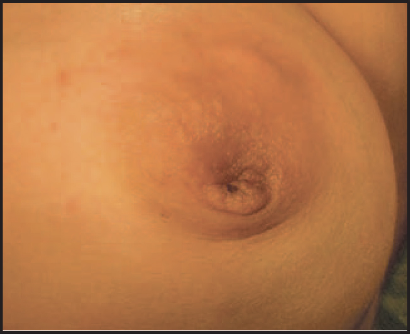
A 48-year-old Hispanic woman with no significant medical history presented with a rapidly enlarging, palpable, painful left breast mass. On examination, the breast mass was located centrally, measured approximately 10 × 9 cm, and was tender to palpation. The nipple was flattened and hyperpigmented (Figure 1). There was no palpable axillary, cervical, or supraclavicular lymphadenopathy.
IMAGING FINDINGS
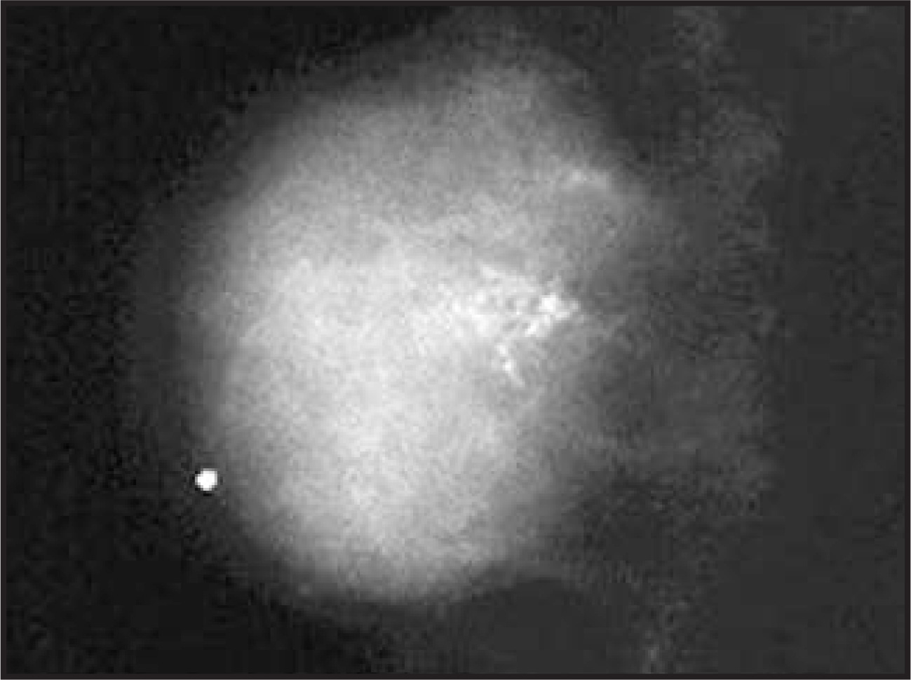
Mammography revealed a large, 4.8-cm, dense, round, well-circumscribed massthat corresponded to the palpable mass (Figure 1). It also showed clustered dystrophic and pleomorphic calcifications at the posterior edge of the mass (Figure 2).
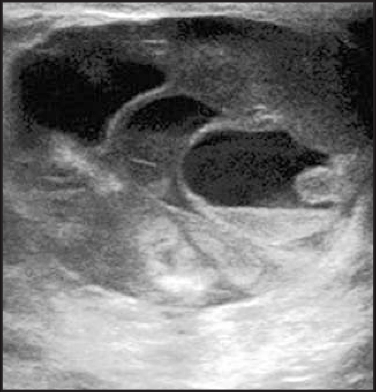
B-mode sonography showed a hypoechoic complex mass at 12 o’clock in the retroareolar position that measured 4.8 × 4.7 × 3.4 cm. The mass had solid and cystic components and multiple fluid-fluid levels. These findings suggested a solid tumor with regions of hemorrhage andcystic necrosis (Figure 3).
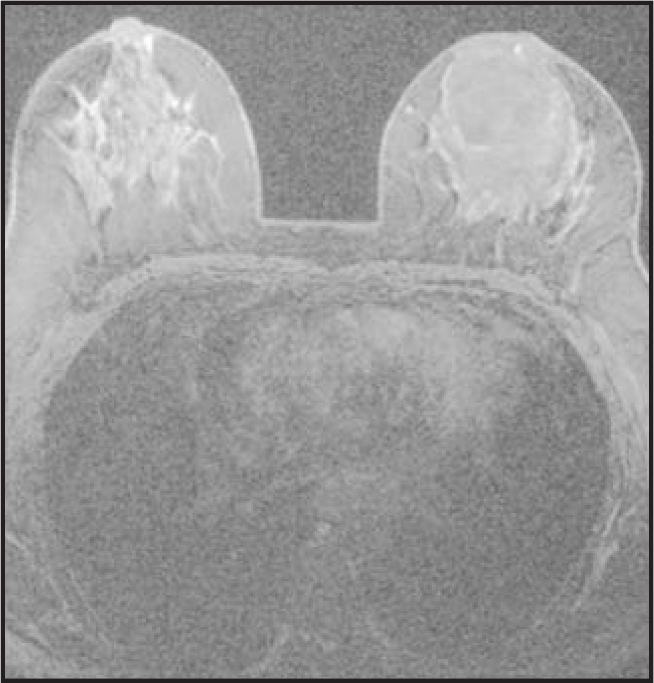
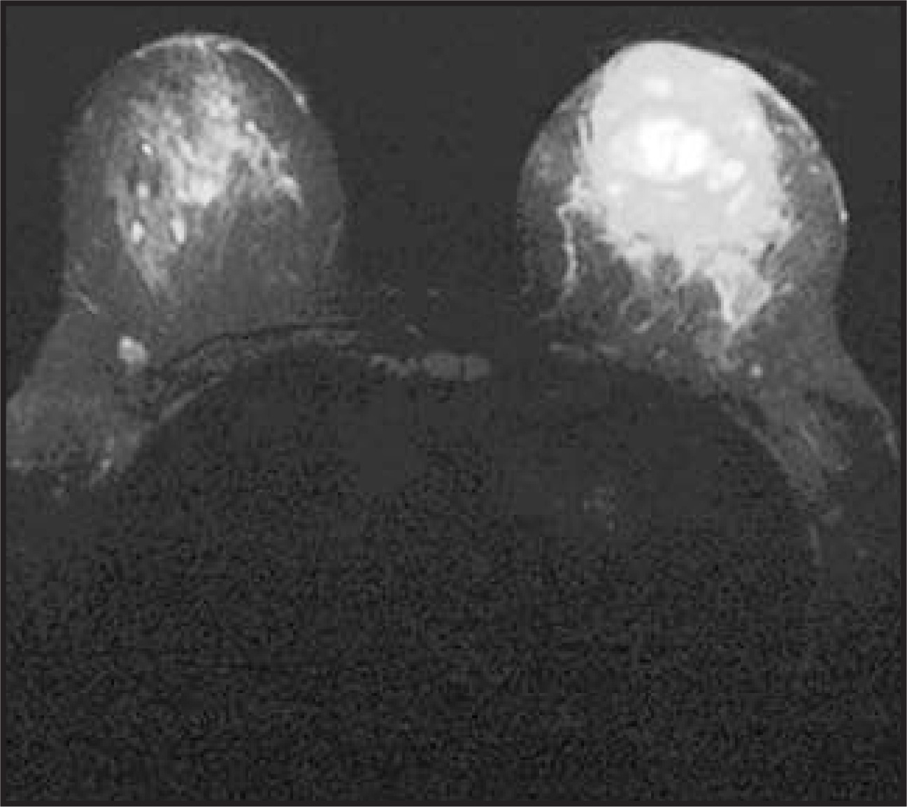
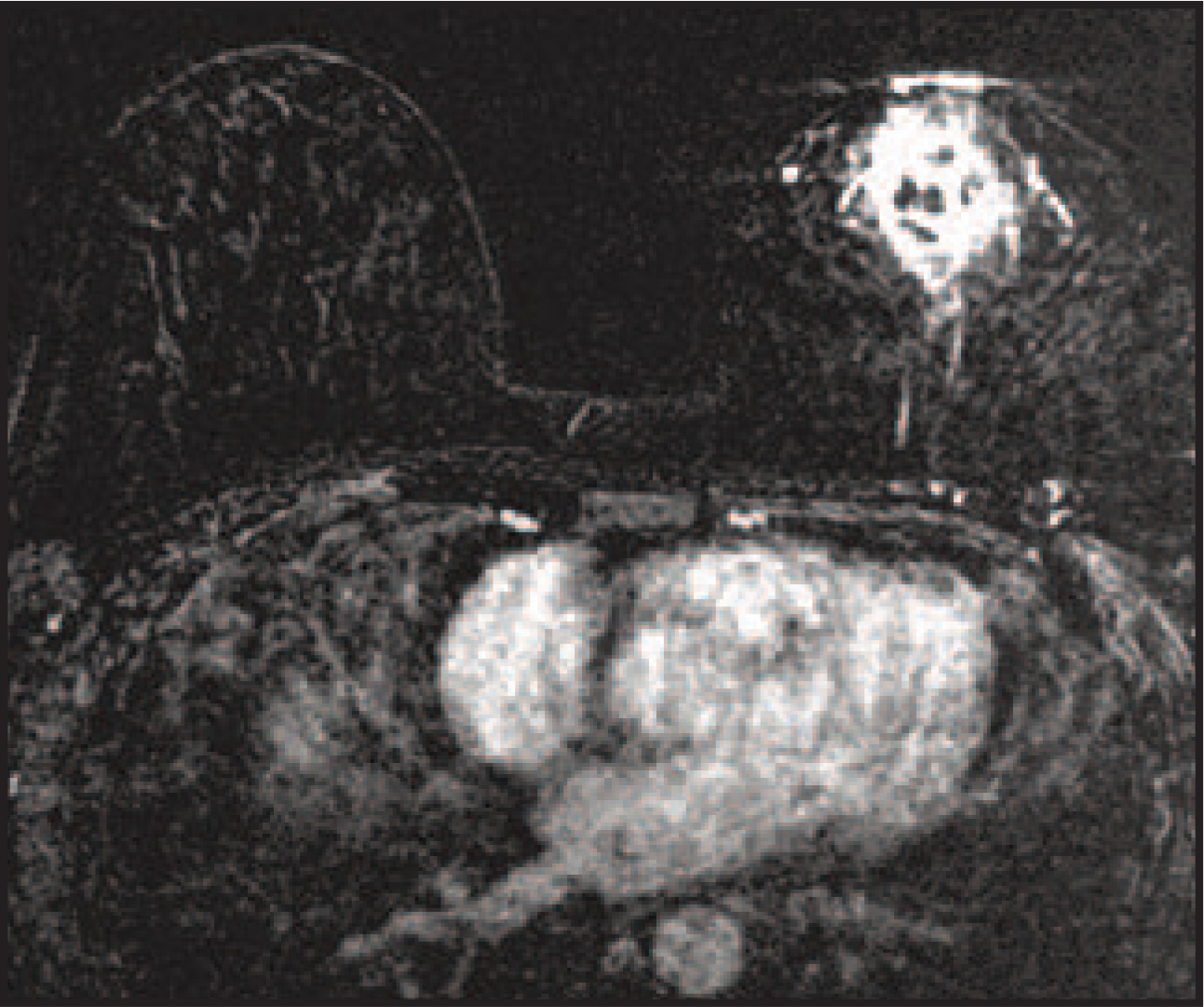
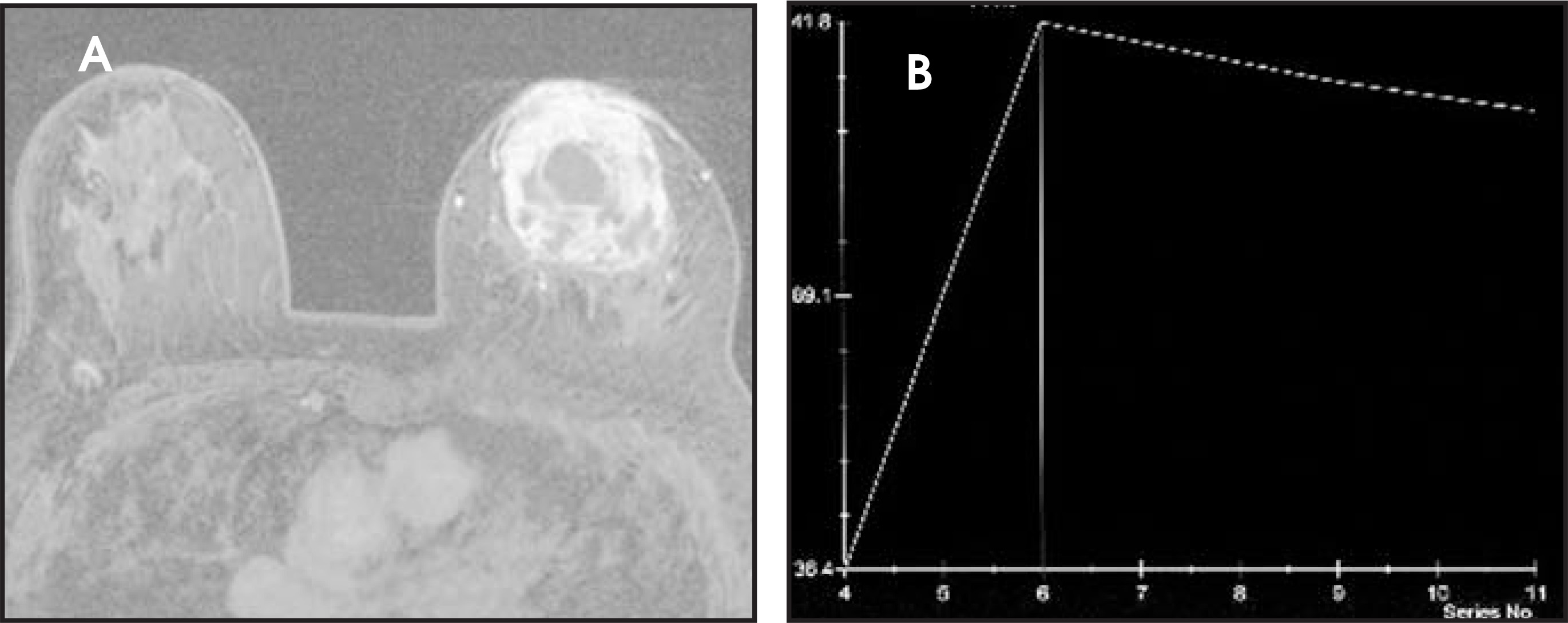
Using a 1.5T magnet with a dedicated breast coil, magnetic resonance imaging (MRI) with gadolinium contrast depicted a large dominant mass in the center of the left breast that measured 5.5 × 6.3 × 6.1 cm. The mass extended anteriorly to the areola. Markedly enlarged vessels circumscribed and traversed this lesion. On T1-weighted (T1W) precontrast fat-suppressed MR images, foci of increased and decreased signal intensity were seen within this hypointense mass. These MRI findings suggested regions of hemorrhage and necrosis, respectively (Figure 4). On short tau inversion recovery (STIR) images, the large mass showed hyperintensity. Areas of especially high signal intensity centrally suggested necrosis (Figure 5). On T1W postcontrast digital subtraction images, this mass exhibited heterogeneous contrast enhancement (Figure 6). Utilizing T1W post contrast images, the mean curve illustrated rapid uptake of contrast followed by rapid washout (Figure 7). These features were all suggestive of malignancy. The breast mass did not extend into the chest wall. No suspicious contralateral breast lesion was identified.
DIAGNOSIS
Metaplastic ductal carcinoma of the breast with a predominant osteosarcomatous differentiation
CASE FOLLOW-UP
Sonographically guided incisional core needle biopsy revealed a malignant spindle cell neoplasm containing osteoclastic-like giantcells with a focal production of osteoid-type matrix.
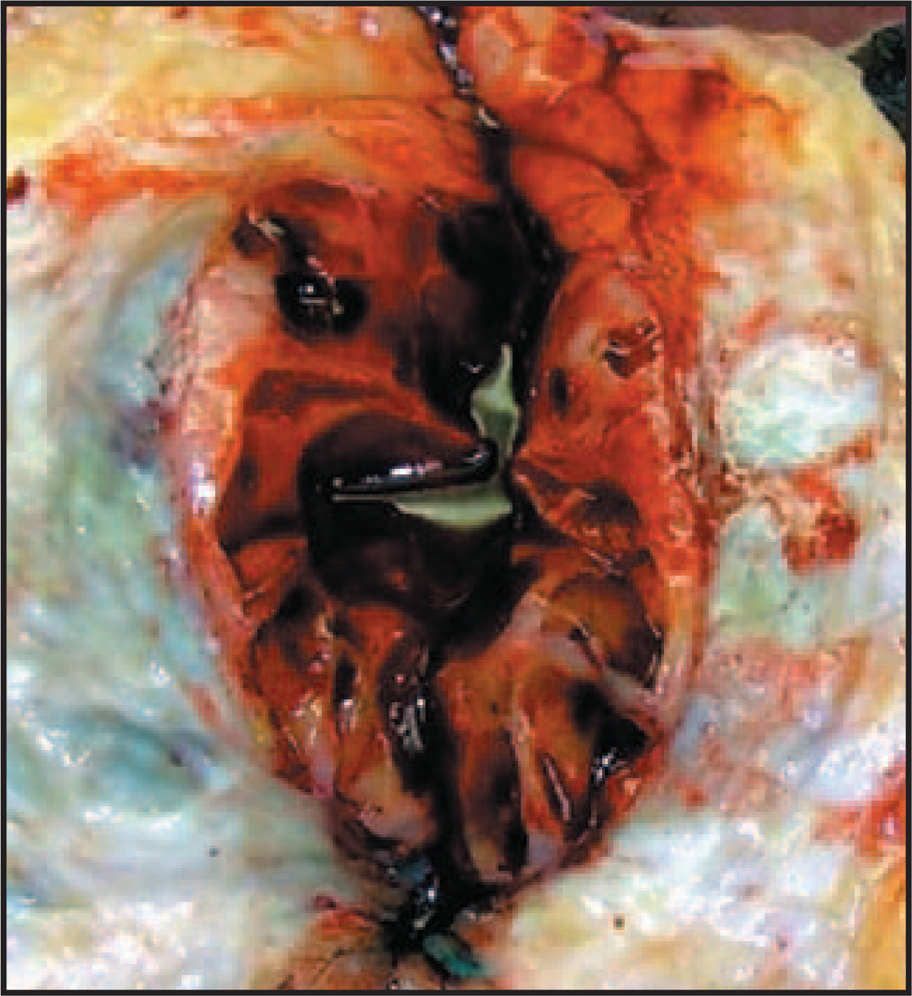
The patient underwent a left total mastectomy. The removed tumor measured 7 cm in greatest dimension and had a significant hemorrhagic component (Figure 8). The surgical margins were negative for malignancy.
The final histopathologic diagnosis was metaplastic carcinoma with a predominant osteosarcomatous differentiation (>99% of thetumor volume) (Figure 9) and minor components of poorly differentiated infiltrating ductal carcinoma, not otherwise specified (Elston Score = 8), and high-grade intraductal carcinoma, cribriform-type with apocrine features (Figure 10).
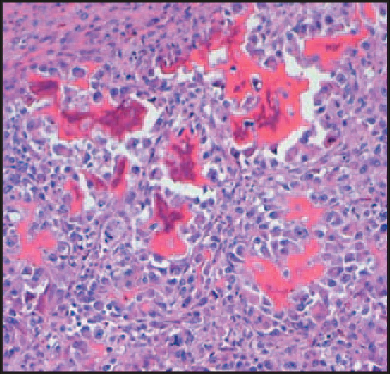
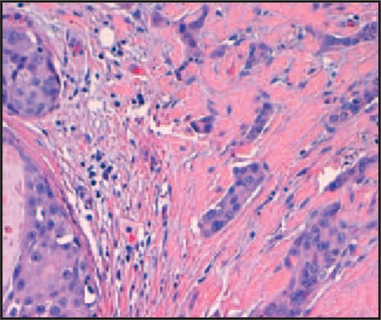
Immunohistochemical stains verified the metaplastic nature of this carcinoma. These tissues were negative for estrogen and progesterone receptors and for the HER-2/neu immunostain.
Left sentinel lymph node excisional biopsy revealed 2 lymph nodes without evidence of local metastatic carcinoma. This finding, combined with the fact that hemorrhage made up much more of the gross tumor volume than actual solid tumor, contributed to the decision not to perform a full axillary dissection. The patient tolerated the surgery well, and received postsurgical chemotherapy and radiation therapy. No distant metastases have been identified, and no local recurrence has been seen.
DISCUSSION
Metaplastic ductal carcinoma of the breast is a well-recognized but rare manifestation of poorly differentiated invasive carcinoma, which contains both epithelial (ductal) and mesenchymal elements, and represents <1% of all breast carcinomas.1-3 Osseous metaplasia is a veryrare manifestation of metaplastic carcinoma, occurring in only 0.2% of all breast carcinomas.4
Clinically, metaplastic carcinoma has similar features to those of breast carcinoma in general.2,3,5,6 As studies of similar cases of metaplastic carcinoma by Günhan-Bilgen et al2(n = 8), Velasco and colleagues3(n = 12), and Patterson and coworkers7(n = 9) found that patients usually present at approximately the age of 50 with a palpable breast mass. Also similar to these series, our patient’s metaplastic tumor was relatively large in size (7 cm), but lacked local or distant metastasis at presentation.2,3,5,7 This case of infiltrating ductal carcinoma with osseous metaplasia is unusual enough to not be definitely identified in any of these larger series.2,3,7 In contrast, the case reported by Evans et al4 of infiltrating ductal carcinoma with osseous metaplasia is most analogous to ours, as their patient exhibited similar clinical features. The 47-year-old woman presented with a palpable mass measuring 5.5 cm.4
Onmammography,well-circumscribedmassestend to indicate benign disease.2,3,5,7 However, metaplastic carcinomas also tend to be well-circumscribed, as in this case. Therefore, it should be included in the differential diagnosis, especially if the mass enlarges rapidly, as in this case, to avoid delay in proper treatment.2-6,8,9 As in the patient reported by Evans et al4 and in all 12 cases in the series from Günhan-Bilgenet al,2 this particular tumor lacked spiculations, a feature that has been previously described for metaplastic carcinoma by Patterson et al7and others.3,8 Such lack of spiculation may result from the fact that this tumor had only a very minor component of infiltrating ductal carcinoma (<1% of tumor volume), which tends to correspond to spiculaton on mammography.7 These findings suggest that shape and border regularity may not be enough to distinguish metaplastic carcinomas mammographically.
Metaplastic carcinoma’s high-density relative to surrounding fibroglandular tissue, as seen in this case, is a mammographic feature with widespread agreement.2,3,7 The especially high density seen here, as well as in Evan’s4 case, likely reflects the significant osseous matrix seen in both cases. Associated calcifications, as seen here, have not been commonly seen, but have been reported by Valasco,3Brenner,8 and Park2,3,7,8,10.
On sonography, this tumor exhibited complexity with both solid and cystic components—features that were also identified in 6 outof 8 ultrasound cases (75%) in the studies by Velasco et al,3 but in only 1 of 8 ultrasound cases (12.5%) in the series reported by Günhan-Bilgen.2 However, even Günhan-Bilgen’s group2 noted that breast masses with solid and cystic components should include metaplastic carcinoma in the differential diagnosis.
On MRI, this highly vascular tumor exhibited heterogeneous contrast enhancement with a rapid uptake of contrast followed by rapid washout, along with associated necrosis and hemorrhage. These features are consistent with a malignant lesion rather than a benign one, which helps to further delineate mammographic and sonographic findings.9 All of the MRI cases reported by Velasco et al3 showed contrast enhancement. Additionally, the necrosis-related T2 hypersignal that is frequently associated with this type of tumor was identified in 11 of 12 of the cases from Velasco et al3 and was also seen in this case. Hence, MRI helps to better characterize metaplastic carcinoma, after mammography and sonography have been performed.3
As Kurian and Al-Nafussi5 note, the main differential pathological diagnosis in this case included primary breast osteosarcoma versus our ultimate diagnosis, metaplastic ductal carcinoma with osteosarcomatous differentiation. This distinction has clinical ramifications affecting surgical treatments and chemotherapy regimens because breast sarcoma metastasizes even less frequently than metaplastic ductal carcinoma.7,11 Sonographically guided core biopsy tissue samples alone could not distinguish between the two. The examination of the much larger surgical specimen revealed that, although the bulk of the tumor represented high-grade osteosarcoma (99% of tumor volume), the presence of adjacent foci of infiltrating ductal carcinoma and high-grade intraductal carcinoma indicated that the tumor was much more likely to be a metaplastic ductal carcinoma with osteosarcomatous differentiation than a primary extraskeletal mammary osteo sarcoma.5
CONCLUSION
Metaplastic ductal carcinoma of the breast accounts for <1% of all breast carcinomas. This case, which involved a predominant osteosarcomatous differentiation, is an even rarer entity. Mammography, sonography, and MRI all play helpful roles in characterizing this tumor before the pathological diagnosis is ultimately made.
References
- Catroppo J, Lara J. Metastatic metaplastic carcinoma of the breast (MCB): An uncharacteristic pattern of presentation with clinicopathologic correlation. Diagn Cytopathol. 2001;25:285-291.
- Günhan-Bilgen I, Memis A, Ustün E. Metaplastic carcinoma of the breast: Clinical, mammograph ic, and sonographic findings with histopathologic correlation. AJR Am J Roentgenol. 2002;178:1421-1425.
- Velasco M, Santamaria G, Ganau S. MRI of metaplastic carcinoma of the breast. AJR Am J Roentgenol. 2005;184:1274-1278.
- Evans H, Shaughnessy E, Nikiforov Y. Infiltrating ductal carcinoma of the breast with osseous metaplasia: Imaging findings with pathologic correlation. AJR Am J Roentgenol. 1999;172:1420-1422.
- Kurian K, Al-Nafussi A. Sarcomatoid/metaplastic carcinoma of the breast: A clinicopathological study of 12 cases. Histopathology. 2002;40(1):58-64.
- Oberman H. Metaplastic carcinoma of the breast: A clinicopathologic study of 29 patients. Am J Surg Pathol. 1987;11:918-929.
- Patterson S, Tworek J, Roubidoux M. Metaplastic carcinoma of the breast: Mammographic appearance with pathologic correlation. AJR Am J Roentgenol. 1997;169:709-712.
- Brenner R, Turner R, Schiller V. Metaplastic carcinoma of the breast: Report of three cases. Cancer. 1998;82:1082-1087.
- Buadu L, Murakami J, Murayama S. Breast lesions: Correlation of contrast medium enhancement patterns on MR images with histopathologic findings and tumor angiogenesis. Radiology. 1996;200:639-649.
- Park J, Han B, Moon W. Metaplastic carcinoma of the breast: Mammographic and sonographic findings. J Clin Ultrasound. 2000;28:179-186.
- Moore M, Kinne D. Breast sarcoma. Surg Clin North Am. 1996;76:383-392.
Citation
. Metaplastic ductal carcinoma of the breast. Applied Radiology. 2008;37(10):32-36. doi:10.37549/AR1651.